 Movie
Movie Controller


+ Open data

- Basic information
| Entry | Database: EMDB / ID: EMD-6914 | |||||||||
|---|---|---|---|---|---|---|---|---|---|---|
| Title | Serial cryotomogram of a metaphase budding yeast cell. | |||||||||
 Map data Map data | Serial cryotomogram of a metaphase budding yeast cell | |||||||||
 Sample Sample |
| |||||||||
| Biological species |  | |||||||||
| Method | electron tomography / cryo EM | |||||||||
 Authors Authors | Ng C / Deng L / Chen C / Lim H / Shi J / Surana U / Gan L | |||||||||
 Citation Citation | Journal: J Cell Biol / Year: 2019 Title: Electron cryotomography analysis of Dam1C/DASH at the kinetochore-spindle interface in situ. Authors: Cai Tong Ng / Li Deng / Chen Chen / Hong Hwa Lim / Jian Shi / Uttam Surana / Lu Gan /  Abstract: In dividing cells, depolymerizing spindle microtubules move chromosomes by pulling at their kinetochores. While kinetochore subcomplexes have been studied extensively in vitro, little is known about ...In dividing cells, depolymerizing spindle microtubules move chromosomes by pulling at their kinetochores. While kinetochore subcomplexes have been studied extensively in vitro, little is known about their in vivo structure and interactions with microtubules or their response to spindle damage. Here we combine electron cryotomography of serial cryosections with genetic and pharmacological perturbation to study the yeast chromosome segregation machinery in vivo. Each kinetochore microtubule has one (rarely, two) Dam1C/DASH outer kinetochore assemblies. Dam1C/DASH contacts the microtubule walls and does so with its flexible "bridges"; there are no contacts with the protofilaments' curved tips. In metaphase, ∼40% of the Dam1C/DASH assemblies are complete rings; the rest are partial rings. Ring completeness and binding position along the microtubule are sensitive to kinetochore attachment and tension, respectively. Our study and those of others support a model in which each kinetochore must undergo cycles of conformational change to couple microtubule depolymerization to chromosome movement. | |||||||||
| History |
|
- Structure visualization
| Movie |
 Movie viewer Movie viewer |
|---|---|
| Supplemental images |

- Downloads & links
-EMDB archive
| Map data | emd_6914.map.gz | 5.1 GB | EMDB map data format | |
|---|---|---|---|---|
| Header (meta data) | emd-6914-v30.xmlemd-6914.xml | 10.8 KB 10.8 KB | Display Display | EMDB header |
| Images | 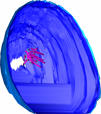 emd_6914.png emd_6914.png | 133.2 KB | ||
| Archive directory |  http://ftp.pdbj.org/pub/emdb/structures/EMD-6914ftp://ftp.pdbj.org/pub/emdb/structures/EMD-6914 http://ftp.pdbj.org/pub/emdb/structures/EMD-6914ftp://ftp.pdbj.org/pub/emdb/structures/EMD-6914 | HTTPS FTP |
-Related structure data
| Related structure data |  6912C C: citing same article ( |
|---|---|
| EM raw data | EMPIAR-10159 (Title: A multi-scale model of the yeast chromosome-segregation system Data size: 95.4 Data #1: Tilt series, tomograms, and models of cryosections of mitotic budding yeast and reconstituted DASH rings around microtubules [tilt series]) |
-Links
| EMDB pages | EMDB (EBI/PDBe) / EMDataResource |
|---|
-Map
| File | Download / File: emd_6914.map.gz / Format: CCP4 / Size: 11.4 GB / Type: IMAGE STORED AS SIGNED BYTE | ||||||||||||||||||||||||||||||||||||||||||||||||||||||||||||||||||||
|---|---|---|---|---|---|---|---|---|---|---|---|---|---|---|---|---|---|---|---|---|---|---|---|---|---|---|---|---|---|---|---|---|---|---|---|---|---|---|---|---|---|---|---|---|---|---|---|---|---|---|---|---|---|---|---|---|---|---|---|---|---|---|---|---|---|---|---|---|---|
| Annotation | Serial cryotomogram of a metaphase budding yeast cell | ||||||||||||||||||||||||||||||||||||||||||||||||||||||||||||||||||||
| Voxel size | X=Y=Z: 8.93 Å | ||||||||||||||||||||||||||||||||||||||||||||||||||||||||||||||||||||
| Density |
| ||||||||||||||||||||||||||||||||||||||||||||||||||||||||||||||||||||
| Symmetry | Space group: 1 | ||||||||||||||||||||||||||||||||||||||||||||||||||||||||||||||||||||
| Details | EMDB XML:
CCP4 map header:
| ||||||||||||||||||||||||||||||||||||||||||||||||||||||||||||||||||||
-Supplemental data
- Sample components
-Entire : S. cerevisiae cell, arrested in metaphase
| Entire | Name: S. cerevisiae cell, arrested in metaphase |
|---|---|
| Components |
|
-Supramolecule #1: S. cerevisiae cell, arrested in metaphase
| Supramolecule | Name: S. cerevisiae cell, arrested in metaphase / type: cell / ID: 1 / Parent: 0 |
|---|---|
| Source (natural) | Organism: |
-Experimental details
-Structure determination
| Method | cryo EM |
|---|---|
 Processing Processing | electron tomography |
| Aggregation state | cell |
-Sample preparation
| Buffer | pH: 7 / Details: YEPD |
|---|---|
| Grid | Model: C-flat-2/0.5 4C / Material: COPPER / Mesh: 150 / Support film - Material: CARBON / Support film - topology: CONTINUOUS / Pretreatment - Type: PLASMA CLEANING / Pretreatment - Atmosphere: AIR Details: plasma cleaned at 15 mA for 45 seconds, carbon side pre-coated with BSA-Gold mixture |
| Vitrification | Cryogen name: ETHANE |
| High pressure freezing | Instrument: OTHER Details: Self-pressurized freezing. The value given for _emd_high_pressure_freezing.instrument is Vitrobot cup. This is not in a list of allowed values set(['LEICA EM PACT2', 'LEICA EM PACT', 'EMS- ...Details: Self-pressurized freezing. The value given for _emd_high_pressure_freezing.instrument is Vitrobot cup. This is not in a list of allowed values set(['LEICA EM PACT2', 'LEICA EM PACT', 'EMS-002 RAPID IMMERSION FREEZER', 'OTHER', 'LEICA EM HPM100', 'BAL-TEC HPM 010']) so OTHER is written into the XML file. |
| Cryo protectant | 25% dextran |
| Sectioning | Ultramicrotomy - Instrument: Leica UC7/FC7 / Ultramicrotomy - Temperature: 123 K / Ultramicrotomy - Final thickness: 100 nm |
| Fiducial marker | Manufacturer: BBI / Diameter: 10 nm |
- Electron microscopy
| Microscope | FEI TITAN KRIOS |
|---|---|
| Image recording | Film or detector model: FEI FALCON II (4k x 4k) / Detector mode: INTEGRATING / Digitization - Dimensions - Width: 4096 pixel / Digitization - Dimensions - Height: 4096 pixel / Digitization - Sampling interval: 14.0 µm / Number grids imaged: 1 / Number real images: 61 / Average exposure time: 1.0 sec. / Average electron dose: 1.6 e/Å2 |
| Electron beam | Acceleration voltage: 300 kV / Electron source:  FIELD EMISSION GUN FIELD EMISSION GUN |
| Electron optics | Calibrated magnification: 15678 / Illumination mode: FLOOD BEAM / Imaging mode: BRIGHT FIELD / Cs: 2.7 mm / Nominal defocus min: 10.0 µm / Nominal magnification: 8700 |
| Sample stage | Specimen holder model: FEI TITAN KRIOS AUTOGRID HOLDER / Cooling holder cryogen: NITROGEN |
| Experimental equipment |  Model: Titan Krios / Image courtesy: FEI Company |
-Image processing
| Final reconstruction | Algorithm: BACK PROJECTION / Software - Name: IMOD (ver. 4.10) / Number images used: 61 |
|---|---|
| CTF correction | Software - Name: eTomo (ver. 4.10) / Details: Phase flipping done in Etomo 4.10 |
