Movie
Movie Controller


+ Open data

- Basic information
| Entry | Database: EMDB / ID: EMD-6640 | |||||||||
|---|---|---|---|---|---|---|---|---|---|---|
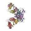
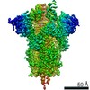
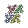
| Title | cryo-EM map of the full-length human NPC1 at 4.4 angstrom | |||||||||
 Map data Map data | Reconstruction of a memebrane protein | |||||||||
 Sample Sample |
| |||||||||
 Keywords Keywords | membrane protein | |||||||||
| Function / homology |  Function and homology information Function and homology informationcyclodextrin metabolic process / cholesterol storage / membrane raft organization / intracellular cholesterol transport / intracellular lipid transport / sterol transport / intestinal cholesterol absorption / LDL clearance / negative regulation of epithelial cell apoptotic process / glycoprotein biosynthetic process ...cyclodextrin metabolic process / cholesterol storage / membrane raft organization / intracellular cholesterol transport / intracellular lipid transport / sterol transport / intestinal cholesterol absorption / LDL clearance / negative regulation of epithelial cell apoptotic process / glycoprotein biosynthetic process / cholesterol transport / cholesterol transfer activity / programmed cell death / bile acid metabolic process / establishment of protein localization to membrane / cholesterol efflux / adult walking behavior / lysosomal transport / cholesterol binding / cellular response to steroid hormone stimulus / negative regulation of macroautophagy / cellular response to low-density lipoprotein particle stimulus / response to cadmium ion / cholesterol metabolic process / negative regulation of TORC1 signaling / neurogenesis / cholesterol homeostasis / macroautophagy / liver development / autophagy / endocytosis / transmembrane signaling receptor activity / nuclear envelope / late endosome membrane / virus receptor activity / signaling receptor activity / gene expression / lysosome / membrane raft / response to xenobiotic stimulus / lysosomal membrane / symbiont entry into host cell / perinuclear region of cytoplasm / Golgi apparatus / endoplasmic reticulum / extracellular exosome / extracellular region / membrane / plasma membrane Similarity search - Function | |||||||||
| Biological species |  Homo sapiens (human) Homo sapiens (human) | |||||||||
| Method | single particle reconstruction / cryo EM / Resolution: 4.43 Å | |||||||||
 Authors Authors | Gong X / Qiang HW / Zhou XH / Wu JP / Zhou Q / Yan N | |||||||||
 Citation Citation | Journal: Cell / Year: 2016 Title: Structural Insights into the Niemann-Pick C1 (NPC1)-Mediated Cholesterol Transfer and Ebola Infection. Authors: Xin Gong / Hongwu Qian / Xinhui Zhou / Jianping Wu / Tao Wan / Pingping Cao / Weiyun Huang / Xin Zhao / Xudong Wang / Peiyi Wang / Yi Shi / George F Gao / Qiang Zhou / Nieng Yan /   Abstract: Niemann-Pick disease type C (NPC) is associated with mutations in NPC1 and NPC2, whose gene products are key players in the endosomal/lysosomal egress of low-density lipoprotein-derived cholesterol. ...Niemann-Pick disease type C (NPC) is associated with mutations in NPC1 and NPC2, whose gene products are key players in the endosomal/lysosomal egress of low-density lipoprotein-derived cholesterol. NPC1 is also the intracellular receptor for Ebola virus (EBOV). Here, we present a 4.4 Å structure of full-length human NPC1 and a low-resolution reconstruction of NPC1 in complex with the cleaved glycoprotein (GPcl) of EBOV, both determined by single-particle electron cryomicroscopy. NPC1 contains 13 transmembrane segments (TMs) and three distinct lumenal domains A (also designated NTD), C, and I. TMs 2-13 exhibit a typical resistance-nodulation-cell division fold, among which TMs 3-7 constitute the sterol-sensing domain conserved in several proteins involved in cholesterol metabolism and signaling. A trimeric EBOV-GPcl binds to one NPC1 monomer through the domain C. Our structural and biochemical characterizations provide an important framework for mechanistic understanding of NPC1-mediated intracellular cholesterol trafficking and Ebola virus infection. | |||||||||
| History |
|
- Structure visualization
| Movie |
Movie viewer |
|---|---|
| Structure viewer | EM map: SurfViewMolmilJmol/JSmol |
| Supplemental images |

- Downloads & links
-EMDB archive
| Map data | emd_6640.map.gz | 28.4 MB | EMDB map data format | |
|---|---|---|---|---|
| Header (meta data) | emd-6640-v30.xmlemd-6640.xml | 12.5 KB 12.5 KB | Display Display | EMDB header |
| Images | 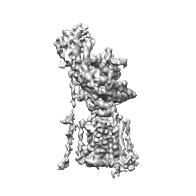 400_6640.gif 400_6640.gif 80_6640.gif 80_6640.gif | 41 KB 2.4 KB | ||
| Archive directory |  http://ftp.pdbj.org/pub/emdb/structures/EMD-6640ftp://ftp.pdbj.org/pub/emdb/structures/EMD-6640 http://ftp.pdbj.org/pub/emdb/structures/EMD-6640ftp://ftp.pdbj.org/pub/emdb/structures/EMD-6640 | HTTPS FTP |
-Related structure data
| Related structure data |  3jd8MC 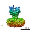 6641C 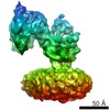 8169C  5jnxC C: citing same article ( M: atomic model generated by this map |
|---|---|
| Similar structure data |


-Links
| EMDB pages | EMDB (EBI/PDBe) / EMDataResource |
|---|---|
| Related items in Molecule of the Month |


-Map
| File | Download / File: emd_6640.map.gz / Format: CCP4 / Size: 29.8 MB / Type: IMAGE STORED AS FLOATING POINT NUMBER (4 BYTES) | ||||||||||||||||||||||||||||||||||||||||||||||||||||||||||||
|---|---|---|---|---|---|---|---|---|---|---|---|---|---|---|---|---|---|---|---|---|---|---|---|---|---|---|---|---|---|---|---|---|---|---|---|---|---|---|---|---|---|---|---|---|---|---|---|---|---|---|---|---|---|---|---|---|---|---|---|---|---|
| Annotation | Reconstruction of a memebrane protein | ||||||||||||||||||||||||||||||||||||||||||||||||||||||||||||
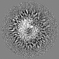
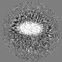
| Projections & slices | Image control
Images are generated by Spider. | ||||||||||||||||||||||||||||||||||||||||||||||||||||||||||||
| Voxel size | X=Y=Z: 1.30654 Å | ||||||||||||||||||||||||||||||||||||||||||||||||||||||||||||
| Density |
| ||||||||||||||||||||||||||||||||||||||||||||||||||||||||||||
| Symmetry | Space group: 1 | ||||||||||||||||||||||||||||||||||||||||||||||||||||||||||||
| Details | EMDB XML:
CCP4 map header:
| ||||||||||||||||||||||||||||||||||||||||||||||||||||||||||||
 Z (Sec.)
 Y (Row.)
 X (Col.)


-Supplemental data
- Sample components
-Entire : NPC1
| Entire | Name: NPC1 |
|---|---|
| Components |
|
-Supramolecule #1000: NPC1
| Supramolecule | Name: NPC1 / type: sample / ID: 1000 / Details: The sample was monodisperse / Number unique components: 1 |
|---|---|
| Molecular weight | Experimental: 140 KDa / Theoretical: 140 KDa |
-Macromolecule #1: Niemann-Pick C1 protein
| Macromolecule | Name: Niemann-Pick C1 protein / type: protein_or_peptide / ID: 1 / Name.synonym: NPC1 / Number of copies: 1 / Oligomeric state: monomer / Recombinant expression: Yes |
|---|---|
| Source (natural) | Organism: Homo sapiens (human) / Organelle: lysosome / Location in cell: lysosome |
| Molecular weight | Theoretical: 140 KDa |
| Recombinant expression | Organism: Homo sapiens (human) / Recombinant cell: HEK 293F / Recombinant plasmid: pCAG |
| Sequence | UniProtKB: NPC intracellular cholesterol transporter 1 |
-Experimental details
-Structure determination
| Method | cryo EM |
|---|---|
 Processing Processing | single particle reconstruction |
| Aggregation state | particle |
-Sample preparation
| Concentration | 15 mg/mL |
|---|---|
| Buffer | pH: 8 / Details: 25mM Tris pH 8.0,150mM NaCl, 0.1% digitonin |
| Grid | Details: Quantifoil R1.2/1.3 copper grid, 200 mesh |
| Vitrification | Cryogen name: ETHANE / Chamber humidity: 100 % / Chamber temperature: 120 K / Instrument: FEI VITROBOT MARK IV / Method: Blot for 3-3.5 seconds before plunging |
- Electron microscopy
| Microscope | FEI TITAN KRIOS |
|---|---|
| Temperature | Min: 80 K / Max: 105 K / Average: 100 K |
| Alignment procedure | Legacy - Astigmatism: Objective lens astigmatism was corrected at 22,500 times magnification |
| Date | Jan 10, 2016 |
| Image recording | Category: CCD / Film or detector model: GATAN K2 SUMMIT (4k x 4k) / Digitization - Sampling interval: 5 µm / Number real images: 4026 / Average electron dose: 50 e/Å2 Details: Every image is the average of 32 frames recorded by the direct electron detector |
| Electron beam | Acceleration voltage: 300 kV / Electron source:  FIELD EMISSION GUN FIELD EMISSION GUN |
| Electron optics | Calibrated magnification: 38270 / Illumination mode: FLOOD BEAM / Imaging mode: BRIGHT FIELD / Cs: 2.7 mm / Nominal defocus max: 3.0 µm / Nominal defocus min: 1.5 µm / Nominal magnification: 22500 |
| Sample stage | Specimen holder: Nitrogen cooled / Specimen holder model: FEI TITAN KRIOS AUTOGRID HOLDER |
| Experimental equipment |  Model: Titan Krios / Image courtesy: FEI Company |
-Image processing
| Details | Iamges were processed using RELION1.4. |
|---|---|
| CTF correction | Details: Each micrograph |
| Final reconstruction | Algorithm: OTHER / Resolution.type: BY AUTHOR / Resolution: 4.43 Å / Resolution method: OTHER / Software - Name: RELION1.4 / Number images used: 102731 |
| Final two d classification | Number classes: 2 |
-Atomic model buiding 1
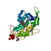
| Initial model | PDB ID: Chain - Chain ID: A |
|---|---|
| Software | Name: Chimera |
| Details | The domains were separately fitted by manual docking |
| Refinement | Space: REAL / Protocol: RIGID BODY FIT |
| Output model | PDB-3jd8: |