Movie
Movie Controller

[English] 日本語
 Yorodumi
- EMDB-66368: CryoEM structure of quinol dependent Nitric Oxide Reductase with BRIL -
+ Open data

- Basic information
| Entry |  | |||||||||
|---|---|---|---|---|---|---|---|---|---|---|
| Title | CryoEM structure of quinol dependent Nitric Oxide Reductase with BRIL | |||||||||
 Map data Map data | ||||||||||
 Sample Sample |
| |||||||||
 Keywords Keywords | quinol-dependent Nitric Oxide Reductase / MEMBRANE PROTEIN | |||||||||
| Function / homology |  Function and homology information Function and homology informationnitric oxide reductase (cytochrome c) / nitric oxide reductase activity / electron transport chain / electron transfer activity / periplasmic space / iron ion binding / heme binding Similarity search - Function | |||||||||
| Biological species |  Achromobacter xylosoxidans (bacteria) Achromobacter xylosoxidans (bacteria) | |||||||||
| Method | single particle reconstruction / cryo EM / Resolution: 3.4 Å | |||||||||
 Authors Authors | Khaja F / Mboukou A / Antonyuk SV / Muench SP / Hasnain SS | |||||||||
| Funding support |  United Kingdom, 1 items United Kingdom, 1 items
| |||||||||
 Citation Citation | Journal: ACS Bio Med Chem Au / Year: 2026 Title: CryoEM Structures of Native Quinol-Dependent Nitric Oxide Reductase in Resting and Quinol-Bound States. Authors: Faisal T Khaja / Allegra Mboukou / Louie P Aspinall / Charlotte E Hawksworth / Robert R Eady / Svetlana V Antonyuk / Stephen P Muench / S Samar Hasnain / Abstract: The membrane-bound quinol-dependent nitric oxide reductases (qNORs), which are members of the respiratory heme-copper oxidase superfamily, are of major importance to food production, environment, and ...The membrane-bound quinol-dependent nitric oxide reductases (qNORs), which are members of the respiratory heme-copper oxidase superfamily, are of major importance to food production, environment, and human health. They are unique to bacteria and catalyze N-N bond formation, converting nitric oxide (NO) to generate the enzymatic product, nitrous oxide (NO), in agricultural and pathogenic conditions. High-resolution qNOR structures have been reported from two bacterial species, in which the molecular size of the protein was increased by the insertion of apocytochrome b (BRIL) at the C-terminus to facilitate cryoEM structure determination. However, it remains uncertain how BRIL fusion alters the native structure of these metalloenzymes. Here, we present the first high-resolution structure of qNOR (qNOR) determined without a fusion tag at two different pH values, revealing structural differences near the catalytic core as well as overall conformational changes between the native and fusion-tagged structures. The native enzyme shows a bell-shaped pH dependence of enzymatic activity, like nitrite reductase, the preceding enzyme in the denitrification pathway, which generates the substrate NO. In addition, we report structures of qNOR bound to quinol and hydroxyquinol that provide valuable insight into the potential electron transfer pathway originating from Trp718 to the redox centers. | |||||||||
| History |
|
- Structure visualization
| Supplemental images |
|---|

- Downloads & links
-EMDB archive
| Map data | emd_66368.map.gz | 141.1 MB | EMDB map data format | |
|---|---|---|---|---|
| Header (meta data) | emd-66368-v30.xmlemd-66368.xml | 20.5 KB 20.5 KB | Display Display | EMDB header |
| FSC (resolution estimation) | emd_66368_fsc.xml | 13.9 KB | Display | FSC data file |
| Images |  emd_66368.png emd_66368.png | 25.7 KB | ||
| Masks | emd_66368_msk_1.map | 282.6 MB | Mask map | |
| Filedesc metadata | emd-66368.cif.gz | 6.5 KB | ||
| Others | emd_66368_additional_1.map.gzemd_66368_half_map_1.map.gzemd_66368_half_map_2.map.gz | 266.7 MB 262.3 MB 262.3 MB | ||
| Archive directory |  http://ftp.pdbj.org/pub/emdb/structures/EMD-66368ftp://ftp.pdbj.org/pub/emdb/structures/EMD-66368 http://ftp.pdbj.org/pub/emdb/structures/EMD-66368ftp://ftp.pdbj.org/pub/emdb/structures/EMD-66368 | HTTPS FTP |
-Related structure data
| Related structure data |  9wylMC  28pnC  28ppC  28pqC  28prC  9st9C  9staC  9wykC  9wymC M: atomic model generated by this map C: citing same article ( |
|---|---|
| Similar structure data |


-Links
| EMDB pages | EMDB (EBI/PDBe) / EMDataResource |
|---|---|
| Related items in Molecule of the Month |


-Map
| File | Download / File: emd_66368.map.gz / Format: CCP4 / Size: 282.6 MB / Type: IMAGE STORED AS FLOATING POINT NUMBER (4 BYTES) | ||||||||||||||||||||||||||||||||||||
|---|---|---|---|---|---|---|---|---|---|---|---|---|---|---|---|---|---|---|---|---|---|---|---|---|---|---|---|---|---|---|---|---|---|---|---|---|---|
| Projections & slices | Image control
Images are generated by Spider. | ||||||||||||||||||||||||||||||||||||
| Voxel size | X=Y=Z: 1.07 Å | ||||||||||||||||||||||||||||||||||||
| Density |
| ||||||||||||||||||||||||||||||||||||
| Symmetry | Space group: 1 | ||||||||||||||||||||||||||||||||||||
| Details | EMDB XML:
|
 Z (Sec.)
 Y (Row.)
 X (Col.)


-Supplemental data
-Mask #1
| File | emd_66368_msk_1.map | ||||||||||||
|---|---|---|---|---|---|---|---|---|---|---|---|---|---|
| Projections & Slices |
| ||||||||||||
| Density Histograms |


-Additional map: sharpened map
| File | emd_66368_additional_1.map | ||||||||||||
|---|---|---|---|---|---|---|---|---|---|---|---|---|---|
| Annotation | sharpened map | ||||||||||||
| Projections & Slices |
| ||||||||||||
| Density Histograms |


-Half map: #2
| File | emd_66368_half_map_1.map | ||||||||||||
|---|---|---|---|---|---|---|---|---|---|---|---|---|---|
| Projections & Slices |
| ||||||||||||
| Density Histograms |


-Half map: #1
| File | emd_66368_half_map_2.map | ||||||||||||
|---|---|---|---|---|---|---|---|---|---|---|---|---|---|
| Projections & Slices |
| ||||||||||||
| Density Histograms |


- Sample components
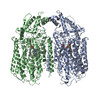
-Entire : quinol-dependent Nitric Oxide Reductase with BRIL
| Entire | Name: quinol-dependent Nitric Oxide Reductase with BRIL |
|---|---|
| Components |
|
-Supramolecule #1: quinol-dependent Nitric Oxide Reductase with BRIL
| Supramolecule | Name: quinol-dependent Nitric Oxide Reductase with BRIL / type: complex / ID: 1 / Parent: 0 / Macromolecule list: #1 Details: BRIL-AxqNOR. Two HEM and one Fe are the cofactors covalently bound to this protein. |
|---|---|
| Source (natural) | Organism: Achromobacter xylosoxidans (bacteria) |
-Macromolecule #1: Nitric oxide reductase subunit B,Soluble cytochrome b562
| Macromolecule | Name: Nitric oxide reductase subunit B,Soluble cytochrome b562 type: protein_or_peptide / ID: 1 / Number of copies: 2 / Enantiomer: LEVO / EC number: nitric oxide reductase (cytochrome c) |
|---|---|
| Source (natural) | Organism: Achromobacter xylosoxidans (bacteria) |
| Molecular weight | Theoretical: 94.903242 KDa |
| Recombinant expression | Organism: |
| Sequence | String: MGPYRRLWFT LIAVLAVTFA LLGFYGGEVY RQAPPIPEEV ASADGTRLFG RDDILDGQTA WQSIGGMQLG SIWGHGAYQA PDWTADWLH RELMAWLDLA ARDAHGRDYG QLDAPAQAAL REQLKAEYRA NRADAAGGKL TLSPRRAQAV AQTEAYYDQL F SDAPALHR ...String: MGPYRRLWFT LIAVLAVTFA LLGFYGGEVY RQAPPIPEEV ASADGTRLFG RDDILDGQTA WQSIGGMQLG SIWGHGAYQA PDWTADWLH RELMAWLDLA ARDAHGRDYG QLDAPAQAAL REQLKAEYRA NRADAAGGKL TLSPRRAQAV AQTEAYYDQL F SDAPALHR SRENYAMKEN TLPDANRRRQ MTHFFFWTAW AAATEREGTS VTYTNNWPHE PLIGNHPSSE NVMWSIISVV VL LAGIGLL IWAWAFLRGK EEDEPPAPAR DPLTTFALTP SQRALGKYLF LVVALFGFQV LLGGFTAHYT VEGQKFYGID LSQ WFPYSL VRTWHIQSAL FWIATGFLAA GLFLAPLING GRDPKYQKAG VDILFWALVL VVVGSFAGNY LAIAQIMPPD LNFW LGHQG YEYVDLGRLW QIGKFAGICF WLVLMLRGIV PALRTPGGDK NLLALLTASV GAIGLFYGAG FFYGERTHLT VMEYW RWWI VHLWVEGFFE VFATTALAFI FSTLGLVSRR MATTASLASA SLFMLGGIPG TFHHLYFAGT TTPVMAVGAS FSALEV VPL IVLGHEAWEN WRLKTRAPWM ENLKWPLMCF VAVAFWNMLG AGVFGFMINP PVSLYYIQGL NTTPVHAHAA LFGVYGF LA LGFTLLVLRY IRPQYALSPG LMKLAFWGLN LGLALMIFTS LLPIGLIQFH ASVSEGMWYA RSEAFMQQDI LKTLRWGR T FGDVVFLLGA LAMVVQVILG LLSGKADLED NWETLNDNLK VIEKADNAAQ VKDALTKMRA AALDAQKATP PKLEDKSPD SPEMKDFRHG FDILVGQIDD ALKLANEGKV KEAQAAAEQL KTTRNAYIQK YL UniProtKB: Nitric oxide reductase subunit B, Soluble cytochrome b562 |
-Macromolecule #2: PROTOPORPHYRIN IX CONTAINING FE
| Macromolecule | Name: PROTOPORPHYRIN IX CONTAINING FE / type: ligand / ID: 2 / Number of copies: 4 / Formula: HEM |
|---|---|
| Molecular weight | Theoretical: 616.487 Da |
| Chemical component information |  ChemComp-HEM: |
-Macromolecule #3: FE (III) ION
| Macromolecule | Name: FE (III) ION / type: ligand / ID: 3 / Number of copies: 2 / Formula: FE |
|---|---|
| Molecular weight | Theoretical: 55.845 Da |
-Macromolecule #4: CALCIUM ION
| Macromolecule | Name: CALCIUM ION / type: ligand / ID: 4 / Number of copies: 2 / Formula: CA |
|---|---|
| Molecular weight | Theoretical: 40.078 Da |
-Experimental details
-Structure determination
| Method | cryo EM |
|---|---|
 Processing Processing | single particle reconstruction |
| Aggregation state | particle |
-Sample preparation
| Concentration | 3 mg/mL |
|---|---|
| Buffer | pH: 7.5 |
| Vitrification | Cryogen name: ETHANE |
- Electron microscopy
| Microscope | TFS KRIOS |
|---|---|
| Image recording | Film or detector model: GATAN K2 SUMMIT (4k x 4k) / Average electron dose: 65.0 e/Å2 |
| Electron beam | Acceleration voltage: 300 kV / Electron source:  FIELD EMISSION GUN FIELD EMISSION GUN |
| Electron optics | Illumination mode: FLOOD BEAM / Imaging mode: BRIGHT FIELD / Nominal defocus max: 3.5 µm / Nominal defocus min: 1.5 µm |
| Experimental equipment |  Model: Titan Krios / Image courtesy: FEI Company |