 Movie
Movie Controller


+ Open data

- Basic information
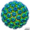
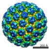
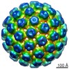
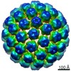
| Entry | Database: EMDB / ID: EMD-6619 | |||||||||
|---|---|---|---|---|---|---|---|---|---|---|
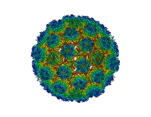
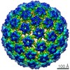
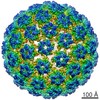
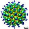
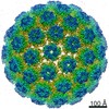
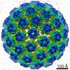
| Title | Electron cryo-microscopy of Human Papillomavirus | |||||||||
 Map data Map data | Reconstruction of HPV16 complex with Heparin | |||||||||
 Sample Sample |
| |||||||||
 Keywords Keywords | DED high resolution map / L1 plus L2 / quasivirus / Heparin | |||||||||
| Function / homology |  Function and homology information Function and homology informationT=7 icosahedral viral capsid / endocytosis involved in viral entry into host cell / virion attachment to host cell / host cell nucleus / structural molecule activity Similarity search - Function | |||||||||
| Biological species |  Human papillomavirus 16 Human papillomavirus 16 | |||||||||
| Method | single particle reconstruction / cryo EM / Resolution: 4.3 Å | |||||||||
 Authors Authors | Guan J / Bywaters SM / Brendle SA / Ashley RE / Makhov AM / Conway JF / Christensen ND / Hafenstein S | |||||||||
 Citation Citation | Journal: Structure / Year: 2017 Title: Cryoelectron Microscopy Maps of Human Papillomavirus 16 Reveal L2 Densities and Heparin Binding Site. Authors: Jian Guan / Stephanie M Bywaters / Sarah A Brendle / Robert E Ashley / Alexander M Makhov / James F Conway / Neil D Christensen / Susan Hafenstein /  Abstract: Human papillomavirus (HPV) is a significant health burden and leading cause of virus-induced cancers. The current commercial vaccines are genotype specific and provide little therapeutic benefit to ...Human papillomavirus (HPV) is a significant health burden and leading cause of virus-induced cancers. The current commercial vaccines are genotype specific and provide little therapeutic benefit to patients with existing HPV infections. Host entry mechanisms represent an excellent target for alternative therapeutics, but HPV receptor use, the details of cell attachment, and host entry are inadequately understood. Here we present near-atomic resolution structures of the HPV16 capsid and HPV16 in complex with heparin, both determined from cryoelectron micrographs collected with direct electron detection technology. The structures clarify details of capsid architecture for the first time, including variation in L1 major capsid protein conformation and putative location of L2 minor protein. Heparin binds specifically around the capsid icosahedral vertices and may recapitulate the earliest stage of infection, providing a framework for continuing biochemical, genetic, and biophysical studies. | |||||||||
| History |
|
- Structure visualization
| Movie |
Movie viewer |
|---|---|
| Structure viewer | EM map: SurfViewMolmilJmol/JSmol |
| Supplemental images |
- Downloads & links
-EMDB archive
| Map data | emd_6619.map.gz | 1 GB | EMDB map data format | |
|---|---|---|---|---|
| Header (meta data) | emd-6619-v30.xmlemd-6619.xml | 9.6 KB 9.6 KB | Display Display | EMDB header |
| Images |  400_6619.gif 400_6619.gif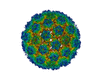 80_6619.gif 80_6619.gif | 89.1 KB 4.6 KB | ||
| Archive directory |  http://ftp.pdbj.org/pub/emdb/structures/EMD-6619ftp://ftp.pdbj.org/pub/emdb/structures/EMD-6619 http://ftp.pdbj.org/pub/emdb/structures/EMD-6619ftp://ftp.pdbj.org/pub/emdb/structures/EMD-6619 | HTTPS FTP |
-Validation report
| Summary document | emd_6619_validation.pdf.gz | 403.3 KB | Display | EMDB validaton report |
|---|---|---|---|---|
| Full document | emd_6619_full_validation.pdf.gz | 402.8 KB | Display | |
| Data in XML | emd_6619_validation.xml.gz | 9 KB | Display | |
| Arichive directory | https://ftp.pdbj.org/pub/emdb/validation_reports/EMD-6619ftp://ftp.pdbj.org/pub/emdb/validation_reports/EMD-6619 | HTTPS FTP |
-Related structure data
| Related structure data |  5keqMC 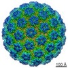 6620C  5kepC M: atomic model generated by this map C: citing same article ( |
|---|---|
| Similar structure data |

-Links
| EMDB pages | EMDB (EBI/PDBe) / EMDataResource |
|---|---|
| Related items in Molecule of the Month |


-Map
| File | Download / File: emd_6619.map.gz / Format: CCP4 / Size: 1.1 GB / Type: IMAGE STORED AS FLOATING POINT NUMBER (4 BYTES) | ||||||||||||||||||||||||||||||||||||||||||||||||||||||||||||
|---|---|---|---|---|---|---|---|---|---|---|---|---|---|---|---|---|---|---|---|---|---|---|---|---|---|---|---|---|---|---|---|---|---|---|---|---|---|---|---|---|---|---|---|---|---|---|---|---|---|---|---|---|---|---|---|---|---|---|---|---|---|
| Annotation | Reconstruction of HPV16 complex with Heparin | ||||||||||||||||||||||||||||||||||||||||||||||||||||||||||||
| Projections & slices | Image control
Images are generated by Spider. | ||||||||||||||||||||||||||||||||||||||||||||||||||||||||||||
| Voxel size | X=Y=Z: 1.147 Å | ||||||||||||||||||||||||||||||||||||||||||||||||||||||||||||
| Density |
| ||||||||||||||||||||||||||||||||||||||||||||||||||||||||||||
| Symmetry | Space group: 1 | ||||||||||||||||||||||||||||||||||||||||||||||||||||||||||||
| Details | EMDB XML:
CCP4 map header:
| ||||||||||||||||||||||||||||||||||||||||||||||||||||||||||||
 Z (Sec.)
 Y (Row.)
 X (Col.)


-Supplemental data
- Sample components
-Entire : Human papillomavirus bound to heparin
| Entire | Name: Human papillomavirus bound to heparin |
|---|---|
| Components |
|
-Supramolecule #1000: Human papillomavirus bound to heparin
| Supramolecule | Name: Human papillomavirus bound to heparin / type: sample / ID: 1000 / Number unique components: 1 |
|---|
-Supramolecule #1: Human papillomavirus 16
| Supramolecule | Name: Human papillomavirus 16 / type: virus / ID: 1 / Name.synonym: HPV16 / NCBI-ID: 337041 / Sci species name: Human papillomavirus 16 / Database: NCBI / Virus type: VIRUS-LIKE PARTICLE / Virus isolate: SEROTYPE / Virus enveloped: No / Virus empty: No / Syn species name: HPV16 / Sci species serotype: HPV16 |
|---|---|
| Host (natural) | Organism:  Homo sapiens (human) / synonym: VERTEBRATES Homo sapiens (human) / synonym: VERTEBRATES |
| Virus shell | Shell ID: 1 / Name: L1 plus L2 / Diameter: 580 Å / T number (triangulation number): 7 |
-Experimental details
-Structure determination
| Method | cryo EM |
|---|---|
 Processing Processing | single particle reconstruction |
| Aggregation state | particle |
-Sample preparation
| Concentration | 1.2 mg/mL |
|---|---|
| Buffer | pH: 7.4 / Details: 1 M NaCl, 200 mM Tris |
| Grid | Details: glow-discharged holey carbon support grid |
| Vitrification | Cryogen name: ETHANE / Chamber humidity: 90 % / Chamber temperature: 102 K / Instrument: GATAN CRYOPLUNGE 3 |
- Electron microscopy
| Microscope | FEI POLARA 300 |
|---|---|
| Date | Jan 20, 2015 |
| Image recording | Category: CCD / Film or detector model: FEI FALCON II (4k x 4k) / Number real images: 6175 |
| Electron beam | Acceleration voltage: 300 kV / Electron source:  FIELD EMISSION GUN FIELD EMISSION GUN |
| Electron optics | Illumination mode: FLOOD BEAM / Imaging mode: BRIGHT FIELD / Nominal magnification: 93000 |
| Sample stage | Specimen holder model: GATAN LIQUID NITROGEN |
| Experimental equipment |  Model: Tecnai Polara / Image courtesy: FEI Company |
-Image processing
| Details | The particles were selected using semi-automatic program e2boxer.py. |
|---|---|
| CTF correction | Details: ctffind for whole image |
| Final reconstruction | Algorithm: OTHER / Resolution.type: BY AUTHOR / Resolution: 4.3 Å / Resolution method: OTHER / Software - Name: auto3dem / Number images used: 51422 |
| Final two d classification | Number classes: 10 |
-Atomic model buiding 1
| Initial model | PDB ID:  3oae |
|---|---|
| Refinement | Space: REAL |
| Output model | PDB-5keq: |

