 Movie
Movie Controller

[English] 日本語
 Yorodumi
- EMDB-6610: Cryo-electron microscopy structure of influenza H1 hemagglutinin ... -
+ Open data

- Basic information
| Entry | Database: EMDB / ID: EMD-6610 | |||||||||
|---|---|---|---|---|---|---|---|---|---|---|
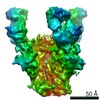
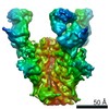
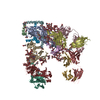
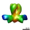
| Title | Cryo-electron microscopy structure of influenza H1 hemagglutinin bound to stalk-targeting antibody, 6F12 | |||||||||
 Map data Map data | Tomographic subvolume average of virus-bound influenza H1 HA in complex with the stalk-binding antibody, 6F12 | |||||||||
 Sample Sample |
| |||||||||
 Keywords Keywords | Influenza / HA | |||||||||
| Biological species |  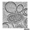 unidentified influenza virus / unidentified influenza virus /  | |||||||||
| Method | subtomogram averaging / cryo EM / Resolution: 25.0 Å | |||||||||
 Authors Authors | Tran EEH / Podolsky KA / Bartesaghi A / Kuybeda O / Grandinetti G / Wohlbold TJ / Tan GS / Nachbagauer R / Palese P / Krammer F / Subramaniam S | |||||||||
 Citation Citation | Journal: mBio / Year: 2016 Title: Cryo-electron Microscopy Structures of Chimeric Hemagglutinin Displayed on a Universal Influenza Vaccine Candidate. Authors: Erin E H Tran / Kira A Podolsky / Alberto Bartesaghi / Oleg Kuybeda / Giovanna Grandinetti / Teddy John Wohlbold / Gene S Tan / Raffael Nachbagauer / Peter Palese / Florian Krammer / Sriram Subramaniam /   Abstract: Influenza viruses expressing chimeric hemagglutinins (HAs) are important tools in the quest for a universal vaccine. Using cryo-electron tomography, we have determined the structures of a chimeric HA ...Influenza viruses expressing chimeric hemagglutinins (HAs) are important tools in the quest for a universal vaccine. Using cryo-electron tomography, we have determined the structures of a chimeric HA variant that comprises an H1 stalk and an H5 globular head domain (cH5/1 HA) in native and antibody-bound states. We show that cH5/1 HA is structurally different from native HA, displaying a 60° rotation between the stalk and head groups, leading to a novel and unexpected "open" arrangement of HA trimers. cH5/1N1 viruses also display higher glycoprotein density than pH1N1 or H5N1 viruses, but despite these differences, antibodies that target either the stalk or head domains of hemagglutinins still bind to cH5/1 HA with the same consequences as those observed with native H1 or H5 HA. Our results show that a large range of structural plasticity can be tolerated in the chimeric spike scaffold without disrupting structural and geometric aspects of antibody binding. IMPORTANCE: Chimeric hemagglutinin proteins are set to undergo human clinical trials as a universal influenza vaccine candidate, yet no structural information for these proteins is available. Using ...IMPORTANCE: Chimeric hemagglutinin proteins are set to undergo human clinical trials as a universal influenza vaccine candidate, yet no structural information for these proteins is available. Using cryo-electron tomography, we report the first three-dimensional (3D) visualization of chimeric hemagglutinin proteins displayed on the surface of the influenza virus. We show that, unexpectedly, the chimeric hemagglutinin structure differs from those of naturally occurring hemagglutinins by displaying a more open head domain and a dramatically twisted head/stalk arrangement. Despite this unusual spatial relationship between head and stalk regions, virus preparations expressing the chimeric hemagglutinin are fully infectious and display a high glycoprotein density, which likely helps induction of a broadly protective immune response. | |||||||||
| History |
|
- Structure visualization
| Movie |
 Movie viewer Movie viewer |
|---|---|
| Structure viewer | EM map: SurfViewMolmilJmol/JSmol |
| Supplemental images |

- Downloads & links
-EMDB archive
| Map data | emd_6610.map.gz | 814 KB | EMDB map data format | |
|---|---|---|---|---|
| Header (meta data) | emd-6610-v30.xmlemd-6610.xml | 12.1 KB 12.1 KB | Display Display | EMDB header |
| Images | 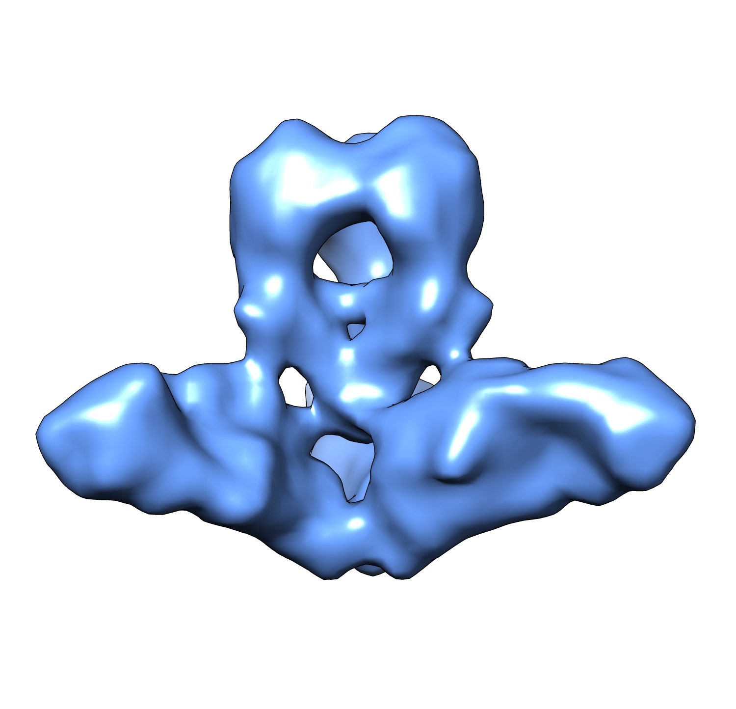 emd_6610.png emd_6610.png | 443.8 KB | ||
| Archive directory |  http://ftp.pdbj.org/pub/emdb/structures/EMD-6610ftp://ftp.pdbj.org/pub/emdb/structures/EMD-6610 http://ftp.pdbj.org/pub/emdb/structures/EMD-6610ftp://ftp.pdbj.org/pub/emdb/structures/EMD-6610 | HTTPS FTP |
-Related structure data
| Related structure data | 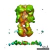 6607C 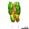 6608C 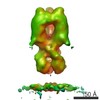 6609C 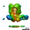 6611C 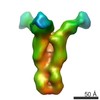 6612C 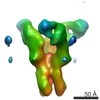 6613C 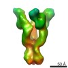 6614C C: citing same article ( |
|---|---|
| Similar structure data |


-Links
| EMDB pages | EMDB (EBI/PDBe) / EMDataResource |
|---|
-Map
| File | Download / File: emd_6610.map.gz / Format: CCP4 / Size: 908.2 KB / Type: IMAGE STORED AS FLOATING POINT NUMBER (4 BYTES) | ||||||||||||||||||||||||||||||||||||||||||||||||||||||||||||||||||||
|---|---|---|---|---|---|---|---|---|---|---|---|---|---|---|---|---|---|---|---|---|---|---|---|---|---|---|---|---|---|---|---|---|---|---|---|---|---|---|---|---|---|---|---|---|---|---|---|---|---|---|---|---|---|---|---|---|---|---|---|---|---|---|---|---|---|---|---|---|---|
| Annotation | Tomographic subvolume average of virus-bound influenza H1 HA in complex with the stalk-binding antibody, 6F12 | ||||||||||||||||||||||||||||||||||||||||||||||||||||||||||||||||||||
| Projections & slices | Image control
Images are generated by Spider. generated in cubic-lattice coordinate | ||||||||||||||||||||||||||||||||||||||||||||||||||||||||||||||||||||
| Voxel size | X=Y=Z: 4.1 Å | ||||||||||||||||||||||||||||||||||||||||||||||||||||||||||||||||||||
| Density |
| ||||||||||||||||||||||||||||||||||||||||||||||||||||||||||||||||||||
| Symmetry | Space group: 1 | ||||||||||||||||||||||||||||||||||||||||||||||||||||||||||||||||||||
| Details | EMDB XML:
CCP4 map header:
| ||||||||||||||||||||||||||||||||||||||||||||||||||||||||||||||||||||
 Z (Sec.)
 Y (Row.)
 X (Col.)


-Supplemental data
- Sample components
-Entire : Tomographic subvolume average of virus-bound influenza H1 HA in c...
| Entire | Name: Tomographic subvolume average of virus-bound influenza H1 HA in complex with the stalk-binding antibody, 6F12 |
|---|---|
| Components |
|
-Supramolecule #1000: Tomographic subvolume average of virus-bound influenza H1 HA in c...
| Supramolecule | Name: Tomographic subvolume average of virus-bound influenza H1 HA in complex with the stalk-binding antibody, 6F12 type: sample / ID: 1000 / Number unique components: 2 |
|---|
-Macromolecule #1: H1 hemagglutinin
| Macromolecule | Name: H1 hemagglutinin / type: protein_or_peptide / ID: 1 / Oligomeric state: trimer / Recombinant expression: No / Database: NCBI |
|---|---|
| Source (natural) | Organism: unidentified influenza virus / Strain: pH1N1 |
-Macromolecule #2: Immunoglobulin 6F12
| Macromolecule | Name: Immunoglobulin 6F12 / type: protein_or_peptide / ID: 2 / Recombinant expression: No / Database: NCBI |
|---|---|
| Source (natural) | Organism: |
-Experimental details
-Structure determination
| Method | cryo EM |
|---|---|
 Processing Processing | subtomogram averaging |
| Aggregation state | particle |
-Sample preparation
| Buffer | pH: 7.4 / Details: 25 mM Tris, 100 mM NaCl |
|---|---|
| Grid | Details: Quantifoil Multi-A 200 mesh carbon grids |
| Vitrification | Cryogen name: ETHANE / Chamber humidity: 95 % / Chamber temperature: 90 K / Instrument: LEICA EM GP Timed resolved state: Virus was incubated with antibody for ~30 minutes on ice before solution was plunge-frozen Method: Blot for 6 seconds before plunging. |
- Electron microscopy
| Microscope | FEI TITAN KRIOS |
|---|---|
| Specialist optics | Energy filter - Name: Gatan, Inc / Energy filter - Lower energy threshold: 0.0 eV / Energy filter - Upper energy threshold: 20.0 eV |
| Date | Apr 24, 2015 |
| Image recording | Category: CCD / Film or detector model: GATAN K2 SUMMIT (4k x 4k) / Number real images: 10 / Average electron dose: 120 e/Å2 Details: Every image is the average of two frames recorded by the direct electron detector. |
| Electron beam | Acceleration voltage: 300 kV / Electron source:  FIELD EMISSION GUN FIELD EMISSION GUN |
| Electron optics | Calibrated magnification: 23000 / Illumination mode: FLOOD BEAM / Imaging mode: BRIGHT FIELD / Nominal defocus max: 3.0 µm / Nominal defocus min: 2.0 µm / Nominal magnification: 64000 |
| Sample stage | Specimen holder: Liquid nitrogen-cooled / Specimen holder model: FEI TITAN KRIOS AUTOGRID HOLDER / Tilt series - Axis1 - Min angle: -60 ° / Tilt series - Axis1 - Max angle: 60 ° |
| Experimental equipment |  Model: Titan Krios / Image courtesy: FEI Company |
-Image processing
| Details | Subtomograms were selected using an automatic selection program. |
|---|---|
| Final reconstruction | Resolution.type: BY AUTHOR / Resolution: 25.0 Å / Resolution method: OTHER / Software - Name: IMOD / Number subtomograms used: 85 |
