ムービー
ムービー コントローラー


+ データを開く

- 基本情報
基本情報
| 登録情報 |  | |||||||||
|---|---|---|---|---|---|---|---|---|---|---|
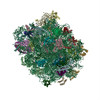
| タイトル | Cryo- EM structure of ribosomal large subunit (LSU) from Entamoeba histolytica at 2.8 angstrom resolution | |||||||||
 マップデータ マップデータ | ||||||||||
 試料 試料 |
| |||||||||
 キーワード キーワード | LSU / Entamoeba histolytica / 53S / Ribosome | |||||||||
| 機能・相同性 |  機能・相同性情報 機能・相同性情報protein-RNA complex assembly / maturation of LSU-rRNA / ribosomal large subunit biogenesis / maturation of LSU-rRNA from tricistronic rRNA transcript (SSU-rRNA, 5.8S rRNA, LSU-rRNA) / ribosome biogenesis / 5S rRNA binding / ribosomal large subunit assembly / large ribosomal subunit rRNA binding / cytosolic large ribosomal subunit / cytoplasmic translation ...protein-RNA complex assembly / maturation of LSU-rRNA / ribosomal large subunit biogenesis / maturation of LSU-rRNA from tricistronic rRNA transcript (SSU-rRNA, 5.8S rRNA, LSU-rRNA) / ribosome biogenesis / 5S rRNA binding / ribosomal large subunit assembly / large ribosomal subunit rRNA binding / cytosolic large ribosomal subunit / cytoplasmic translation / negative regulation of translation / rRNA binding / structural constituent of ribosome / ribosome / translation / ribonucleoprotein complex / mRNA binding / endoplasmic reticulum / RNA binding / zinc ion binding 類似検索 - 分子機能 | |||||||||
| 生物種 |  Entamoeba histolytica HM-1:IMSS (赤痢アメーバ) Entamoeba histolytica HM-1:IMSS (赤痢アメーバ) | |||||||||
| 手法 | 単粒子再構成法 / クライオ電子顕微鏡法 / 解像度: 2.8 Å | |||||||||
 データ登録者 データ登録者 | Sharma S / Mishra S / Gourinath S / Kaushal PS | |||||||||
| 資金援助 |  インド, 1件 インド, 1件
| |||||||||
 引用 引用 | ジャーナル: Nat Commun / 年: 2025 タイトル: Cryo-EM structure of ribosome from pathogenic protozoa Entamoeba histolytica reveals unique features of its architecture. 著者: Shivani Sharma / Shalini Mishra / Samudrala Gourinath / Prem S Kaushal / 要旨: Entamoeba histolytica, an anaerobic protozoan parasite, is the causative agent of amoebiasis, bloody diarrhea, and liver abscesses in humans. Amoebiasis is more predominant in tropical areas with ...Entamoeba histolytica, an anaerobic protozoan parasite, is the causative agent of amoebiasis, bloody diarrhea, and liver abscesses in humans. Amoebiasis is more predominant in tropical areas with poor sanitation conditions, and it remains the fourth leading cause of death due to a protozoan infection. E. histolytica life cycle spans between an infective 'cyst stage' and an active disease-causing 'trophozoite stage'. We have isolated ribosomes from the trophozoite stage of E. histolytica. Here, we report single particle cryo-EM structures of the 53S ribosome large subunit (LSU), and 75S associated ribosomes, with P-tRNA, A/P and P/E tRNAs, and with paromomycin antibiotic, at 2.8 Å to 3.4 Å resolution. The E. histolytica possesses a reduced ribosome with a conserved core, and the periphery evolved with species-specific unique features. The most notable features are the presence of the rRNA triple helix near the peptide exit tunnel, the expansion segment H88ES102 near the exit site on LSU, and unique insertions in r-proteins. Furthermore, the 75S ribosome paromomycin complex structure provides the atomic details of its interactions. These structures provide insights into the evolutionary adaptation of the E. histolytica translational machinery and may be explored further for amoebicidal therapeutic intervention. | |||||||||
| 履歴 |
|
- 構造の表示
構造の表示
| 添付画像 |
|---|

- ダウンロードとリンク
-EMDBアーカイブ
| マップデータ | emd_64694.map.gz | 91.2 MB | EMDBマップデータ形式 | |
|---|---|---|---|---|
| ヘッダ (付随情報) | emd-64694-v30.xmlemd-64694.xml | 63.9 KB 63.9 KB | 表示 表示 | EMDBヘッダ |
| FSC (解像度算出) | emd_64694_fsc.xml | 10.6 KB | 表示 | FSCデータファイル |
| 画像 |  emd_64694.png emd_64694.png | 139.8 KB | ||
| Filedesc metadata | emd-64694.cif.gz | 13.7 KB | ||
| その他 | emd_64694_half_map_1.map.gzemd_64694_half_map_2.map.gz | 79.8 MB 79.8 MB | ||
| アーカイブディレクトリ |  http://ftp.pdbj.org/pub/emdb/structures/EMD-64694ftp://ftp.pdbj.org/pub/emdb/structures/EMD-64694 http://ftp.pdbj.org/pub/emdb/structures/EMD-64694ftp://ftp.pdbj.org/pub/emdb/structures/EMD-64694 | HTTPS FTP |
-関連構造データ
| 関連構造データ |  9v1iMC  9v1jC  9v1kC  9v1lC  9v21C  9v24C  9v25C  9v26C  9v27C  9v28C  9v29C M: このマップから作成された原子モデル C: 同じ文献を引用 ( |
|---|---|
| 類似構造データ |


-リンク
| EMDBのページ | EMDB (EBI/PDBe) / EMDataResource |
|---|---|
| 「今月の分子」の関連する項目 |


-マップ
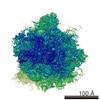
| ファイル | ダウンロード / ファイル: emd_64694.map.gz / 形式: CCP4 / 大きさ: 103 MB / タイプ: IMAGE STORED AS FLOATING POINT NUMBER (4 BYTES) | ||||||||||||||||||||||||||||||||||||
|---|---|---|---|---|---|---|---|---|---|---|---|---|---|---|---|---|---|---|---|---|---|---|---|---|---|---|---|---|---|---|---|---|---|---|---|---|---|
| 投影像・断面図 | 画像のコントロール
画像は Spider により作成 | ||||||||||||||||||||||||||||||||||||
| ボクセルのサイズ | X=Y=Z: 1.07 Å | ||||||||||||||||||||||||||||||||||||
| 密度 |
| ||||||||||||||||||||||||||||||||||||
| 対称性 | 空間群: 1 | ||||||||||||||||||||||||||||||||||||
| 詳細 | EMDB XML:
|
 X (Sec.)
 Y (Row.)
 Z (Col.)


-添付データ
-ハーフマップ: #2
| ファイル | emd_64694_half_map_1.map | ||||||||||||
|---|---|---|---|---|---|---|---|---|---|---|---|---|---|
| 投影像・断面図 |
| ||||||||||||
| 密度ヒストグラム |


-ハーフマップ: #1
| ファイル | emd_64694_half_map_2.map | ||||||||||||
|---|---|---|---|---|---|---|---|---|---|---|---|---|---|
| 投影像・断面図 |
| ||||||||||||
| 密度ヒストグラム |


- 試料の構成要素
+全体 : Entamoeba histolytica Ribosome Large Subunit
+超分子 #1: Entamoeba histolytica Ribosome Large Subunit
+分子 #1: 25S rRNA (3068-MER)
+分子 #2: 5.8S rRNA (145-MER)
+分子 #3: 5S rRNA (117-MER)
+分子 #4: Large ribosomal subunit protein uL2
+分子 #5: 60S ribosomal protein L3, putative
+分子 #6: 60S ribosomal protein L4, putative
+分子 #7: 60S ribosomal protein L5, putative
+分子 #8: 60S ribosomal protein L6, putative
+分子 #9: 60S ribosomal protein L7, putative
+分子 #10: 60S ribosomal protein L7a
+分子 #11: 60S ribosomal protein L9, putative
+分子 #12: Ribosomal protein L10, putative
+分子 #13: 60S ribosomal protein L11, putative
+分子 #14: 60S ribosomal protein L13, putative
+分子 #15: 60S ribosomal protein L13, putative
+分子 #16: 60S ribosomal protein L14, putative
+分子 #17: Ribosomal protein L15
+分子 #18: 60S ribosomal protein L17, putative
+分子 #19: 60S ribosomal protein L18, putative
+分子 #20: 60S ribosomal protein L18a
+分子 #21: Ribosomal protein L19
+分子 #22: 60S ribosomal protein L21, putative
+分子 #23: Large ribosomal subunit protein eL22
+分子 #24: 60S ribosomal protein L23, putative
+分子 #25: Ribosomal protein L23A, putative
+分子 #26: 60S ribosomal protein L24, putative
+分子 #27: 60S ribosomal protein L26, putative
+分子 #28: 60S ribosomal protein L27, putative
+分子 #29: Large ribosomal subunit protein uL15A
+分子 #30: 60S ribosomal protein L29
+分子 #31: 60S ribosomal protein L30, putative
+分子 #32: 60S ribosomal protein L31, putative
+分子 #33: 60S ribosomal protein L32, putative
+分子 #34: 60S ribosomal protein L34, putative
+分子 #35: uL29
+分子 #36: 60S ribosomal protein L35a, putative
+分子 #37: 60S ribosomal protein L36, putative
+分子 #38: 60S ribosomal protein L37-A, putative
+分子 #39: 60S ribosomal protein L37A, putative
+分子 #40: 60S ribosomal protein L38, putative
+分子 #41: Ribosomal protein L39, putative
+分子 #42: 60S ribosomal protein L40, putative
+分子 #43: 60S ribosomal protein L44, putative
+分子 #44: Unknown peptide
-実験情報
-構造解析
| 手法 | クライオ電子顕微鏡法 |
|---|---|
 解析 解析 | 単粒子再構成法 |
| 試料の集合状態 | particle |
-試料調製
| 濃度 | 1 mg/mL |
|---|---|
| 緩衝液 | pH: 7.4 詳細: 20nM HEPES-sodium salt buffer pH 7.4, 100mM Potassium acetate, 10mM Magnesium acetate, 10mm Ammonium acetate, 3mM DTT |
| グリッド | モデル: Quantifoil R1.2/1.3 / 材質: GOLD / メッシュ: 300 / 支持フィルム - 材質: CARBON / 支持フィルム - トポロジー: HOLEY / 前処理 - タイプ: GLOW DISCHARGE / 前処理 - 時間: 50 sec. |
| 凍結 | 凍結剤: ETHANE / チャンバー内湿度: 100 % / チャンバー内温度: 290 K / 装置: FEI VITROBOT MARK IV |
- 電子顕微鏡法
| 顕微鏡 | TFS KRIOS |
|---|---|
| 撮影 | フィルム・検出器のモデル: FEI FALCON III (4k x 4k) 検出モード: INTEGRATING / 実像数: 3470 / 平均露光時間: 2.0 sec. / 平均電子線量: 1.34 e/Å2 |
| 電子線 | 加速電圧: 300 kV / 電子線源:  FIELD EMISSION GUN FIELD EMISSION GUN |
| 電子光学系 | 倍率(補正後): 75000 / 照射モード: FLOOD BEAM / 撮影モード: BRIGHT FIELD / 最大 デフォーカス(公称値): 3.0 µm / 最小 デフォーカス(公称値): 1.8 µm |
| 試料ステージ | 試料ホルダーモデル: FEI TITAN KRIOS AUTOGRID HOLDER ホルダー冷却材: NITROGEN |
| 実験機器 |  モデル: Titan Krios / 画像提供: FEI Company |
+画像解析

-原子モデル構築 1
| 初期モデル |
| ||||||
|---|---|---|---|---|---|---|---|
| 詳細 | phenix.real_space_refinement | ||||||
| 精密化 | 空間: REAL / プロトコル: FLEXIBLE FIT / 温度因子: 67.76 | ||||||
| 得られたモデル | PDB-9v1i: |