Movie
Movie Controller

[English] 日本語
 Yorodumi
- EMDB-64694: Cryo- EM structure of ribosomal large subunit (LSU) from Entamoeb... -
+ Open data

- Basic information
| Entry |  | |||||||||
|---|---|---|---|---|---|---|---|---|---|---|
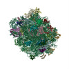
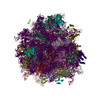
| Title | Cryo- EM structure of ribosomal large subunit (LSU) from Entamoeba histolytica at 2.8 angstrom resolution | |||||||||
 Map data Map data | ||||||||||
 Sample Sample |
| |||||||||
 Keywords Keywords | LSU / Entamoeba histolytica / 53S / Ribosome | |||||||||
| Function / homology |  Function and homology information Function and homology informationprotein-RNA complex assembly / maturation of LSU-rRNA / ribosomal large subunit biogenesis / maturation of LSU-rRNA from tricistronic rRNA transcript (SSU-rRNA, 5.8S rRNA, LSU-rRNA) / ribosome biogenesis / 5S rRNA binding / ribosomal large subunit assembly / large ribosomal subunit rRNA binding / cytosolic large ribosomal subunit / cytoplasmic translation ...protein-RNA complex assembly / maturation of LSU-rRNA / ribosomal large subunit biogenesis / maturation of LSU-rRNA from tricistronic rRNA transcript (SSU-rRNA, 5.8S rRNA, LSU-rRNA) / ribosome biogenesis / 5S rRNA binding / ribosomal large subunit assembly / large ribosomal subunit rRNA binding / cytosolic large ribosomal subunit / cytoplasmic translation / negative regulation of translation / rRNA binding / structural constituent of ribosome / ribosome / translation / ribonucleoprotein complex / mRNA binding / endoplasmic reticulum / RNA binding / zinc ion binding Similarity search - Function | |||||||||
| Biological species |  Entamoeba histolytica HM-1:IMSS (eukaryote) Entamoeba histolytica HM-1:IMSS (eukaryote) | |||||||||
| Method | single particle reconstruction / cryo EM / Resolution: 2.8 Å | |||||||||
 Authors Authors | Sharma S / Mishra S / Gourinath S / Kaushal PS | |||||||||
| Funding support |  India, 1 items India, 1 items
| |||||||||
 Citation Citation | Journal: Nat Commun / Year: 2025 Title: Cryo-EM structure of ribosome from pathogenic protozoa Entamoeba histolytica reveals unique features of its architecture. Authors: Shivani Sharma / Shalini Mishra / Samudrala Gourinath / Prem S Kaushal / Abstract: Entamoeba histolytica, an anaerobic protozoan parasite, is the causative agent of amoebiasis, bloody diarrhea, and liver abscesses in humans. Amoebiasis is more predominant in tropical areas with ...Entamoeba histolytica, an anaerobic protozoan parasite, is the causative agent of amoebiasis, bloody diarrhea, and liver abscesses in humans. Amoebiasis is more predominant in tropical areas with poor sanitation conditions, and it remains the fourth leading cause of death due to a protozoan infection. E. histolytica life cycle spans between an infective 'cyst stage' and an active disease-causing 'trophozoite stage'. We have isolated ribosomes from the trophozoite stage of E. histolytica. Here, we report single particle cryo-EM structures of the 53S ribosome large subunit (LSU), and 75S associated ribosomes, with P-tRNA, A/P and P/E tRNAs, and with paromomycin antibiotic, at 2.8 Å to 3.4 Å resolution. The E. histolytica possesses a reduced ribosome with a conserved core, and the periphery evolved with species-specific unique features. The most notable features are the presence of the rRNA triple helix near the peptide exit tunnel, the expansion segment H88ES102 near the exit site on LSU, and unique insertions in r-proteins. Furthermore, the 75S ribosome paromomycin complex structure provides the atomic details of its interactions. These structures provide insights into the evolutionary adaptation of the E. histolytica translational machinery and may be explored further for amoebicidal therapeutic intervention. | |||||||||
| History |
|
- Structure visualization
| Supplemental images |
|---|

- Downloads & links
-EMDB archive
| Map data | emd_64694.map.gz | 91.2 MB | EMDB map data format | |
|---|---|---|---|---|
| Header (meta data) | emd-64694-v30.xmlemd-64694.xml | 63.9 KB 63.9 KB | Display Display | EMDB header |
| FSC (resolution estimation) | emd_64694_fsc.xml | 10.6 KB | Display | FSC data file |
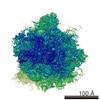
| Images |  emd_64694.png emd_64694.png | 139.8 KB | ||
| Filedesc metadata | emd-64694.cif.gz | 13.7 KB | ||
| Others | emd_64694_half_map_1.map.gzemd_64694_half_map_2.map.gz | 79.8 MB 79.8 MB | ||
| Archive directory |  http://ftp.pdbj.org/pub/emdb/structures/EMD-64694ftp://ftp.pdbj.org/pub/emdb/structures/EMD-64694 http://ftp.pdbj.org/pub/emdb/structures/EMD-64694ftp://ftp.pdbj.org/pub/emdb/structures/EMD-64694 | HTTPS FTP |
-Related structure data
| Related structure data |  9v1iMC  9v1jC  9v1kC  9v1lC  9v21C  9v24C  9v25C  9v26C  9v27C  9v28C  9v29C M: atomic model generated by this map C: citing same article ( |
|---|---|
| Similar structure data |


-Links
| EMDB pages | EMDB (EBI/PDBe) / EMDataResource |
|---|---|
| Related items in Molecule of the Month |


-Map
| File | Download / File: emd_64694.map.gz / Format: CCP4 / Size: 103 MB / Type: IMAGE STORED AS FLOATING POINT NUMBER (4 BYTES) | ||||||||||||||||||||||||||||||||||||
|---|---|---|---|---|---|---|---|---|---|---|---|---|---|---|---|---|---|---|---|---|---|---|---|---|---|---|---|---|---|---|---|---|---|---|---|---|---|
| Projections & slices | Image control
Images are generated by Spider. | ||||||||||||||||||||||||||||||||||||
| Voxel size | X=Y=Z: 1.07 Å | ||||||||||||||||||||||||||||||||||||
| Density |
| ||||||||||||||||||||||||||||||||||||
| Symmetry | Space group: 1 | ||||||||||||||||||||||||||||||||||||
| Details | EMDB XML:
|
 X (Sec.)
 Y (Row.)
 Z (Col.)


-Supplemental data
-Half map: #2
| File | emd_64694_half_map_1.map | ||||||||||||
|---|---|---|---|---|---|---|---|---|---|---|---|---|---|
| Projections & Slices |
| ||||||||||||
| Density Histograms |


-Half map: #1
| File | emd_64694_half_map_2.map | ||||||||||||
|---|---|---|---|---|---|---|---|---|---|---|---|---|---|
| Projections & Slices |
| ||||||||||||
| Density Histograms |


- Sample components
+Entire : Entamoeba histolytica Ribosome Large Subunit
+Supramolecule #1: Entamoeba histolytica Ribosome Large Subunit
+Macromolecule #1: 25S rRNA (3068-MER)
+Macromolecule #2: 5.8S rRNA (145-MER)
+Macromolecule #3: 5S rRNA (117-MER)
+Macromolecule #4: Large ribosomal subunit protein uL2
+Macromolecule #5: 60S ribosomal protein L3, putative
+Macromolecule #6: 60S ribosomal protein L4, putative
+Macromolecule #7: 60S ribosomal protein L5, putative
+Macromolecule #8: 60S ribosomal protein L6, putative
+Macromolecule #9: 60S ribosomal protein L7, putative
+Macromolecule #10: 60S ribosomal protein L7a
+Macromolecule #11: 60S ribosomal protein L9, putative
+Macromolecule #12: Ribosomal protein L10, putative
+Macromolecule #13: 60S ribosomal protein L11, putative
+Macromolecule #14: 60S ribosomal protein L13, putative
+Macromolecule #15: 60S ribosomal protein L13, putative
+Macromolecule #16: 60S ribosomal protein L14, putative
+Macromolecule #17: Ribosomal protein L15
+Macromolecule #18: 60S ribosomal protein L17, putative
+Macromolecule #19: 60S ribosomal protein L18, putative
+Macromolecule #20: 60S ribosomal protein L18a
+Macromolecule #21: Ribosomal protein L19
+Macromolecule #22: 60S ribosomal protein L21, putative
+Macromolecule #23: Large ribosomal subunit protein eL22
+Macromolecule #24: 60S ribosomal protein L23, putative
+Macromolecule #25: Ribosomal protein L23A, putative
+Macromolecule #26: 60S ribosomal protein L24, putative
+Macromolecule #27: 60S ribosomal protein L26, putative
+Macromolecule #28: 60S ribosomal protein L27, putative
+Macromolecule #29: Large ribosomal subunit protein uL15A
+Macromolecule #30: 60S ribosomal protein L29
+Macromolecule #31: 60S ribosomal protein L30, putative
+Macromolecule #32: 60S ribosomal protein L31, putative
+Macromolecule #33: 60S ribosomal protein L32, putative
+Macromolecule #34: 60S ribosomal protein L34, putative
+Macromolecule #35: uL29
+Macromolecule #36: 60S ribosomal protein L35a, putative
+Macromolecule #37: 60S ribosomal protein L36, putative
+Macromolecule #38: 60S ribosomal protein L37-A, putative
+Macromolecule #39: 60S ribosomal protein L37A, putative
+Macromolecule #40: 60S ribosomal protein L38, putative
+Macromolecule #41: Ribosomal protein L39, putative
+Macromolecule #42: 60S ribosomal protein L40, putative
+Macromolecule #43: 60S ribosomal protein L44, putative
+Macromolecule #44: Unknown peptide
-Experimental details
-Structure determination
| Method | cryo EM |
|---|---|
 Processing Processing | single particle reconstruction |
| Aggregation state | particle |
-Sample preparation
| Concentration | 1 mg/mL |
|---|---|
| Buffer | pH: 7.4 Details: 20nM HEPES-sodium salt buffer pH 7.4, 100mM Potassium acetate, 10mM Magnesium acetate, 10mm Ammonium acetate, 3mM DTT |
| Grid | Model: Quantifoil R1.2/1.3 / Material: GOLD / Mesh: 300 / Support film - Material: CARBON / Support film - topology: HOLEY / Pretreatment - Type: GLOW DISCHARGE / Pretreatment - Time: 50 sec. |
| Vitrification | Cryogen name: ETHANE / Chamber humidity: 100 % / Chamber temperature: 290 K / Instrument: FEI VITROBOT MARK IV |
- Electron microscopy
| Microscope | TFS KRIOS |
|---|---|
| Image recording | Film or detector model: FEI FALCON III (4k x 4k) / Detector mode: INTEGRATING / Number real images: 3470 / Average exposure time: 2.0 sec. / Average electron dose: 1.34 e/Å2 |
| Electron beam | Acceleration voltage: 300 kV / Electron source:  FIELD EMISSION GUN FIELD EMISSION GUN |
| Electron optics | Calibrated magnification: 75000 / Illumination mode: FLOOD BEAM / Imaging mode: BRIGHT FIELD / Nominal defocus max: 3.0 µm / Nominal defocus min: 1.8 µm |
| Sample stage | Specimen holder model: FEI TITAN KRIOS AUTOGRID HOLDER / Cooling holder cryogen: NITROGEN |
| Experimental equipment |  Model: Titan Krios / Image courtesy: FEI Company |
+Image processing

-Atomic model buiding 1
| Initial model |
| ||||||
|---|---|---|---|---|---|---|---|
| Details | phenix.real_space_refinement | ||||||
| Refinement | Space: REAL / Protocol: FLEXIBLE FIT / Overall B value: 67.76 | ||||||
| Output model | PDB-9v1i: |