 Movie
Movie Controller

[English] 日本語
 Yorodumi
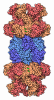
- EMDB-63817: Substrate-engaged human 26S proteasome bound to midnolin with RPT... -
+ Open data

- Basic information
| Entry |  | |||||||||
|---|---|---|---|---|---|---|---|---|---|---|
| Title | Substrate-engaged human 26S proteasome bound to midnolin with RPT2 at top of spiral staircase | |||||||||
 Map data Map data | ||||||||||
 Sample Sample |
| |||||||||
 Keywords Keywords | Midnolin / 26S proteasome / Complex / HYDROLASE | |||||||||
| Function / homology |  Function and homology information Function and homology informationpositive regulation of inclusion body assembly / thyrotropin-releasing hormone receptor binding / nuclear proteasome complex / host-mediated perturbation of viral transcription / Impaired BRCA2 translocation to the nucleus / Impaired BRCA2 binding to SEM1 (DSS1) / proteasome accessory complex / purine ribonucleoside triphosphate binding / integrator complex / cytosolic proteasome complex ...positive regulation of inclusion body assembly / thyrotropin-releasing hormone receptor binding / nuclear proteasome complex / host-mediated perturbation of viral transcription / Impaired BRCA2 translocation to the nucleus / Impaired BRCA2 binding to SEM1 (DSS1) / proteasome accessory complex / purine ribonucleoside triphosphate binding / integrator complex / cytosolic proteasome complex / proteasome regulatory particle / positive regulation of proteasomal protein catabolic process / transcription factor binding / CD8-positive, alpha-beta T cell differentiation / CD8-positive, alpha-beta T cell homeostasis / thymic T cell selection / Antigen processing: Ub, ATP-independent proteasomal degradation / transcription export complex 2 / proteasome-activating activity / structural constituent of proteasome / spermatoproteasome complex / proteasome regulatory particle, lid subcomplex / negative regulation of regulatory T cell differentiation / T-helper 1 cell differentiation / metal-dependent deubiquitinase activity / Regulation of ornithine decarboxylase (ODC) / proteasome core complex / Proteasome assembly / cellular response to type I interferon / T-helper 17 cell differentiation / Cross-presentation of soluble exogenous antigens (endosomes) / retrograde vesicle-mediated transport, Golgi to endoplasmic reticulum / Somitogenesis / protein K63-linked deubiquitination / K63-linked deubiquitinase activity / Homologous DNA Pairing and Strand Exchange / Defective homologous recombination repair (HRR) due to BRCA1 loss of function / Defective HDR through Homologous Recombination Repair (HRR) due to PALB2 loss of BRCA1 binding function / Defective HDR through Homologous Recombination Repair (HRR) due to PALB2 loss of BRCA2/RAD51/RAD51C binding function / Resolution of D-loop Structures through Synthesis-Dependent Strand Annealing (SDSA) / flagellated sperm motility / Resolution of D-loop Structures through Holliday Junction Intermediates / proteasome binding / sperm end piece / immune system process / Impaired BRCA2 binding to RAD51 / myofibril / proteasomal ubiquitin-independent protein catabolic process / general transcription initiation factor binding / ciliary tip / Presynaptic phase of homologous DNA pairing and strand exchange / protein deubiquitination / proteasome endopeptidase complex / NF-kappaB binding / proteasome core complex, beta-subunit complex / negative regulation of programmed cell death / endopeptidase activator activity / threonine-type endopeptidase activity / proteasome core complex, alpha-subunit complex / mRNA export from nucleus / proteasome assembly / enzyme regulator activity / regulation of G1/S transition of mitotic cell cycle / regulation of macroautophagy / stem cell differentiation / positive regulation of interleukin-2 production / proteasome storage granule / response to type II interferon / inclusion body / proteasome complex / TBP-class protein binding / regulation of proteasomal protein catabolic process / meiotic cell cycle / sarcomere / double-strand break repair via nonhomologous end joining / Regulation of activated PAK-2p34 by proteasome mediated degradation / ubiquitin binding / negative regulation of inflammatory response to antigenic stimulus / proteasomal protein catabolic process / Autodegradation of Cdh1 by Cdh1:APC/C / APC/C:Cdc20 mediated degradation of Securin / N-glycan trimming in the ER and Calnexin/Calreticulin cycle / lipopolysaccharide binding / Asymmetric localization of PCP proteins / Ubiquitin-dependent degradation of Cyclin D / positive regulation of type II interferon production / SCF-beta-TrCP mediated degradation of Emi1 / AUF1 (hnRNP D0) binds and destabilizes mRNA / NIK-->noncanonical NF-kB signaling / P-body / TNFR2 non-canonical NF-kB pathway / protein catabolic process / Assembly of the pre-replicative complex / Vpu mediated degradation of CD4 / Cdc20:Phospho-APC/C mediated degradation of Cyclin A / double-strand break repair via homologous recombination / Dectin-1 mediated noncanonical NF-kB signaling / Degradation of DVL / Degradation of AXIN / Degradation of CRY and PER proteins Similarity search - Function | |||||||||
| Biological species |  Homo sapiens (human) Homo sapiens (human) | |||||||||
| Method | single particle reconstruction / cryo EM / Resolution: 2.91 Å | |||||||||
 Authors Authors | Zhu C / Qin L / Liang L | |||||||||
| Funding support |  China, 1 items China, 1 items
| |||||||||
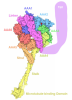
 Citation Citation | Journal: Nat Commun / Year: 2026 Title: Structural dynamics of the midnolin-proteasome during ubiquitin-independent substrate turnover. Authors: Chuanda Zhu / Lu Qin / Zonglin Dai / Peng Zuo / Ao Yang / Lijun Zhong / Zhiqiang Lin / Ling Liang / Abstract: The 26S proteasome typically degrades proteins marked by ubiquitin chains. However, a distinct, ubiquitin-independent degradation pathway for nuclear proteins exists, mediated by the adaptor protein ...The 26S proteasome typically degrades proteins marked by ubiquitin chains. However, a distinct, ubiquitin-independent degradation pathway for nuclear proteins exists, mediated by the adaptor protein midnolin, yet its molecular mechanism remains poorly understood. Here, we present nine cryo-electron microscopy structures of the human 26S proteasome in complex with midnolin, which collectively delineate a near-complete catalytic cycle. Our structures reveal that midnolin binds to the proteasome via the RPN1 subunit by its C-terminal helix. Unexpectedly, its ubiquitin-like domain interacts with the RPN11 deubiquitinase in a non-catalytic role. This interaction positions the adjacent Catch domain, which is responsible for substrate binding, directly above the proteasomal entrance, potentially facilitating substrate entry into the proteasome. Furthermore, we observe four consecutive spiral staircase conformations of the AAA+ ATPase hexamer during substrate translocation. These findings provide insights into the mechanisms underlying ubiquitin-independent nuclear protein degradation and may help develop strategies for targeting nuclear proteins via direct proteasomal degradation. | |||||||||
| History |
|
- Structure visualization
| Supplemental images |
|---|

- Downloads & links
-EMDB archive
| Map data | emd_63817.map.gz | 778.8 MB | EMDB map data format | |
|---|---|---|---|---|
| Header (meta data) | emd-63817-v30.xmlemd-63817.xml | 55.1 KB 55.1 KB | Display Display | EMDB header |
| Images |  emd_63817.png emd_63817.png | 52.4 KB | ||
| Filedesc metadata | emd-63817.cif.gz | 14.6 KB | ||
| Archive directory |  https://data.pdbj.org/pub/emdb/structures/EMD-63817ftp://data.pdbj.org/pub/emdb/structures/EMD-63817 https://data.pdbj.org/pub/emdb/structures/EMD-63817ftp://data.pdbj.org/pub/emdb/structures/EMD-63817 | HTTPS FTP |
-Related structure data
| Related structure data |  9u3lMC  22mmC  9mboC  9mbpC  9mbqC  9u4mC  9u7rC  9w39C  9wbgC C: citing same article ( M: atomic model generated by this map |
|---|---|
| Similar structure data |


-Links
| EMDB pages | EMDB (EBI/PDBe) / EMDataResource |
|---|---|
| Related items in Molecule of the Month |








-Map
| File | Download / File: emd_63817.map.gz / Format: CCP4 / Size: 824 MB / Type: IMAGE STORED AS FLOATING POINT NUMBER (4 BYTES) | ||||||||||||||||||||||||||||||||||||
|---|---|---|---|---|---|---|---|---|---|---|---|---|---|---|---|---|---|---|---|---|---|---|---|---|---|---|---|---|---|---|---|---|---|---|---|---|---|
| Projections & slices | Image control
Images are generated by Spider. | ||||||||||||||||||||||||||||||||||||
| Voxel size | X=Y=Z: 0.85 Å | ||||||||||||||||||||||||||||||||||||
| Density |
| ||||||||||||||||||||||||||||||||||||
| Symmetry | Space group: 1 | ||||||||||||||||||||||||||||||||||||
| Details | EMDB XML:
|
 Z (Sec.)
 Y (Row.)
 X (Col.)


-Supplemental data
- Sample components
+Entire : 26S proteasome complexed with midnolin
+Supramolecule #1: 26S proteasome complexed with midnolin
+Macromolecule #1: 26S proteasome regulatory subunit 7
+Macromolecule #2: 26S proteasome regulatory subunit 4
+Macromolecule #3: 26S proteasome regulatory subunit 8
+Macromolecule #4: 26S proteasome regulatory subunit 6B
+Macromolecule #5: Proteasome 26S subunit, ATPase 6
+Macromolecule #6: Proteasome subunit alpha type-6
+Macromolecule #7: Proteasome subunit alpha type-2
+Macromolecule #8: Proteasome subunit alpha type-4
+Macromolecule #9: Proteasome subunit alpha type-7
+Macromolecule #10: Isoform Long of Proteasome subunit alpha type-1
+Macromolecule #11: Proteasome subunit alpha type-3
+Macromolecule #12: Proteasome subunit beta type-6
+Macromolecule #13: Proteasome subunit beta type-7
+Macromolecule #14: Proteasome subunit beta type-3
+Macromolecule #15: Proteasome subunit beta type-2
+Macromolecule #16: Proteasome subunit beta type-5
+Macromolecule #17: Proteasome subunit beta type-1
+Macromolecule #18: Proteasome subunit beta type-4
+Macromolecule #19: 26S proteasome non-ATPase regulatory subunit 3
+Macromolecule #20: 26S proteasome non-ATPase regulatory subunit 12
+Macromolecule #21: 26S proteasome non-ATPase regulatory subunit 11
+Macromolecule #22: 26S proteasome non-ATPase regulatory subunit 6
+Macromolecule #23: 26S proteasome non-ATPase regulatory subunit 7
+Macromolecule #24: 26S proteasome non-ATPase regulatory subunit 13
+Macromolecule #25: 26S proteasome non-ATPase regulatory subunit 4
+Macromolecule #26: 26S proteasome non-ATPase regulatory subunit 14
+Macromolecule #27: 26S proteasome non-ATPase regulatory subunit 8
+Macromolecule #28: 26S proteasome non-ATPase regulatory subunit 2
+Macromolecule #29: 26S proteasome non-ATPase regulatory subunit 1
+Macromolecule #30: 26S proteasome complex subunit SEM1
+Macromolecule #31: 26S proteasome regulatory subunit 6A
+Macromolecule #32: Proteasome subunit alpha type-5
+Macromolecule #33: substrate peptide
+Macromolecule #34: ADENOSINE-5'-DIPHOSPHATE

+Macromolecule #35: ADENOSINE-5'-TRIPHOSPHATE
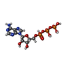
+Macromolecule #36: MAGNESIUM ION
+Macromolecule #37: N-[(benzyloxy)carbonyl]-L-leucyl-N-[(2S)-4-methyl-1-oxopentan-2-y...

+Macromolecule #38: ZINC ION
-Experimental details
-Structure determination
| Method | cryo EM |
|---|---|
 Processing Processing | single particle reconstruction |
| Aggregation state | particle |
-Sample preparation
| Buffer | pH: 7.4 |
|---|---|
| Vitrification | Cryogen name: ETHANE |
- Electron microscopy
| Microscope | TFS KRIOS |
|---|---|
| Image recording | Film or detector model: GATAN K3 (6k x 4k) / Average electron dose: 40.0 e/Å2 |
| Electron beam | Acceleration voltage: 300 kV / Electron source:  FIELD EMISSION GUN FIELD EMISSION GUN |
| Electron optics | Illumination mode: FLOOD BEAM / Imaging mode: BRIGHT FIELD / Nominal defocus max: 1.8 µm / Nominal defocus min: 0.8 µm |
| Experimental equipment |  Model: Titan Krios / Image courtesy: FEI Company |
