 Movie
Movie Controller


+ Open data

- Basic information
| Entry |  | ||||||||||||
|---|---|---|---|---|---|---|---|---|---|---|---|---|---|
| Title | Cryo-EM structure of bacteriophage T1 stopper-tail terminator | ||||||||||||
 Map data Map data | |||||||||||||
 Sample Sample |
| ||||||||||||
 Keywords Keywords | stopper / tail terminator / phage / VIRUS PROTEIN / VIRAL PROTEIN | ||||||||||||
| Function / homology | Protein of unknown function DUF4128 / Phage tail terminator protein / Tail protein / Uncharacterized protein Function and homology information Function and homology information | ||||||||||||
| Biological species |  Escherichia phage T1 (virus) Escherichia phage T1 (virus) | ||||||||||||
| Method | single particle reconstruction / cryo EM / Resolution: 3.6 Å | ||||||||||||
 Authors Authors | Chen Y / Liu HR | ||||||||||||
| Funding support |  China, 3 items China, 3 items
| ||||||||||||
 Citation Citation | Journal: Viruses / Year: 2025 Title: The In Situ Structure of T-Series T1 Reveals a Conserved Lambda-Like Tail Tip. Authors: Yuan Chen / Hao Xiao / Junquan Zhou / Zeng Peng / Yuning Peng / Jingdong Song / Jing Zheng / Hongrong Liu / Abstract: It is estimated that over 60% of known tailed phages are siphophages, which are characterized by a long, flexible, and non-contractile tail. Nevertheless, entire high-resolution structures of ...It is estimated that over 60% of known tailed phages are siphophages, which are characterized by a long, flexible, and non-contractile tail. Nevertheless, entire high-resolution structures of siphophages remain scarce. Using cryo-EM, we resolved the structures of T-series siphophage T1, encompassing its head, connector complex, tail tube, and tail tip, at near-atomic resolution. The density maps enabled us to build the atomic models for the majority of T1 proteins. The T1 head comprises 415 copies of the major capsid protein gp47, arranged into an icosahedron with a triangulation number of seven, decorated with 80 homologous trimers and 60 heterotrimers along the threefold and quasi-threefold axes of the icosahedron. The T1 connector complex is composed of two dodecamers (a portal and an adaptor) and two hexamers (a stopper and a tail terminator). The flexible tail tube comprises approximately 34 hexameric rings of tail tube. The extensive disulfide bond network along the successive tail rings may mediate the flexible bending. The distal tip of T1, which is cone-shaped and assembled by proteins gp33, gp34, gp36, gp37, and gp38, displays structural similarity to that of phage lambda. In conjunction with previous studies of lambda-like siphophages, our structure will facilitate further exploration of the structural and mechanistic aspects of lambda-like siphophages. | ||||||||||||
| History |
|
- Structure visualization
| Supplemental images |
|---|

- Downloads & links
-EMDB archive
| Map data | emd_62698.map.gz | 115.4 MB | EMDB map data format | |
|---|---|---|---|---|
| Header (meta data) | emd-62698-v30.xmlemd-62698.xml | 16.9 KB 16.9 KB | Display Display | EMDB header |
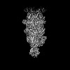
| Images |  emd_62698.png emd_62698.png | 32 KB | ||
| Filedesc metadata | emd-62698.cif.gz | 5.3 KB | ||
| Others | emd_62698_half_map_1.map.gzemd_62698_half_map_2.map.gz | 115.7 MB 115.7 MB | ||
| Archive directory |  http://ftp.pdbj.org/pub/emdb/structures/EMD-62698ftp://ftp.pdbj.org/pub/emdb/structures/EMD-62698 http://ftp.pdbj.org/pub/emdb/structures/EMD-62698ftp://ftp.pdbj.org/pub/emdb/structures/EMD-62698 | HTTPS FTP |
-Validation report
| Summary document | emd_62698_validation.pdf.gz | 1 MB | Display | EMDB validaton report |
|---|---|---|---|---|
| Full document | emd_62698_full_validation.pdf.gz | 1 MB | Display | |
| Data in XML | emd_62698_validation.xml.gz | 14.3 KB | Display | |
| Data in CIF | emd_62698_validation.cif.gz | 17 KB | Display | |
| Arichive directory | https://ftp.pdbj.org/pub/emdb/validation_reports/EMD-62698ftp://ftp.pdbj.org/pub/emdb/validation_reports/EMD-62698 | HTTPS FTP |
-Related structure data
| Related structure data |  9l0eMC  9kzjC  9l01C  9l0fC  9l9pC M: atomic model generated by this map C: citing same article ( |
|---|---|
| Similar structure data |


-Links
| EMDB pages | EMDB (EBI/PDBe) / EMDataResource |
|---|
-Map
| File | Download / File: emd_62698.map.gz / Format: CCP4 / Size: 125 MB / Type: IMAGE STORED AS FLOATING POINT NUMBER (4 BYTES) | ||||||||||||||||||||||||||||||||||||
|---|---|---|---|---|---|---|---|---|---|---|---|---|---|---|---|---|---|---|---|---|---|---|---|---|---|---|---|---|---|---|---|---|---|---|---|---|---|
| Projections & slices | Image control
Images are generated by Spider. | ||||||||||||||||||||||||||||||||||||
| Voxel size | X=Y=Z: 1.36 Å | ||||||||||||||||||||||||||||||||||||
| Density |
| ||||||||||||||||||||||||||||||||||||
| Symmetry | Space group: 1 | ||||||||||||||||||||||||||||||||||||
| Details | EMDB XML:
|
 Z (Sec.)
 Y (Row.)
 X (Col.)


-Supplemental data
-Half map: #2
| File | emd_62698_half_map_1.map | ||||||||||||
|---|---|---|---|---|---|---|---|---|---|---|---|---|---|
| Projections & Slices |
| ||||||||||||
| Density Histograms |


-Half map: #1
| File | emd_62698_half_map_2.map | ||||||||||||
|---|---|---|---|---|---|---|---|---|---|---|---|---|---|
| Projections & Slices |
| ||||||||||||
| Density Histograms |


- Sample components
-Entire : Escherichia phage T1
| Entire | Name: Escherichia phage T1 (virus) |
|---|---|
| Components |
|
-Supramolecule #1: Escherichia phage T1
| Supramolecule | Name: Escherichia phage T1 / type: complex / ID: 1 / Parent: 0 / Macromolecule list: all |
|---|---|
| Source (natural) | Organism: Escherichia phage T1 (virus) |
-Macromolecule #1: Tail protein
| Macromolecule | Name: Tail protein / type: protein_or_peptide / ID: 1 / Number of copies: 6 / Enantiomer: LEVO |
|---|---|
| Source (natural) | Organism: Escherichia phage T1 (virus) |
| Molecular weight | Theoretical: 15.191521 KDa |
| Sequence | String: MHYELSAAAR AAFLSKYRDF PHYMENRNFT PPKDGGMWLR FNYIEGDTLY LSIDRKCKSY IAIVQIGVVF PPGSGVDEAR LKAKEIADF FKDGKMLNVG YIFEGAIVHQ IVKHESGWMI PVRFTVRVDT KET UniProtKB: Tail protein |
-Macromolecule #2: stopper protein
| Macromolecule | Name: stopper protein / type: protein_or_peptide / ID: 2 / Number of copies: 6 / Enantiomer: LEVO |
|---|---|
| Source (natural) | Organism: Escherichia phage T1 (virus) |
| Molecular weight | Theoretical: 13.831523 KDa |
| Sequence | String: MNYSQIERMA RKGVAFFTDP SRPMNLIKQG EYGYDENGFE IPPMEQVIPI SGATRRPNAR EIDGETIRAS DILGIFNNDH EINEGDYIE IDGIRHVVVD ARPVQASLEP VAYRPVLRRV SVGG UniProtKB: Uncharacterized protein |
-Experimental details
-Structure determination
| Method | cryo EM |
|---|---|
 Processing Processing | single particle reconstruction |
| Aggregation state | particle |
-Sample preparation
| Buffer | pH: 7.6 |
|---|---|
| Vitrification | Cryogen name: ETHANE |
- Electron microscopy
| Microscope | TFS KRIOS |
|---|---|
| Image recording | Film or detector model: GATAN K3 (6k x 4k) / Average electron dose: 32.0 e/Å2 |
| Electron beam | Acceleration voltage: 300 kV / Electron source:  FIELD EMISSION GUN FIELD EMISSION GUN |
| Electron optics | Illumination mode: FLOOD BEAM / Imaging mode: BRIGHT FIELD / Nominal defocus max: 4.0 µm / Nominal defocus min: 2.0 µm |
| Experimental equipment |  Model: Titan Krios / Image courtesy: FEI Company |
