 Movie
Movie Controller


+ Open data

- Basic information
| Entry | Database: PDB / ID: 9l9p | |||||||||||||||||||||
|---|---|---|---|---|---|---|---|---|---|---|---|---|---|---|---|---|---|---|---|---|---|---|
| Title | Cryo-EM structure of bacteriophage T1 tail tip complex | |||||||||||||||||||||
 Components Components |
| |||||||||||||||||||||
 Keywords Keywords | VIRAL PROTEIN / tail tip complex | |||||||||||||||||||||
| Function / homology |  Function and homology information Function and homology informationsymbiont genome ejection through host cell envelope, long flexible tail mechanism / viral tail assembly / virus tail / iron-sulfur cluster binding / host cell cytoplasm / symbiont entry into host cell Similarity search - Function | |||||||||||||||||||||
| Biological species |  Escherichia phage T1 (virus) Escherichia phage T1 (virus) | |||||||||||||||||||||
| Method | ELECTRON MICROSCOPY / single particle reconstruction / cryo EM / Resolution: 4.3 Å | |||||||||||||||||||||
 Authors Authors | Chen, Y. / Liu, H.R. | |||||||||||||||||||||
| Funding support |  China, 3items China, 3items
| |||||||||||||||||||||
 Citation Citation | Journal: Viruses / Year: 2025 Title: The In Situ Structure of T-Series T1 Reveals a Conserved Lambda-Like Tail Tip. Authors: Yuan Chen / Hao Xiao / Junquan Zhou / Zeng Peng / Yuning Peng / Jingdong Song / Jing Zheng / Hongrong Liu / Abstract: It is estimated that over 60% of known tailed phages are siphophages, which are characterized by a long, flexible, and non-contractile tail. Nevertheless, entire high-resolution structures of ...It is estimated that over 60% of known tailed phages are siphophages, which are characterized by a long, flexible, and non-contractile tail. Nevertheless, entire high-resolution structures of siphophages remain scarce. Using cryo-EM, we resolved the structures of T-series siphophage T1, encompassing its head, connector complex, tail tube, and tail tip, at near-atomic resolution. The density maps enabled us to build the atomic models for the majority of T1 proteins. The T1 head comprises 415 copies of the major capsid protein gp47, arranged into an icosahedron with a triangulation number of seven, decorated with 80 homologous trimers and 60 heterotrimers along the threefold and quasi-threefold axes of the icosahedron. The T1 connector complex is composed of two dodecamers (a portal and an adaptor) and two hexamers (a stopper and a tail terminator). The flexible tail tube comprises approximately 34 hexameric rings of tail tube. The extensive disulfide bond network along the successive tail rings may mediate the flexible bending. The distal tip of T1, which is cone-shaped and assembled by proteins gp33, gp34, gp36, gp37, and gp38, displays structural similarity to that of phage lambda. In conjunction with previous studies of lambda-like siphophages, our structure will facilitate further exploration of the structural and mechanistic aspects of lambda-like siphophages. | |||||||||||||||||||||
| History |
|
- Structure visualization
| Structure viewer | Molecule: MolmilJmol/JSmol |
|---|
- Downloads & links
-Download
| PDBx/mmCIF format | 9l9p.cif.gz | 265.2 KB | Display | PDBx/mmCIF format |
|---|---|---|---|---|
| PDB format | pdb9l9p.ent.gz | 187 KB | Display | PDB format |
| PDBx/mmJSON format | 9l9p.json.gz | Tree view | PDBx/mmJSON format | |
| Others |  Other downloads Other downloads |
-Validation report
| Arichive directory | https://data.pdbj.org/pub/pdb/validation_reports/l9/9l9pftp://data.pdbj.org/pub/pdb/validation_reports/l9/9l9p | HTTPS FTP |
|---|
-Related structure data
| Related structure data | 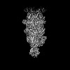 62912MC  9kzjC  9l01C  9l0eC  9l0fC M: map data used to model this data C: citing same article ( |
|---|---|
| Similar structure data |


-Links
 PDBj
PDBj- Assembly
| Deposited unit | 
|
|---|---|
| 1 |
|
-Components
-Putative tail ... , 2 types, 2 molecules SA
| #1: Protein | Mass: 130265.047 Da / Num. of mol.: 1 / Source method: isolated from a natural source / Source: (natural) Escherichia phage T1 (virus) / References: UniProt: A0A3S9W0W9 |
|---|---|
| #2: Protein | Mass: 20916.922 Da / Num. of mol.: 1 / Source method: isolated from a natural source / Source: (natural) Escherichia phage T1 (virus) / References: UniProt: Q6XQC0 |
-Putative minor tail ... , 2 types, 2 molecules JH
| #3: Protein | Mass: 29058.648 Da / Num. of mol.: 1 / Source method: isolated from a natural source / Source: (natural) Escherichia phage T1 (virus) / References: UniProt: A0A3S9W0Y3 |
|---|---|
| #4: Protein | Mass: 12992.562 Da / Num. of mol.: 1 / Source method: isolated from a natural source / Source: (natural) Escherichia phage T1 (virus) / References: UniProt: Q6XQC3 |
-Protein / Non-polymers , 2 types, 2 molecules B
| #5: Protein | Mass: 103784.391 Da / Num. of mol.: 1 / Source method: isolated from a natural source / Source: (natural) Escherichia phage T1 (virus) / References: UniProt: Q6XQC4 |
|---|---|
| #6: Chemical | ChemComp-SF4 /  Mass: 351.640 Da / Num. of mol.: 1 / Source method: obtained synthetically / Formula: Fe4S4 / Feature type: SUBJECT OF INVESTIGATION Mass: 351.640 Da / Num. of mol.: 1 / Source method: obtained synthetically / Formula: Fe4S4 / Feature type: SUBJECT OF INVESTIGATION |
-Details
| Has ligand of interest | Y |
|---|---|
| Has protein modification | N |
-Experimental details
-Experiment
| Experiment | Method: ELECTRON MICROSCOPY |
|---|---|
| EM experiment | Aggregation state: PARTICLE / 3D reconstruction method: single particle reconstruction |
- Sample preparation
| Component | Name: Escherichia phage T1 / Type: COMPLEX / Entity ID: #1-#5 / Source: NATURAL |
|---|---|
| Source (natural) | Organism: Escherichia phage T1 (virus) |
| Buffer solution | pH: 7.6 |
| Specimen | Embedding applied: NO / Shadowing applied: NO / Staining applied: NO / Vitrification applied: YES |
| Vitrification | Cryogen name: ETHANE |
- Electron microscopy imaging
| Experimental equipment |  Model: Titan Krios / Image courtesy: FEI Company |
|---|---|
| Microscopy | Model: TFS KRIOS |
| Electron gun | Electron source:  FIELD EMISSION GUN / Accelerating voltage: 300 kV / Illumination mode: FLOOD BEAM FIELD EMISSION GUN / Accelerating voltage: 300 kV / Illumination mode: FLOOD BEAM |
| Electron lens | Mode: BRIGHT FIELD / Nominal defocus max: 4000 nm / Nominal defocus min: 2000 nm |
| Image recording | Electron dose: 32 e/Å2 / Film or detector model: GATAN K3 (6k x 4k) |
- Processing
| EM software | Name: PHENIX / Version: 1.20.1_4487 / Category: model refinement | ||||||||||||||||||||||||
|---|---|---|---|---|---|---|---|---|---|---|---|---|---|---|---|---|---|---|---|---|---|---|---|---|---|
| CTF correction | Type: NONE | ||||||||||||||||||||||||
| 3D reconstruction | Resolution: 4.3 Å / Resolution method: FSC 0.143 CUT-OFF / Num. of particles: 4467 / Symmetry type: POINT | ||||||||||||||||||||||||
| Refinement | Highest resolution: 4.3 Å Stereochemistry target values: REAL-SPACE (WEIGHTED MAP SUM AT ATOM CENTERS) | ||||||||||||||||||||||||
| Refine LS restraints |
|
