 Movie
Movie Controller


+ Open data

- Basic information
| Entry | 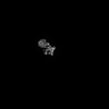 | |||||||||
|---|---|---|---|---|---|---|---|---|---|---|
| Title | RpoC2 SI domain of the spinach plastid-encoded RNA polymerase | |||||||||
 Map data Map data | Author said This map is a locally refined map of 8XZV/EMD-38799. Author said they masked a flexible domain of the map to get some pose information for model building. Consequently, the density of the unmasked region is fragmented and looks like noise. All the calculations were performed in cryoSPARC, which may have led to an overestimation of the resolution, especially in the case of intrinsic flexibility. Author said they can either adopt the resolution calculated by the EMDB and make corresponding revisions to the article, or we can provide the Auto-tightening mask used during the CryoSPARC refinement. | |||||||||
 Sample Sample |
| |||||||||
 Keywords Keywords | plastid-encoded RNA polymerase / chloroplast / spinach / TRANSCRIPTION | |||||||||
| Biological species |  Spinacia oleracea (spinach) Spinacia oleracea (spinach) | |||||||||
| Method | single particle reconstruction / cryo EM / Resolution: 3.24 Å | |||||||||
 Authors Authors | Wang G-L / Yu L-J / Lu C | |||||||||
| Funding support |  China, 1 items China, 1 items
| |||||||||
 Citation Citation | Journal: Nat Commun / Year: 2024 Title: Architecture of the spinach plastid-encoded RNA polymerase. Authors: Tongtong Wang / Guang-Lei Wang / Ying Fang / Yi Zhang / Wenxin Peng / Yue Zhou / Aihong Zhang / Long-Jiang Yu / Congming Lu / Abstract: The plastid-encoded RNA polymerase serves as the principal transcription machinery within chloroplasts, transcribing over 80% of all primary plastid transcripts. This polymerase consists of a ...The plastid-encoded RNA polymerase serves as the principal transcription machinery within chloroplasts, transcribing over 80% of all primary plastid transcripts. This polymerase consists of a prokaryotic-like core enzyme known as the plastid-encoded RNA polymerase core, and is supplemented by newly evolved associated proteins known as PAPs. However, the architecture of the plastid-encoded RNA polymerase and the possible functions of PAPs remain unknown. Here, we present the cryo-electron microscopy structure of a 19-subunit plastid-encoded RNA polymerase complex derived from spinach (Spinacia oleracea). The structure shows that the plastid-encoded RNA polymerase core resembles bacterial RNA polymerase. Twelve PAPs and two additional proteins (FLN2 and pTAC18) bind at the periphery of the plastid-encoded RNA polymerase core, forming extensive interactions that may facilitate complex assembly and stability. PAPs may also protect the complex against oxidative damage and has potential functions in transcriptional regulation. This research offers a structural basis for future investigations into the functions and regulatory mechanisms governing the transcription of plastid genes. | |||||||||
| History |
|
- Structure visualization
| Supplemental images |
|---|

- Downloads & links
-EMDB archive
| Map data | emd_61149.map.gz | 484 MB |  EMDB map data format EMDB map data format | |
|---|---|---|---|---|
| Header (meta data) | emd-61149-v30.xmlemd-61149.xml | 15.1 KB 15.1 KB | Display Display | EMDB header |
| FSC (resolution estimation) | emd_61149_fsc.xml | 16.9 KB | Display | FSC data file |
| Images |  emd_61149.png emd_61149.png | 54.6 KB | ||
| Masks | emd_61149_msk_1.map | 512 MB | Mask map | |
| Filedesc metadata | emd-61149.cif.gz | 5.5 KB | ||
| Others | emd_61149_half_map_1.map.gzemd_61149_half_map_2.map.gz | 475.1 MB 475 MB | ||
| Archive directory |  http://ftp.pdbj.org/pub/emdb/structures/EMD-61149ftp://ftp.pdbj.org/pub/emdb/structures/EMD-61149 http://ftp.pdbj.org/pub/emdb/structures/EMD-61149ftp://ftp.pdbj.org/pub/emdb/structures/EMD-61149 | HTTPS FTP |
-Related structure data


-Links
| EMDB pages | EMDB (EBI/PDBe) / EMDataResource |
|---|
-Map
| File | Download / File: emd_61149.map.gz / Format: CCP4 / Size: 512 MB / Type: IMAGE STORED AS FLOATING POINT NUMBER (4 BYTES) | ||||||||||||||||||||||||||||||||||||
|---|---|---|---|---|---|---|---|---|---|---|---|---|---|---|---|---|---|---|---|---|---|---|---|---|---|---|---|---|---|---|---|---|---|---|---|---|---|
| Annotation | Author said This map is a locally refined map of 8XZV/EMD-38799. Author said they masked a flexible domain of the map to get some pose information for model building. Consequently, the density of the unmasked region is fragmented and looks like noise. All the calculations were performed in cryoSPARC, which may have led to an overestimation of the resolution, especially in the case of intrinsic flexibility. Author said they can either adopt the resolution calculated by the EMDB and make corresponding revisions to the article, or we can provide the Auto-tightening mask used during the CryoSPARC refinement. | ||||||||||||||||||||||||||||||||||||
| Projections & slices | Image control
Images are generated by Spider. | ||||||||||||||||||||||||||||||||||||
| Voxel size | X=Y=Z: 1.1 Å | ||||||||||||||||||||||||||||||||||||
| Density |
| ||||||||||||||||||||||||||||||||||||
| Symmetry | Space group: 1 | ||||||||||||||||||||||||||||||||||||
| Details | EMDB XML:
|
 Z (Sec.)
 Y (Row.)
 X (Col.)


-Supplemental data
-Mask #1
| File | emd_61149_msk_1.map | ||||||||||||
|---|---|---|---|---|---|---|---|---|---|---|---|---|---|
| Projections & Slices |
| ||||||||||||
| Density Histograms |


-Half map: #1
| File | emd_61149_half_map_1.map | ||||||||||||
|---|---|---|---|---|---|---|---|---|---|---|---|---|---|
| Projections & Slices |
| ||||||||||||
| Density Histograms |


-Half map: #2
| File | emd_61149_half_map_2.map | ||||||||||||
|---|---|---|---|---|---|---|---|---|---|---|---|---|---|
| Projections & Slices |
| ||||||||||||
| Density Histograms |


- Sample components
-Entire : DNA-directed RNA polymerase subunit beta''
| Entire | Name: DNA-directed RNA polymerase subunit beta'' |
|---|---|
| Components |
|
-Supramolecule #1: DNA-directed RNA polymerase subunit beta''
| Supramolecule | Name: DNA-directed RNA polymerase subunit beta'' / type: complex / ID: 1 / Parent: 0 / Macromolecule list: all |
|---|---|
| Source (natural) | Organism: Spinacia oleracea (spinach) |
-Macromolecule #1: DNA-directed RNA polymerase subunit beta''
| Macromolecule | Name: DNA-directed RNA polymerase subunit beta'' / type: protein_or_peptide / ID: 1 / Enantiomer: LEVO / EC number: DNA-directed RNA polymerase |
|---|---|
| Source (natural) | Organism: Spinacia oleracea (spinach) |
| Sequence | String: MAERANLVFH NKAIDGTAMK RLISRLIDHF GMAYTSHILD QLKTLGFQQA TATSISLGID DLLTIPSKGW LVQDAEQQSL ILEKHHHYG NVHAVEKLRQ SIEIWYSTSE YLRQEMNPNF RMTDPYNPVH IMSFSGARGN VSQVHQLVGM RGLMSDPQGQ M IDLPIQSN ...String: MAERANLVFH NKAIDGTAMK RLISRLIDHF GMAYTSHILD QLKTLGFQQA TATSISLGID DLLTIPSKGW LVQDAEQQSL ILEKHHHYG NVHAVEKLRQ SIEIWYSTSE YLRQEMNPNF RMTDPYNPVH IMSFSGARGN VSQVHQLVGM RGLMSDPQGQ M IDLPIQSN LREGLSLTEY IISCYGARKG VVDTAVRTSD AGYLTRRLVE VVQHIVVRRR DCGTIRGISV SPQNSTMPER IL IQTLIGR VLADDIYMGS RCIATRNQDI GVGLVNRFIT LRTQLISIRT PFTCRSASWI CRLCYGRSPT HGGLVELGEA VGI IAGQSI GEPGTQLTLR TFHTGGVFTG GTAEHVRAPS NGKIQFNEDL VHPTRTRHGH PAFLCYIDLY VTIESDDILH NVNI PPKSF LLVQNDQYVE SEQVIAEIRA GTSTLNFKER VRKHIYSDSE GEMHWSTDVY HAPEFTYGNV HLLPKTSHLW VLSGK PYRS SVVPFSLSKD QDQMNTHSLS FEQIYISNPS VTNDQVKDKL SDSFSKKEDR ITDYSELNRI GHCNLIYPAK NLDLLA KKR RNRFIIPFQG SQERKKELMS LSGISIEIPI NGIFRKNSIF AYFDDPRYRR KSSGITKYGT IEMHSIVKKE DLIEYRG VK EFRPKYQMKV DRFFFIPEEV HILAGSSSIM VRNNSIIGVD TWITLNTRSR IGGVVRVERK KKKIELTIFS GDIHFPGE T DKISRHSGIL IPPSRKNSKD SKNLKKWIYV QRITPTKKKY FVLVRPVVPY EITDGINLAT LFPQDLLQER DNVQLRVVN YILYGNGKVT RGISDTSIQL VRTCLVLNWN QDKKGSSIEE ARGSFVEVRT NGMIQDFLKV NLVKPAISYI SKRNDPSSEK KEGSDHTNM NPFYSIYIYP KTKLQKSFNQ NQGTVRTLLG INKECQFFLI LSSSNCFRIG PFKGVKYPKE LIKKDPLIPI R NSFGPLGT ALQIANFFSF YYLITHNQIL VTNYLQLDNL KQTFQPFKFQ YYLMDENGRI YNPDPCSNII FNPFKLNWYF LH YHFCEET STKIDLGQFV CENVCITKKG THLKSGQVLI VQFDSVVIRS AKPYLATPGA TLHGHYGEII YEGDTLVTFI YEK SRSGDI TQGLPKVEQV LEVRSIDSIS INLEKRIDSW NERITRILGS PWGFLIGAEL TIAQSRISLV NKIQKVYRSQ GVQI HNRHI EIIVRQITSK VLVSEDGMSN VFLPGELIGL FRAERTGRAL EEAICYRATL LGITRASLNT QSFISEASFQ ETARV LAKA ALRGRIDWLK GLKENVVLGG MIPVGTGFKG FVHHSSQHKD IPLKTKKQNL FEGEMGDILF YHRELFESCL SKN |
-Experimental details
-Structure determination
| Method | cryo EM |
|---|---|
 Processing Processing | single particle reconstruction |
| Aggregation state | particle |
-Sample preparation
| Concentration | 2 mg/mL |
|---|---|
| Buffer | pH: 7.6 |
| Vitrification | Cryogen name: ETHANE / Chamber humidity: 100 % / Chamber temperature: 298 K / Instrument: FEI VITROBOT MARK IV |
- Electron microscopy
| Microscope | FEI TITAN KRIOS |
|---|---|
| Image recording | Film or detector model: GATAN K3 (6k x 4k) / Average electron dose: 50.0 e/Å2 |
| Electron beam | Acceleration voltage: 300 kV / Electron source:  FIELD EMISSION GUN FIELD EMISSION GUN |
| Electron optics | Illumination mode: FLOOD BEAM / Imaging mode: BRIGHT FIELD / Nominal defocus max: 2.0 µm / Nominal defocus min: 0.8 µm |
| Experimental equipment |  Model: Titan Krios / Image courtesy: FEI Company |

