 Movie
Movie Controller

[English] 日本語
 Yorodumi
- EMDB-5744: Zernike Phase Contrast electron cryo-tomography of cyanophage Syn... -
+ Open data

- Basic information
| Entry | Database: EMDB / ID: EMD-5744 | |||||||||
|---|---|---|---|---|---|---|---|---|---|---|
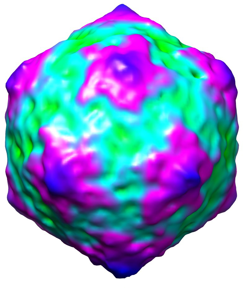
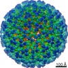
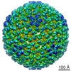
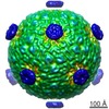
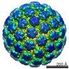
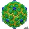
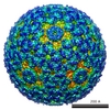
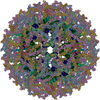
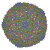
| Title | Zernike Phase Contrast electron cryo-tomography of cyanophage Syn5 assembly intermediates | |||||||||
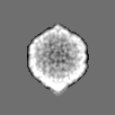
 Map data Map data | Cyanophage Syn5 assembly intermediate - DNA-containing capsid without tail or horn | |||||||||
 Sample Sample |
| |||||||||
 Keywords Keywords | Zernike phase contrast / electron cryo-tomography / cyanobacteria / dsDNA cyanophage / phage assembly / capsid shell conformational change | |||||||||
| Biological species |  Synechococcus phage Syn5 (virus) Synechococcus phage Syn5 (virus) | |||||||||
| Method | subtomogram averaging / cryo EM / Resolution: 57.0 Å | |||||||||
 Authors Authors | Dai W / Fu C / Raytcheva D / Flanagan J / Khant HA / Liu X / Rochat RH / Haase-Pettingell C / Piret J / Ludtke SJ ...Dai W / Fu C / Raytcheva D / Flanagan J / Khant HA / Liu X / Rochat RH / Haase-Pettingell C / Piret J / Ludtke SJ / Nagayama K / Schmid MF / King JA / Chiu W | |||||||||
 Citation Citation | Journal: Nature / Year: 2013 Title: Visualizing virus assembly intermediates inside marine cyanobacteria. Authors: Wei Dai / Caroline Fu / Desislava Raytcheva / John Flanagan / Htet A Khant / Xiangan Liu / Ryan H Rochat / Cameron Haase-Pettingell / Jacqueline Piret / Steve J Ludtke / Kuniaki Nagayama / ...Authors: Wei Dai / Caroline Fu / Desislava Raytcheva / John Flanagan / Htet A Khant / Xiangan Liu / Ryan H Rochat / Cameron Haase-Pettingell / Jacqueline Piret / Steve J Ludtke / Kuniaki Nagayama / Michael F Schmid / Jonathan A King / Wah Chiu /  Abstract: Cyanobacteria are photosynthetic organisms responsible for ∼25% of organic carbon fixation on the Earth. These bacteria began to convert solar energy and carbon dioxide into bioenergy and oxygen ...Cyanobacteria are photosynthetic organisms responsible for ∼25% of organic carbon fixation on the Earth. These bacteria began to convert solar energy and carbon dioxide into bioenergy and oxygen more than two billion years ago. Cyanophages, which infect these bacteria, have an important role in regulating the marine ecosystem by controlling cyanobacteria community organization and mediating lateral gene transfer. Here we visualize the maturation process of cyanophage Syn5 inside its host cell, Synechococcus, using Zernike phase contrast electron cryo-tomography (cryoET). This imaging modality yields dramatic enhancement of image contrast over conventional cryoET and thus facilitates the direct identification of subcellular components, including thylakoid membranes, carboxysomes and polyribosomes, as well as phages, inside the congested cytosol of the infected cell. By correlating the structural features and relative abundance of viral progeny within cells at different stages of infection, we identify distinct Syn5 assembly intermediates. Our results indicate that the procapsid releases scaffolding proteins and expands its volume at an early stage of genome packaging. Later in the assembly process, we detected full particles with a tail either with or without an additional horn. The morphogenetic pathway we describe here is highly conserved and was probably established long before that of double-stranded DNA viruses infecting more complex organisms. | |||||||||
| History |
|
- Structure visualization
| Movie |
 Movie viewer Movie viewer |
|---|---|
| Structure viewer | EM map: SurfViewMolmilJmol/JSmol |
| Supplemental images |
- Downloads & links
-EMDB archive
| Map data | emd_5744.map.gz | 770.1 KB | EMDB map data format | |
|---|---|---|---|---|
| Header (meta data) | emd-5744-v30.xmlemd-5744.xml | 11.4 KB 11.4 KB | Display Display | EMDB header |
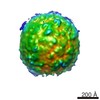
| Images |  emd_5744.jpg emd_5744.jpg | 332.1 KB | ||
| Archive directory |  http://ftp.pdbj.org/pub/emdb/structures/EMD-5744ftp://ftp.pdbj.org/pub/emdb/structures/EMD-5744 http://ftp.pdbj.org/pub/emdb/structures/EMD-5744ftp://ftp.pdbj.org/pub/emdb/structures/EMD-5744 | HTTPS FTP |
-Validation report
| Summary document | emd_5744_validation.pdf.gz | 78.6 KB | Display | EMDB validaton report |
|---|---|---|---|---|
| Full document | emd_5744_full_validation.pdf.gz | 77.7 KB | Display | |
| Data in XML | emd_5744_validation.xml.gz | 494 B | Display | |
| Arichive directory | https://ftp.pdbj.org/pub/emdb/validation_reports/EMD-5744ftp://ftp.pdbj.org/pub/emdb/validation_reports/EMD-5744 | HTTPS FTP |
-Related structure data

-Links
| EMDB pages | EMDB (EBI/PDBe) / EMDataResource |
|---|
-Map
| File | Download / File: emd_5744.map.gz / Format: CCP4 / Size: 7.8 MB / Type: IMAGE STORED AS FLOATING POINT NUMBER (4 BYTES) | ||||||||||||||||||||||||||||||||||||||||||||||||||||||||||||||||||||
|---|---|---|---|---|---|---|---|---|---|---|---|---|---|---|---|---|---|---|---|---|---|---|---|---|---|---|---|---|---|---|---|---|---|---|---|---|---|---|---|---|---|---|---|---|---|---|---|---|---|---|---|---|---|---|---|---|---|---|---|---|---|---|---|---|---|---|---|---|---|
| Annotation | Cyanophage Syn5 assembly intermediate - DNA-containing capsid without tail or horn | ||||||||||||||||||||||||||||||||||||||||||||||||||||||||||||||||||||
| Projections & slices | Image control
Images are generated by Spider. | ||||||||||||||||||||||||||||||||||||||||||||||||||||||||||||||||||||
| Voxel size | X=Y=Z: 9.04 Å | ||||||||||||||||||||||||||||||||||||||||||||||||||||||||||||||||||||
| Density |
| ||||||||||||||||||||||||||||||||||||||||||||||||||||||||||||||||||||
| Symmetry | Space group: 1 | ||||||||||||||||||||||||||||||||||||||||||||||||||||||||||||||||||||
| Details | EMDB XML:
CCP4 map header:
| ||||||||||||||||||||||||||||||||||||||||||||||||||||||||||||||||||||
 Z (Sec.)
 Y (Row.)
 X (Col.)


-Supplemental data
- Sample components
-Entire : Cyanophage Syn5 assembly intermediate inside cyanobacteria Synech...
| Entire | Name: Cyanophage Syn5 assembly intermediate inside cyanobacteria Synechococcus WH8109 cells |
|---|---|
| Components |
|
-Supramolecule #1000: Cyanophage Syn5 assembly intermediate inside cyanobacteria Synech...
| Supramolecule | Name: Cyanophage Syn5 assembly intermediate inside cyanobacteria Synechococcus WH8109 cells type: sample / ID: 1000 / Number unique components: 1 |
|---|
-Supramolecule #1: Synechococcus phage Syn5
| Supramolecule | Name: Synechococcus phage Syn5 / type: virus / ID: 1 Details: DNA-containing capsid without tail or horn - assembly intermediate NCBI-ID: 438482 / Sci species name: Synechococcus phage Syn5 / Sci species strain: Syn5 / Database: NCBI / Virus type: VIRION / Virus isolate: STRAIN / Virus enveloped: No / Virus empty: No |
|---|---|
| Host (natural) | Organism:  Synechococcus sp. WH 8109 (bacteria) / Strain: WH8109 / synonym: BACTERIA(EUBACTERIA) Synechococcus sp. WH 8109 (bacteria) / Strain: WH8109 / synonym: BACTERIA(EUBACTERIA) |
| Virus shell | Shell ID: 1 / Diameter: 660 Å |
-Experimental details
-Structure determination
| Method | cryo EM |
|---|---|
 Processing Processing | subtomogram averaging |
| Aggregation state | particle |
-Sample preparation
| Buffer | pH: 8 / Details: artificial sea water |
|---|---|
| Grid | Details: 200 mesh Quantifoil holey grids, glow discharged |
| Vitrification | Cryogen name: ETHANE / Chamber humidity: 95 % / Chamber temperature: 85 K / Instrument: FEI VITROBOT MARK III / Method: Blot for 3 seconds before plunging |
- Electron microscopy
| Microscope | JEOL 2200FS |
|---|---|
| Temperature | Min: 93 K / Max: 105 K / Average: 103 K |
| Alignment procedure | Legacy - Astigmatism: Objective lens astigmatism was corrected at 100,000 times magnification |
| Specialist optics | Energy filter - Name: in-column GIF energy filter / Energy filter - Lower energy threshold: 15.0 eV / Energy filter - Upper energy threshold: 20.0 eV |
| Date | Jun 1, 2011 |
| Image recording | Category: CCD / Film or detector model: GATAN ULTRASCAN 4000 (4k x 4k) / Number real images: 42 / Average electron dose: 1 e/Å2 / Camera length: 1200 / Bits/pixel: 16 |
| Electron beam | Acceleration voltage: 200 kV / Electron source:  FIELD EMISSION GUN FIELD EMISSION GUN |
| Electron optics | Calibrated magnification: 25000 / Illumination mode: FLOOD BEAM / Imaging mode: BRIGHT FIELD / Cs: 4.1 mm / Nominal defocus max: 1.0 µm / Nominal defocus min: 0.0 µm / Nominal magnification: 25000 |
| Sample stage | Specimen holder model: GATAN LIQUID NITROGEN / Tilt series - Axis1 - Min angle: -60 ° / Tilt series - Axis1 - Max angle: 60 ° |
-Image processing
| Details | The subtomograms were extracted from infected cells. |
|---|---|
| Final reconstruction | Algorithm: OTHER / Resolution.type: BY AUTHOR / Resolution: 57.0 Å / Resolution method: FSC 0.143 CUT-OFF / Software - Name: IMOD / Number subtomograms used: 103 |
| Final 3D classification | Number classes: 1 |
