 Movie
Movie Controller

[English] 日本語
 Yorodumi
- EMDB-5713: Electron microscopy of negatively-stained gp12 tubular protein of... -
+ Open data

- Basic information
| Entry | Database: EMDB / ID: EMD-5713 | |||||||||
|---|---|---|---|---|---|---|---|---|---|---|
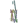
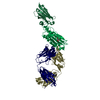
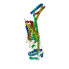
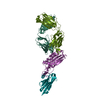
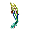
| Title | Electron microscopy of negatively-stained gp12 tubular protein of T7 bacteriophage | |||||||||
 Map data Map data | Reconstruction of gp12 tubular protein of T7 bacteriophage | |||||||||
 Sample Sample |
| |||||||||
 Keywords Keywords | Bacteriophage / DNA ejection / tail structure / tail tubular protein | |||||||||
| Biological species |   Enterobacteria phage T7 (virus) Enterobacteria phage T7 (virus) | |||||||||
| Method | single particle reconstruction / negative staining / Resolution: 26.0 Å | |||||||||
 Authors Authors | Cuervo A / Pulido-Cid M / Chagoyen M / Arranz R / Gonzalez-Garcia VA / Garcia-Doval C / Caston JR / Valpuesta JM / van Raaij MJ / Martin-Benito J / Carrascosa JL | |||||||||
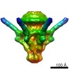
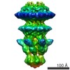
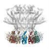
 Citation Citation | Journal: J Biol Chem / Year: 2013 Title: Structural characterization of the bacteriophage T7 tail machinery. Authors: Ana Cuervo / Mar Pulido-Cid / Mónica Chagoyen / Rocío Arranz / Verónica A González-García / Carmela Garcia-Doval / José R Castón / José M Valpuesta / Mark J van Raaij / Jaime Martín- ...Authors: Ana Cuervo / Mar Pulido-Cid / Mónica Chagoyen / Rocío Arranz / Verónica A González-García / Carmela Garcia-Doval / José R Castón / José M Valpuesta / Mark J van Raaij / Jaime Martín-Benito / José L Carrascosa /  Abstract: Most bacterial viruses need a specialized machinery, called "tail," to inject their genomes inside the bacterial cytoplasm without disrupting the cellular integrity. Bacteriophage T7 is a well ...Most bacterial viruses need a specialized machinery, called "tail," to inject their genomes inside the bacterial cytoplasm without disrupting the cellular integrity. Bacteriophage T7 is a well characterized member of the Podoviridae family infecting Escherichia coli, and it has a short noncontractile tail that assembles sequentially on the viral head after DNA packaging. The T7 tail is a complex of around 2.7 MDa composed of at least four proteins as follows: the connector (gene product 8, gp8), the tail tubular proteins gp11 and gp12, and the fibers (gp17). Using cryo-electron microscopy and single particle image reconstruction techniques, we have determined the precise topology of the tail proteins by comparing the structure of the T7 tail extracted from viruses and a complex formed by recombinant gp8, gp11, and gp12 proteins. Furthermore, the order of assembly of the structural components within the complex was deduced from interaction assays with cloned and purified tail proteins. The existence of common folds among similar tail proteins allowed us to obtain pseudo-atomic threaded models of gp8 (connector) and gp11 (gatekeeper) proteins, which were docked into the corresponding cryo-EM volumes of the tail complex. This pseudo-atomic model of the connector-gatekeeper interaction revealed the existence of a common molecular architecture among viruses belonging to the three tailed bacteriophage families, strongly suggesting that a common molecular mechanism has been favored during evolution to coordinate the transition between DNA packaging and tail assembly. | |||||||||
| History |
|
- Structure visualization
| Movie |
 Movie viewer Movie viewer |
|---|---|
| Structure viewer | EM map: SurfViewMolmilJmol/JSmol |
| Supplemental images |

- Downloads & links
-EMDB archive
| Map data | emd_5713.map.gz | 1.6 MB | EMDB map data format | |
|---|---|---|---|---|
| Header (meta data) | emd-5713-v30.xmlemd-5713.xml | 9.9 KB 9.9 KB | Display Display | EMDB header |
| Images | 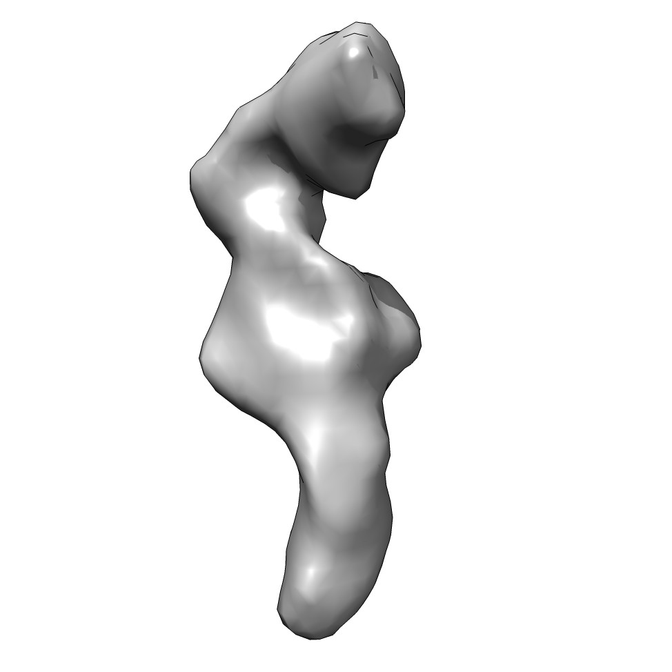 emd_5713_1.jpg emd_5713_1.jpg | 40.8 KB | ||
| Archive directory |  http://ftp.pdbj.org/pub/emdb/structures/EMD-5713ftp://ftp.pdbj.org/pub/emdb/structures/EMD-5713 http://ftp.pdbj.org/pub/emdb/structures/EMD-5713ftp://ftp.pdbj.org/pub/emdb/structures/EMD-5713 | HTTPS FTP |
-Related structure data


-Links
| EMDB pages | EMDB (EBI/PDBe) / EMDataResource |
|---|
-Map
| File | Download / File: emd_5713.map.gz / Format: CCP4 / Size: 1.9 MB / Type: IMAGE STORED AS FLOATING POINT NUMBER (4 BYTES) | ||||||||||||||||||||||||||||||||||||||||||||||||||||||||||||||||||||
|---|---|---|---|---|---|---|---|---|---|---|---|---|---|---|---|---|---|---|---|---|---|---|---|---|---|---|---|---|---|---|---|---|---|---|---|---|---|---|---|---|---|---|---|---|---|---|---|---|---|---|---|---|---|---|---|---|---|---|---|---|---|---|---|---|---|---|---|---|---|
| Annotation | Reconstruction of gp12 tubular protein of T7 bacteriophage | ||||||||||||||||||||||||||||||||||||||||||||||||||||||||||||||||||||
| Projections & slices | Image control
Images are generated by Spider. | ||||||||||||||||||||||||||||||||||||||||||||||||||||||||||||||||||||
| Voxel size | X=Y=Z: 4.14 Å | ||||||||||||||||||||||||||||||||||||||||||||||||||||||||||||||||||||
| Density |
| ||||||||||||||||||||||||||||||||||||||||||||||||||||||||||||||||||||
| Symmetry | Space group: 1 | ||||||||||||||||||||||||||||||||||||||||||||||||||||||||||||||||||||
| Details | EMDB XML:
CCP4 map header:
| ||||||||||||||||||||||||||||||||||||||||||||||||||||||||||||||||||||
 Z (Sec.)
 Y (Row.)
 X (Col.)


-Supplemental data
- Sample components
-Entire : T7 tail tubular protein gp12
| Entire | Name: T7 tail tubular protein gp12 |
|---|---|
| Components |
|
-Supramolecule #1000: T7 tail tubular protein gp12
| Supramolecule | Name: T7 tail tubular protein gp12 / type: sample / ID: 1000 / Oligomeric state: Monomer / Number unique components: 1 |
|---|---|
| Molecular weight | Theoretical: 90 KDa |
-Macromolecule #1: gp12
| Macromolecule | Name: gp12 / type: protein_or_peptide / ID: 1 / Name.synonym: Tail tubular protein / Details: The protein was cloned with a His-tag. / Number of copies: 1 / Oligomeric state: Monomer / Recombinant expression: Yes |
|---|---|
| Source (natural) | Organism: Enterobacteria phage T7 (virus) / synonym: T7 Bacteriophage |
| Molecular weight | Theoretical: 90 KDa |
| Recombinant expression | Organism:  |
-Experimental details
-Structure determination
| Method | negative staining |
|---|---|
 Processing Processing | single particle reconstruction |
| Aggregation state | particle |
-Sample preparation
| Concentration | 0.01 mg/mL |
|---|---|
| Buffer | pH: 7.8 / Details: 50mM Tris-HCl, 10mM MgCl2, 100 mM NaCl |
| Staining | Type: NEGATIVE Details: Samples were applied to grids for 1 min, washed with water, blotted, and stained for 1 min in uranyl acetate. |
| Grid | Details: 400 mesh cupper grid coated with a carbon layer, glow discharged |
| Vitrification | Cryogen name: NONE / Instrument: OTHER |
- Electron microscopy
| Microscope | FEI TECNAI F20 |
|---|---|
| Date | Nov 8, 2012 |
| Image recording | Category: CCD / Film or detector model: FEI EAGLE (4k x 4k) / Digitization - Sampling interval: 15 µm / Number real images: 181 / Average electron dose: 10 e/Å2 |
| Electron beam | Acceleration voltage: 200 kV / Electron source:  FIELD EMISSION GUN FIELD EMISSION GUN |
| Electron optics | Calibrated magnification: 108696 / Illumination mode: FLOOD BEAM / Imaging mode: BRIGHT FIELD / Cs: 2.26 mm / Nominal defocus max: 3.5 µm / Nominal defocus min: 1.5 µm / Nominal magnification: 108696 |
| Sample stage | Specimen holder model: PHILIPS ROTATION HOLDER |
| Experimental equipment |  Model: Tecnai F20 / Image courtesy: FEI Company |
-Image processing
| CTF correction | Details: each micrograph |
|---|---|
| Final reconstruction | Algorithm: OTHER / Resolution.type: BY AUTHOR / Resolution: 26.0 Å / Resolution method: FSC 0.33 CUT-OFF / Software - Name: XMIPP, SPIDER, EMAN / Number images used: 640 |
