 ムービー
ムービー コントローラー


+ データを開く

- 基本情報
基本情報
| 登録情報 | データベース: EMDB / ID: EMD-5672 | |||||||||
|---|---|---|---|---|---|---|---|---|---|---|
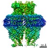
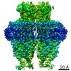
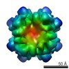
| タイトル | CryoEM Structure of the KaiBC Complex | |||||||||
 マップデータ マップデータ | Reconstruction of KaiB-KaiC(489 deletion) | |||||||||
 試料 試料 |
| |||||||||
 キーワード キーワード | Circadian oscillator / cryoEM / MDFF / protein-protein interface / Synechococcus elongatus | |||||||||
| 機能・相同性 |  機能・相同性情報 機能・相同性情報: / : / negative regulation of phosphorylation / regulation of phosphorelay signal transduction system / negative regulation of circadian rhythm / entrainment of circadian clock / protein serine/threonine/tyrosine kinase activity / circadian rhythm / regulation of circadian rhythm / 加水分解酵素; 酸無水物に作用; 酸無水物に作用・細胞または細胞小器官の運動に関与 ...: / : / negative regulation of phosphorylation / regulation of phosphorelay signal transduction system / negative regulation of circadian rhythm / entrainment of circadian clock / protein serine/threonine/tyrosine kinase activity / circadian rhythm / regulation of circadian rhythm / 加水分解酵素; 酸無水物に作用; 酸無水物に作用・細胞または細胞小器官の運動に関与 / non-specific serine/threonine protein kinase / protein serine kinase activity / protein serine/threonine kinase activity / regulation of DNA-templated transcription / magnesium ion binding / ATP hydrolysis activity / DNA binding / ATP binding / identical protein binding / plasma membrane / cytoplasm 類似検索 - 分子機能 | |||||||||
| 生物種 |  Synechococcus elongatus (バクテリア) Synechococcus elongatus (バクテリア) | |||||||||
| 手法 | 単粒子再構成法 / クライオ電子顕微鏡法 / 解像度: 16.0 Å | |||||||||
 データ登録者 データ登録者 | Villarreal SA / Pattanayek R / Williams DR / Mori T / Qin X / Johnson CH / Egli M / Stewart PL | |||||||||
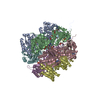
 引用 引用 | ジャーナル: J Mol Biol / 年: 2013 タイトル: CryoEM and molecular dynamics of the circadian KaiB-KaiC complex indicates that KaiB monomers interact with KaiC and block ATP binding clefts. 著者: Seth A Villarreal / Rekha Pattanayek / Dewight R Williams / Tetsuya Mori / Ximing Qin / Carl H Johnson / Martin Egli / Phoebe L Stewart /  要旨: The circadian control of cellular processes in cyanobacteria is regulated by a posttranslational oscillator formed by three Kai proteins. During the oscillator cycle, KaiA serves to promote ...The circadian control of cellular processes in cyanobacteria is regulated by a posttranslational oscillator formed by three Kai proteins. During the oscillator cycle, KaiA serves to promote autophosphorylation of KaiC while KaiB counteracts this effect. Here, we present a crystallographic structure of the wild-type Synechococcus elongatus KaiB and a cryo-electron microscopy (cryoEM) structure of a KaiBC complex. The crystal structure shows the expected dimer core structure and significant conformational variations of the KaiB C-terminal region, which is functionally important in maintaining rhythmicity. The KaiBC sample was formed with a C-terminally truncated form of KaiC, KaiC-Δ489, which is persistently phosphorylated. The KaiB-KaiC-Δ489 structure reveals that the KaiC hexamer can bind six monomers of KaiB, which form a continuous ring of density in the KaiBC complex. We performed cryoEM-guided molecular dynamics flexible fitting simulations with crystal structures of KaiB and KaiC to probe the KaiBC protein-protein interface. This analysis indicated a favorable binding mode for the KaiB monomer on the CII end of KaiC, involving two adjacent KaiC subunits and spanning an ATP binding cleft. A KaiC mutation, R468C, which has been shown to affect the affinity of KaiB for KaiC and lengthen the period in a bioluminescence rhythm assay, is found within the middle of the predicted KaiBC interface. The proposed KaiB binding mode blocks access to the ATP binding cleft in the CII ring of KaiC, which provides insight into how KaiB might influence the phosphorylation status of KaiC. | |||||||||
| 履歴 |
|
- 構造の表示
構造の表示
| ムービー |
ムービービューア |
|---|---|
| 構造ビューア | EMマップ: SurfViewMolmilJmol/JSmol |
| 添付画像 |

- ダウンロードとリンク
-EMDBアーカイブ
| マップデータ | emd_5672.map.gz | 1.5 MB | EMDBマップデータ形式 | |
|---|---|---|---|---|
| ヘッダ (付随情報) | emd-5672-v30.xmlemd-5672.xml | 11.3 KB 11.3 KB | 表示 表示 | EMDBヘッダ |
| 画像 | emd_5672_1.tif | 163.7 KB | ||
| アーカイブディレクトリ |  http://ftp.pdbj.org/pub/emdb/structures/EMD-5672ftp://ftp.pdbj.org/pub/emdb/structures/EMD-5672 http://ftp.pdbj.org/pub/emdb/structures/EMD-5672ftp://ftp.pdbj.org/pub/emdb/structures/EMD-5672 | HTTPS FTP |
-検証レポート
| 文書・要旨 | emd_5672_validation.pdf.gz | 79.2 KB | 表示 | EMDB検証レポート |
|---|---|---|---|---|
| 文書・詳細版 | emd_5672_full_validation.pdf.gz | 78.3 KB | 表示 | |
| XML形式データ | emd_5672_validation.xml.gz | 495 B | 表示 | |
| アーカイブディレクトリ | https://ftp.pdbj.org/pub/emdb/validation_reports/EMD-5672ftp://ftp.pdbj.org/pub/emdb/validation_reports/EMD-5672 | HTTPS FTP |
-関連構造データ


-リンク
| EMDBのページ | EMDB (EBI/PDBe) / EMDataResource |
|---|---|
| 「今月の分子」の関連する項目 |

-マップ
| ファイル | ダウンロード / ファイル: emd_5672.map.gz / 形式: CCP4 / 大きさ: 1.9 MB / タイプ: IMAGE STORED AS FLOATING POINT NUMBER (4 BYTES) | ||||||||||||||||||||||||||||||||||||||||||||||||||||||||||||||||||||
|---|---|---|---|---|---|---|---|---|---|---|---|---|---|---|---|---|---|---|---|---|---|---|---|---|---|---|---|---|---|---|---|---|---|---|---|---|---|---|---|---|---|---|---|---|---|---|---|---|---|---|---|---|---|---|---|---|---|---|---|---|---|---|---|---|---|---|---|---|---|
| 注釈 | Reconstruction of KaiB-KaiC(489 deletion) | ||||||||||||||||||||||||||||||||||||||||||||||||||||||||||||||||||||
| 投影像・断面図 | 画像のコントロール
画像は Spider により作成 | ||||||||||||||||||||||||||||||||||||||||||||||||||||||||||||||||||||
| ボクセルのサイズ | X=Y=Z: 2.32 Å | ||||||||||||||||||||||||||||||||||||||||||||||||||||||||||||||||||||
| 密度 |
| ||||||||||||||||||||||||||||||||||||||||||||||||||||||||||||||||||||
| 対称性 | 空間群: 1 | ||||||||||||||||||||||||||||||||||||||||||||||||||||||||||||||||||||
| 詳細 | EMDB XML:
CCP4マップ ヘッダ情報:
| ||||||||||||||||||||||||||||||||||||||||||||||||||||||||||||||||||||
 Z (Sec.)
 Y (Row.)
 X (Col.)


-添付データ
- 試料の構成要素
-全体 : KaiB in complex with KaiC(489del)
| 全体 | 名称: KaiB in complex with KaiC(489del) |
|---|---|
| 要素 |
|
-超分子 #1000: KaiB in complex with KaiC(489del)
| 超分子 | 名称: KaiB in complex with KaiC(489del) / タイプ: sample / ID: 1000 詳細: Visualization of the sample showed complexes of KaiBC and KaiC. 集合状態: One Hexamer of KaiC binds to 6 monomers of KaiB Number unique components: 2 |
|---|---|
| 分子量 | 理論値: 510 KDa |
-分子 #1: KaiB
| 分子 | 名称: KaiB / タイプ: protein_or_peptide / ID: 1 / Name.synonym: Circadian clock protein KaiB / コピー数: 6 / 集合状態: Dimer / 組換発現: Yes |
|---|---|
| 由来(天然) | 生物種: Synechococcus elongatus (バクテリア) / 株: PCC 7942 / 細胞中の位置: Cytoplasm |
| 分子量 | 理論値: 12 KDa |
| 組換発現 | 生物種: |
| 配列 | UniProtKB: Circadian clock oscillator protein KaiB / GO: GO: 3773505 / InterPro: Circadian clock protein KaiB |
-分子 #2: KaiC
| 分子 | 名称: KaiC / タイプ: protein_or_peptide / ID: 2 / Name.synonym: Circadian clock protein kinase KaiC / 詳細: C terminal truncation of KaiC at 489 / コピー数: 1 / 集合状態: Hexamer / 組換発現: Yes |
|---|---|
| 由来(天然) | 生物種: Synechococcus elongatus (バクテリア) / 株: PCC 7942 / 細胞中の位置: Cytoplasm |
| 分子量 | 理論値: 340 KDa |
| 組換発現 | 生物種: |
| 配列 | UniProtKB: Circadian clock oscillator protein KaiC / GO: GO: 3773504 / InterPro: Circadian clock KaiC, bacteria |
-実験情報
-構造解析
| 手法 | クライオ電子顕微鏡法 |
|---|---|
 解析 解析 | 単粒子再構成法 |
| 試料の集合状態 | particle |
-試料調製
| 緩衝液 | 詳細: 20 mM HEPES-NaOH, pH 8.0, 150 mM NaCl, 5 mM MgCl2, 0.5 mM EDTA |
|---|---|
| グリッド | 詳細: homemade holey carbon film and C-flat grids (Protochips, Inc.) |
| 凍結 | 凍結剤: ETHANE / チャンバー内温度: 184 K / 装置: HOMEMADE PLUNGER |
- 電子顕微鏡法
| 顕微鏡 | FEI POLARA 300 |
|---|---|
| 温度 | 最低: 98 K / 最高: 105 K / 平均: 101 K |
| 日付 | 2010年6月2日 |
| 撮影 | カテゴリ: CCD フィルム・検出器のモデル: GATAN ULTRASCAN 4000 (4k x 4k) 実像数: 11734 / ビット/ピクセル: 16 |
| 電子線 | 加速電圧: 300 kV / 電子線源:  FIELD EMISSION GUN FIELD EMISSION GUN |
| 電子光学系 | 倍率(補正後): 254669 / 照射モード: FLOOD BEAM / 撮影モード: BRIGHT FIELD / Cs: 2.26 mm / 最大 デフォーカス(公称値): -5.0 µm / 最小 デフォーカス(公称値): -1.0 µm / 倍率(公称値): 200000 |
| 試料ステージ | 試料ホルダーモデル: OTHER |
| 実験機器 |  モデル: Tecnai Polara / 画像提供: FEI Company |
-画像解析
| 詳細 | The particles were selected manually. |
|---|---|
| CTF補正 | 詳細: Each micrograph |
| 最終 再構成 | アルゴリズム: OTHER / 解像度のタイプ: BY AUTHOR / 解像度: 16.0 Å / 解像度の算出法: FSC 0.5 CUT-OFF ソフトウェア - 名称: FREALIGN, Rubinstein_2_Refinement_package, IMAGIC 詳細: Final map was calculated from 3 data sets. / 使用した粒子像数: 195226 |
| 最終 2次元分類 | クラス数: 2000 |
