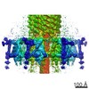
Movie
Movie Controller


+ Open data

- Basic information
| Entry |  | |||||||||
|---|---|---|---|---|---|---|---|---|---|---|
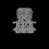
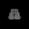
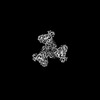
| Title | Phage 812 baseplate in the post-contraction state (C1) | |||||||||
 Map data Map data | Phage 812 baseplate in the post-contraction state (C1); postprocessed map | |||||||||
 Sample Sample |
| |||||||||
 Keywords Keywords | phage / baseplate / VIRUS | |||||||||
| Biological species |  Staphylococcus phage 812K1/420 (virus) Staphylococcus phage 812K1/420 (virus) | |||||||||
| Method | single particle reconstruction / cryo EM / Resolution: 6.6 Å | |||||||||
 Authors Authors | Binovsky J / Plevka P | |||||||||
| Funding support | European Union,  Czech Republic, 2 items Czech Republic, 2 items
| |||||||||
 Citation Citation | Journal: EMBO J / Year: 2026 Title: Conformational changes of the baseplate regulating tail contraction of Staphylococcus phage 812. Authors: Ján Bíňovský / Marta Šiborová / Maryna Zlatohurska / Jiří Nováček / Pavol Bárdy / Roman Baška / Karel Škubník / Tibor Botka / Martin Benešík / Roman Pantůček / Konstantinos ...Authors: Ján Bíňovský / Marta Šiborová / Maryna Zlatohurska / Jiří Nováček / Pavol Bárdy / Roman Baška / Karel Škubník / Tibor Botka / Martin Benešík / Roman Pantůček / Konstantinos Tripsianes / Pavel Plevka / Abstract: Phages with contractile tails employ elaborate mechanisms to penetrate bacterial cell walls and deliver their genomes into the host cytoplasm. Here, we used cryo-EM to show that the baseplate of ...Phages with contractile tails employ elaborate mechanisms to penetrate bacterial cell walls and deliver their genomes into the host cytoplasm. Here, we used cryo-EM to show that the baseplate of phage 812, a member of the Kayvirus genus, which infects Gram-positive Staphylococcus strains, is formed of a core, wedge modules, and baseplate arms carrying receptor-binding proteins 1 and 2 and tripod complexes. Upon binding to a host cell, the receptor-binding proteins of phage 812 baseplate reorient and undergo conformational changes. The changes to the tripod complexes trigger the release of the central spike and weld proteins, which expose peptidoglycan-degrading domains of the hub proteins. Changes in the positions of baseplate arms are transmitted through wedge modules to tail sheath initiator proteins. The ring of the tail sheath initiator proteins expands and triggers the contraction of the tail sheath, which shortens to 50% and pushes the tail tube 10-30 nm into the bacterial cytoplasm. Homologous molecular mechanisms are probably shared by phages of the Herelleviridae family with contractile tails to infect Gram-positive bacteria. | |||||||||
| History |
|
- Structure visualization
| Supplemental images |
|---|

- Downloads & links
-EMDB archive
| Map data | emd_55966.map.gz | 83 MB |  EMDB map data format EMDB map data format | |
|---|---|---|---|---|
| Header (meta data) | emd-55966-v30.xmlemd-55966.xml | 19.3 KB 19.3 KB | Display Display | EMDB header |
| FSC (resolution estimation) | emd_55966_fsc.xml | 22.5 KB | Display | FSC data file |
| Images |  emd_55966.png emd_55966.png | 129.2 KB | ||
| Masks | emd_55966_msk_1.map | 1000 MB | Mask map | |
| Filedesc metadata | emd-55966.cif.gz | 4.8 KB | ||
| Others | emd_55966_half_map_1.map.gzemd_55966_half_map_2.map.gz | 810.6 MB 809.9 MB | ||
| Archive directory |  https://data.pdbj.org/pub/emdb/structures/EMD-55966ftp://data.pdbj.org/pub/emdb/structures/EMD-55966 https://data.pdbj.org/pub/emdb/structures/EMD-55966ftp://data.pdbj.org/pub/emdb/structures/EMD-55966 | HTTPS FTP |
-Related structure data
| Related structure data | 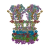 9eujC 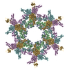 9eukC 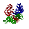 9eulC  9eumC  9f04C  9f05C  9f06C  9fkoC  9ticC  9tidC  9tieC  9tifC  9tigC  9tihC  9tiiC  9tijC  9tikC  9tilC  9timC  9tinC  9tioC  9tipC  9tirC  9tisC  9titC  9tiwC C: citing same article ( |
|---|


-Links
| EMDB pages | EMDB (EBI/PDBe) / EMDataResource |
|---|
-Map
| File | Download / File: emd_55966.map.gz / Format: CCP4 / Size: 1000 MB / Type: IMAGE STORED AS FLOATING POINT NUMBER (4 BYTES) | ||||||||||||||||||||||||||||||||||||
|---|---|---|---|---|---|---|---|---|---|---|---|---|---|---|---|---|---|---|---|---|---|---|---|---|---|---|---|---|---|---|---|---|---|---|---|---|---|
| Annotation | Phage 812 baseplate in the post-contraction state (C1); postprocessed map | ||||||||||||||||||||||||||||||||||||
| Projections & slices | Image control
Images are generated by Spider. | ||||||||||||||||||||||||||||||||||||
| Voxel size | X=Y=Z: 2.11 Å | ||||||||||||||||||||||||||||||||||||
| Density |
| ||||||||||||||||||||||||||||||||||||
| Symmetry | Space group: 1 | ||||||||||||||||||||||||||||||||||||
| Details | EMDB XML:
|
 Z (Sec.)
 Y (Row.)
 X (Col.)


-Supplemental data
-Mask #1
| File | emd_55966_msk_1.map | ||||||||||||
|---|---|---|---|---|---|---|---|---|---|---|---|---|---|
| Projections & Slices |
| ||||||||||||
| Density Histograms |


-Half map: Phage 812 baseplate in the post-contraction state (C1); half map1
| File | emd_55966_half_map_1.map | ||||||||||||
|---|---|---|---|---|---|---|---|---|---|---|---|---|---|
| Annotation | Phage 812 baseplate in the post-contraction state (C1); half map1 | ||||||||||||
| Projections & Slices |
| ||||||||||||
| Density Histograms |


-Half map: Phage 812 baseplate in the post-contraction state (C1); half map2
| File | emd_55966_half_map_2.map | ||||||||||||
|---|---|---|---|---|---|---|---|---|---|---|---|---|---|
| Annotation | Phage 812 baseplate in the post-contraction state (C1); half map2 | ||||||||||||
| Projections & Slices |
| ||||||||||||
| Density Histograms |


- Sample components
-Entire : Staphylococcus phage 812K1/420
| Entire | Name: Staphylococcus phage 812K1/420 (virus) |
|---|---|
| Components |
|
-Supramolecule #1: Staphylococcus phage 812K1/420
| Supramolecule | Name: Staphylococcus phage 812K1/420 / type: virus / ID: 1 / Parent: 0 / Macromolecule list: #1-#10 Details: Propagated in S. aureus SA 812 (CCM 4028) planktonic culture and purified on a CsCl gradient. NCBI-ID: 3434241 / Sci species name: Staphylococcus phage 812K1/420 / Virus type: VIRION / Virus isolate: STRAIN / Virus enveloped: No / Virus empty: No |
|---|
-Experimental details
-Structure determination
| Method | cryo EM |
|---|---|
 Processing Processing | single particle reconstruction |
| Aggregation state | particle |
-Sample preparation
| Buffer | pH: 8 / Details: 50mM Tris, 10mM NaCl, 10mM CaCl2 |
|---|---|
| Grid | Model: Quantifoil R2/1 / Material: COPPER / Mesh: 200 / Support film - Material: CARBON / Support film - topology: HOLEY |
| Vitrification | Cryogen name: ETHANE / Chamber humidity: 100 % / Chamber temperature: 277 K / Instrument: FEI VITROBOT MARK IV |
- Electron microscopy
| Microscope | TFS KRIOS |
|---|---|
| Specialist optics | Energy filter - Name: GIF Quantum LS / Energy filter - Slit width: 20 eV |
| Image recording | Film or detector model: GATAN K2 SUMMIT (4k x 4k) / Detector mode: COUNTING / Digitization - Dimensions - Width: 3838 pixel / Digitization - Dimensions - Height: 3710 pixel / Digitization - Frames/image: 1-40 / Number real images: 15371 / Average exposure time: 7.0 sec. / Average electron dose: 42.0 e/Å2 |
| Electron beam | Acceleration voltage: 300 kV / Electron source:  FIELD EMISSION GUN FIELD EMISSION GUN |
| Electron optics | C2 aperture diameter: 70.0 µm / Illumination mode: FLOOD BEAM / Imaging mode: BRIGHT FIELD / Cs: 2.7 mm / Nominal defocus max: 2.4 µm / Nominal defocus min: 1.0 µm / Nominal magnification: 130000 |
| Experimental equipment |  Model: Titan Krios / Image courtesy: FEI Company |