Movie
 Controller


[English] 日本語
 Yorodumi
- EMDB-55778: Active-state structure of the GPR55-N171Q mutant in complex with ... -
+ Open data

- Basic information
| Entry |  | |||||||||
|---|---|---|---|---|---|---|---|---|---|---|
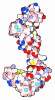
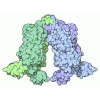
| Title | Active-state structure of the GPR55-N171Q mutant in complex with G13 and BI-6229 | |||||||||
 Map data | ||||||||||
 Sample |
| |||||||||
 Keywords | GPCR / G protein-coupled receptor / GPR55 / G13 / efficacy-switch / MEMBRANE PROTEIN | |||||||||
| Function / homology |  Function and homology information D5 dopamine receptor binding / Rho-activating G protein-coupled receptor signaling pathway / guanyl nucleotide binding / cannabinoid receptor activity / negative regulation of adenylate cyclase-activating adrenergic receptor signaling pathway / negative regulation of calcium ion-dependent exocytosis / negative regulation of osteoclast differentiation / negative regulation of adenylate cyclase activity / G protein-coupled adenosine receptor signaling pathway / Class A/1 (Rhodopsin-like receptors) ...D5 dopamine receptor binding / Rho-activating G protein-coupled receptor signaling pathway / guanyl nucleotide binding / cannabinoid receptor activity / negative regulation of adenylate cyclase-activating adrenergic receptor signaling pathway / negative regulation of calcium ion-dependent exocytosis / negative regulation of osteoclast differentiation / negative regulation of adenylate cyclase activity / G protein-coupled adenosine receptor signaling pathway / Class A/1 (Rhodopsin-like receptors) / regulation of small GTPase mediated signal transduction / positive regulation of neural precursor cell proliferation / negative regulation of synaptic transmission / bone resorption / positive regulation of urine volume / NRAGE signals death through JNK / gamma-aminobutyric acid signaling pathway / positive regulation of Rho protein signal transduction / regulation of calcium ion transport / negative regulation of apoptotic signaling pathway / neuronal dense core vesicle / regulation of postsynapse assembly / CDC42 GTPase cycle / Rho protein signal transduction / positive regulation of vascular associated smooth muscle cell proliferation / positive regulation of superoxide anion generation / RAC1 GTPase cycle / Adenylate cyclase inhibitory pathway / hippocampal mossy fiber to CA3 synapse / guanyl-nucleotide exchange factor activity / response to nutrient / bioluminescence / brush border membrane / regulation of blood pressure / generation of precursor metabolites and energy / Regulation of insulin secretion / G protein-coupled receptor binding / angiogenesis / G protein-coupled receptor activity / adenylate cyclase-modulating G protein-coupled receptor signaling pathway / ciliary basal body / G-protein beta/gamma-subunit complex binding / cell division / adenylate cyclase-inhibiting G protein-coupled receptor signaling pathway / Olfactory Signaling Pathway / Activation of the phototransduction cascade / G protein-coupled acetylcholine receptor signaling pathway / G beta:gamma signalling through PLC beta / Presynaptic function of Kainate receptors / Thromboxane signalling through TP receptor / Activation of G protein gated Potassium channels / Inhibition of voltage gated Ca2+ channels via Gbeta/gamma subunits / phospholipase C-activating G protein-coupled receptor signaling pathway / G-protein activation / melanosome / Glucagon signaling in metabolic regulation / G beta:gamma signalling through CDC42 / Prostacyclin signalling through prostacyclin receptor / Synthesis, secretion, and inactivation of Glucagon-like Peptide-1 (GLP-1) / photoreceptor disc membrane / G beta:gamma signalling through BTK / ADP signalling through P2Y purinoceptor 12 / Sensory perception of sweet, bitter, and umami (glutamate) taste / Glucagon-type ligand receptors / Adrenaline,noradrenaline inhibits insulin secretion / Vasopressin regulates renal water homeostasis via Aquaporins / Glucagon-like Peptide-1 (GLP1) regulates insulin secretion / G alpha (z) signalling events / ADP signalling through P2Y purinoceptor 1 / cellular response to catecholamine stimulus / G beta:gamma signalling through PI3Kgamma / ADORA2B mediated anti-inflammatory cytokines production / adenylate cyclase-activating dopamine receptor signaling pathway / Cooperation of PDCL (PhLP1) and TRiC/CCT in G-protein beta folding / cellular response to prostaglandin E stimulus / heterotrimeric G-protein complex / GPER1 signaling / G alpha (12/13) signalling events / Inactivation, recovery and regulation of the phototransduction cascade / G-protein beta-subunit binding / extracellular vesicle / Thrombin signalling through proteinase activated receptors (PARs) / adenylate cyclase-activating G protein-coupled receptor signaling pathway / signaling receptor complex adaptor activity / cell body / GTPase binding / midbody / Ca2+ pathway / High laminar flow shear stress activates signaling by PIEZO1 and PECAM1:CDH5:KDR in endothelial cells / G alpha (i) signalling events / G alpha (s) signalling events / G alpha (q) signalling events / Hydrolases; Acting on acid anhydrides; Acting on GTP to facilitate cellular and subcellular movement / cytoplasmic side of plasma membrane / postsynapse / Ras protein signal transduction / positive regulation of ERK1 and ERK2 cascade / G protein activity / Extra-nuclear estrogen signaling / positive regulation of cell migration Similarity search - Function | |||||||||
| Biological species |  Homo sapiens (human) / Synthetic construct (others) | |||||||||
| Method | single particle reconstruction / cryo EM / Resolution: 2.88 Å | |||||||||
 Authors | Claff T / Ebenhoch R / Kley JT / Weichert D | |||||||||
| Funding support | 1 items
| |||||||||
 Citation | Journal: Nat Commun / Year: 2026 Title: Molecular insights into GPR55 modulation reveal an atypical activation mechanism and glycosylation-dependent efficacy regulation. Authors: Tobias Claff / Chamalee Demalgiriya-Gamage / Andhika B Mahardhika / Nikolaos Angelos Stamos / Frederik Bangnowski / Xue Sun / Katharina L Dürr / Carol V Robinson / Rainer Haney / Lee Frego ...Authors: Tobias Claff / Chamalee Demalgiriya-Gamage / Andhika B Mahardhika / Nikolaos Angelos Stamos / Frederik Bangnowski / Xue Sun / Katharina L Dürr / Carol V Robinson / Rainer Haney / Lee Frego / Herbert Nar / Christa E Müller / Rebecca Ebenhoch / Jörg T Kley / Dietmar Weichert /    Abstract: GPR55 is a lipid-activated G protein-coupled receptor (GPCR) implicated in metabolism, inflammation, and neurodegeneration. Lysophosphatidylinositol (LPI) species are proposed as endogenous agonists, ...GPR55 is a lipid-activated G protein-coupled receptor (GPCR) implicated in metabolism, inflammation, and neurodegeneration. Lysophosphatidylinositol (LPI) species are proposed as endogenous agonists, but progress in understanding GPR55 function has still been limited by suboptimal chemical probes. Here, we present cryo-electron microscopy (cryo-EM) structures of GPR55 in both inactive and active states. The inactive-state structure bound to the inverse agonist CID16020046 facilitated the rational design of an improved in vitro tool compound, BI-8310. Surprisingly, mutation of a glycosylation site in extracellular loop 2 converted CID16020046 into a potent agonist, enabling co-structure determination of the active, G-bound receptor. Comparative analysis of both states bound to the same ligand provides insights into an atypical activation mechanism that diverges from the typical helix VI outward movement observed in other GPCRs. Functional mutagenesis, N-glycoproteomics, and hydrogen-deuterium exchange mass spectrometry (HDX-MS) substantiate the findings. These results establish a basis for targeted modulation of GPR55 in pathophysiological conditions. | |||||||||
| History |
|
- Structure visualization
| Supplemental images |
|---|

- Downloads & links
-EMDB archive
| Map data | emd_55778.map.gz | 9.6 MB | EMDB map data format | |
|---|---|---|---|---|
| Header (meta data) | emd-55778-v30.xmlemd-55778.xml | 18.3 KB 18.3 KB | Display Display | EMDB header |
| Images |  emd_55778.png | 86 KB | ||
| Filedesc metadata | emd-55778.cif.gz | 7 KB | ||
| Archive directory |  https://data.pdbj.org/pub/emdb/structures/EMD-55778ftp://data.pdbj.org/pub/emdb/structures/EMD-55778 | HTTPS FTP |
-Related structure data
| Related structure data |  9tc9MC  9tc7C  9tc8C C: citing same article ( M: atomic model generated by this map |
|---|---|
| Similar structure data |


-Links
| EMDB pages | EMDB (EBI/PDBe) / EMDataResource |
|---|---|
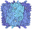
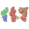
| Related items in Molecule of the Month |
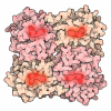
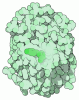
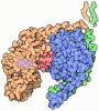
-Map
| File | Download / File: emd_55778.map.gz / Format: CCP4 / Size: 103 MB / Type: IMAGE STORED AS FLOATING POINT NUMBER (4 BYTES) | ||||||||||||||||||||||||||||||||||||
|---|---|---|---|---|---|---|---|---|---|---|---|---|---|---|---|---|---|---|---|---|---|---|---|---|---|---|---|---|---|---|---|---|---|---|---|---|---|
| Projections & slices | Image control
Images are generated by Spider. | ||||||||||||||||||||||||||||||||||||
| Voxel size | X=Y=Z: 0.951 Å | ||||||||||||||||||||||||||||||||||||
| Density |
| ||||||||||||||||||||||||||||||||||||
| Symmetry | Space group: 1 | ||||||||||||||||||||||||||||||||||||
| Details | EMDB XML:
|
 X (Sec.)
 Y (Row.)
 Z (Col.)


-Supplemental data
- Sample components
-Entire : GPR55-N171Q-G13-ScFv16-BI-6229
| Entire | Name: GPR55-N171Q-G13-ScFv16-BI-6229 |
|---|---|
| Components |
|
-Supramolecule #1: GPR55-N171Q-G13-ScFv16-BI-6229
| Supramolecule | Name: GPR55-N171Q-G13-ScFv16-BI-6229 / type: complex / ID: 1 / Parent: 0 / Macromolecule list: #1-#5 |
|---|---|
| Source (natural) | Organism: Homo sapiens (human) |
-Macromolecule #1: Guanine nucleotide-binding protein G(i) subunit alpha-2,Guanine n...
| Macromolecule | Name: Guanine nucleotide-binding protein G(i) subunit alpha-2,Guanine nucleotide-binding protein subunit alpha-13 type: protein_or_peptide / ID: 1 / Number of copies: 1 / Enantiomer: LEVO |
|---|---|
| Source (natural) | Organism: Homo sapiens (human) |
| Molecular weight | Theoretical: 26.5744 KDa |
| Recombinant expression | Organism:  Trichoplusia ni (cabbage looper) |
| Sequence | String: MGSTVSAEDK AAAERSKEID KCLSREKTYV KRLVKILLLG ADNSGKSTFL KQMRIIHGGS GGSGGTKGIH EYDFEIKNVP FKMVDVGGQ RSERKRWFEC FDSVTSILFL VDSSDFNRLT ESLNDFETIV NNRVFSNVSI ILFLNKTDLL EEKVQIVSIK D YFLEFEGD ...String: MGSTVSAEDK AAAERSKEID KCLSREKTYV KRLVKILLLG ADNSGKSTFL KQMRIIHGGS GGSGGTKGIH EYDFEIKNVP FKMVDVGGQ RSERKRWFEC FDSVTSILFL VDSSDFNRLT ESLNDFETIV NNRVFSNVSI ILFLNKTDLL EEKVQIVSIK D YFLEFEGD PHCLRDVQKF LVECFRNKRR DQQQKPLYHH FTTAINTENA RLIFRDVKDT ILHDNLKQLM LQ UniProtKB: Guanine nucleotide-binding protein G(i) subunit alpha-2, Guanine nucleotide-binding protein subunit alpha-13, Guanine nucleotide-binding protein subunit alpha-13 |
-Macromolecule #2: Guanine nucleotide-binding protein G(I)/G(S)/G(T) subunit beta-1
| Macromolecule | Name: Guanine nucleotide-binding protein G(I)/G(S)/G(T) subunit beta-1 type: protein_or_peptide / ID: 2 / Number of copies: 1 / Enantiomer: LEVO |
|---|---|
| Source (natural) | Organism: Homo sapiens (human) |
| Molecular weight | Theoretical: 39.418086 KDa |
| Recombinant expression | Organism: Trichoplusia ni (cabbage looper) |
| Sequence | String: MHHHHHHLEV LFQGPGSSGS ELDQLRQEAE QLKNQIRDAR KACADATLSQ ITNNIDPVGR IQMRTRRTLR GHLAKIYAMH WGTDSRLLV SASQDGKLII WDSYTTNKVH AIPLRSSWVM TCAYAPSGNY VACGGLDNIC SIYNLKTREG NVRVSRELAG H TGYLSCCR ...String: MHHHHHHLEV LFQGPGSSGS ELDQLRQEAE QLKNQIRDAR KACADATLSQ ITNNIDPVGR IQMRTRRTLR GHLAKIYAMH WGTDSRLLV SASQDGKLII WDSYTTNKVH AIPLRSSWVM TCAYAPSGNY VACGGLDNIC SIYNLKTREG NVRVSRELAG H TGYLSCCR FLDDNQIVTS SGDTTCALWD IETGQQTTTF TGHTGDVMSL SLAPDTRLFV SGACDASAKL WDVREGMCRQ TF TGHESDI NAICFFPNGN AFATGSDDAT CRLFDLRADQ ELMTYSHDNI ICGITSVSFS KSGRLLLAGY DDFNCNVWDA LKA DRAGVL AGHDNRVSCL GVTDDGMAVA TGSWDSFLKI WN UniProtKB: Guanine nucleotide-binding protein G(I)/G(S)/G(T) subunit beta-1 |
-Macromolecule #3: ScFv16
| Macromolecule | Name: ScFv16 / type: protein_or_peptide / ID: 3 / Number of copies: 1 / Enantiomer: LEVO |
|---|---|
| Source (natural) | Organism: Synthetic construct (others) |
| Molecular weight | Theoretical: 32.626523 KDa |
| Recombinant expression | Organism: Trichoplusia ni (cabbage looper) |
| Sequence | String: MLLVNQSHQG FNKEHTSKMV SAIVLYVLLA AAAHSAFADV QLVESGGGLV QPGGSRKLSC SASGFAFSSF GMHWVRQAPE KGLEWVAYI SSGSGTIYYA DTVKGRFTIS RDDPKNTLFL QMTSLRSEDT AMYYCVRSIY YYGSSPFDFW GGTTLTVSSG G GGSGGGGS ...String: MLLVNQSHQG FNKEHTSKMV SAIVLYVLLA AAAHSAFADV QLVESGGGLV QPGGSRKLSC SASGFAFSSF GMHWVRQAPE KGLEWVAYI SSGSGTIYYA DTVKGRFTIS RDDPKNTLFL QMTSLRSEDT AMYYCVRSIY YYGSSPFDFW GGTTLTVSSG G GGSGGGGS GGGGSDIVMT QATSSVPVTP GESVSISCRS SKSLLHSNGN TYLYWFLQRP GQSPQLLIYR MSNLASGVPD RF SGSGSGT AFTLTISRLE AEDVGVYYCM QHLEYPLTFG AGTKLELKLE VLFQGPAAAH HHHHHHH |
-Macromolecule #4: Guanine nucleotide-binding protein G(I)/G(S)/G(O) subunit gamma-2
| Macromolecule | Name: Guanine nucleotide-binding protein G(I)/G(S)/G(O) subunit gamma-2 type: protein_or_peptide / ID: 4 / Number of copies: 1 / Enantiomer: LEVO |
|---|---|
| Source (natural) | Organism: Homo sapiens (human) |
| Molecular weight | Theoretical: 8.506765 KDa |
| Recombinant expression | Organism: Trichoplusia ni (cabbage looper) |
| Sequence | String: MASNNTASIA QARKLVEQLK MEANIDRIKV SKAAADLMAY CEAHAKEDPL LTPVPASENP FREKKFFCAI LGSAGSAGSA UniProtKB: Guanine nucleotide-binding protein G(I)/G(S)/G(O) subunit gamma-2 |
-Macromolecule #5: Green fluorescent protein,G-protein coupled receptor 55
| Macromolecule | Name: Green fluorescent protein,G-protein coupled receptor 55 type: protein_or_peptide / ID: 5 / Number of copies: 1 / Enantiomer: LEVO |
|---|---|
| Source (natural) | Organism: Homo sapiens (human) |
| Molecular weight | Theoretical: 73.138836 KDa |
| Recombinant expression | Organism: Trichoplusia ni (cabbage looper) |
| Sequence | String: MKTIIALSYI FCLVFADYKD DDDKKGEELF TGVVPILVEL DGDVNGHKFS VSGEGEGDAT YGKLTLKFIC TTGKLPVPWP TLVTTLTYG VQCFSRYPDH MKRHDFFKSA MPEGYVQERT ISFKDDGNYK TRAEVKFEGD TLVNRIELKG IDFKEDGNIL G HKLEYNYN ...String: MKTIIALSYI FCLVFADYKD DDDKKGEELF TGVVPILVEL DGDVNGHKFS VSGEGEGDAT YGKLTLKFIC TTGKLPVPWP TLVTTLTYG VQCFSRYPDH MKRHDFFKSA MPEGYVQERT ISFKDDGNYK TRAEVKFEGD TLVNRIELKG IDFKEDGNIL G HKLEYNYN SHNVYITADK QKNGIKANFK IRHNIEDGSV QLADHYQQNT PIGDGPVLLP DNHYLSTQSA LSKDPNEKRD HM VLLEFVT AAGITHGMDE LYKAAGSGEF LEVLFQGPGA GSDSMSQQNT SGDCLFDGVN ELMKTLQFAV HIPTFVLGLL LNL LAIHGF STFLKNRWPD YAATSIYMIN LAVFDLLLVL SLPFKMVLSQ VQSPFPSLCT LVECLYFVSM YGSVFTICFI SMDR FLAIR YPLLVSHLRS PRKIFGICCT IWVLVWTGSI PIYSFHGKVE KYMCFHQMSD DTWSAKVFFP LEVFGFLLPM GIMGF CCSR SIHILLGRRD HTQDWVQQKA CIYSIAASLA VFVVSFLPVH LGFFLQFLVR NSFIVECRAK QSISFFLQLS MCFSNV NCC LDVFCYYFVI KEFRMNIRAH RPSRVQLVLQ DTTISRGAGS GAGSAWSHPQ FEKGGGSGGG SGGSAWSHPQ FEKGAGS HH HHHHHHHH UniProtKB: Green fluorescent protein, G protein-coupled receptor 55 |
-Macromolecule #6: 4-[(4~{S})-4-(3-hydroxyphenyl)-3-(4-methylphenyl)-6-oxidanylidene...
| Macromolecule | Name: 4-[(4~{S})-4-(3-hydroxyphenyl)-3-(4-methylphenyl)-6-oxidanylidene-1,4-dihydropyrrolo[3,4-c]pyrazol-5-yl]benzoic acid type: ligand / ID: 6 / Number of copies: 1 / Formula: A1JU1 |
|---|---|
| Molecular weight | Theoretical: 425.436 Da |
-Experimental details
-Structure determination
| Method | cryo EM |
|---|---|
 Processing | single particle reconstruction |
| Aggregation state | particle |
-Sample preparation
| Buffer | pH: 7.4 |
|---|---|
| Vitrification | Cryogen name: ETHANE |
- Electron microscopy
| Microscope | TFS KRIOS |
|---|---|
| Image recording | Film or detector model: FEI FALCON IV (4k x 4k) / Average electron dose: 40.0 e/Å2 |
| Electron beam | Acceleration voltage: 300 kV / Electron source:  FIELD EMISSION GUN |
| Electron optics | Illumination mode: OTHER / Imaging mode: BRIGHT FIELD / Nominal defocus max: 30.0 µm / Nominal defocus min: 3.0 µm |
| Experimental equipment | 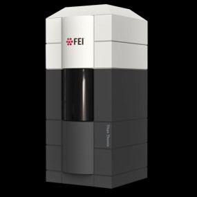 Model: Titan Krios / Image courtesy: FEI Company |