Movie
Movie Controller

[English] 日本語
 Yorodumi
- EMDB-55404: Catalase CryoEM Structure from Rhizobium radiobacter at 1.7A reso... -
+ Open data

- Basic information
| Entry |  | |||||||||
|---|---|---|---|---|---|---|---|---|---|---|
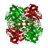
| Title | Catalase CryoEM Structure from Rhizobium radiobacter at 1.7A resolution | |||||||||
 Map data Map data | Contour level is in Volts | |||||||||
 Sample Sample |
| |||||||||
 Keywords Keywords | Catalase / NADPH / PROTOPORPHYRIN IX CONTAINING FE / OXIDOREDUCTASE | |||||||||
| Function / homology |  Function and homology information Function and homology informationcatalase / catalase activity / hydrogen peroxide catabolic process / response to hydrogen peroxide / periplasmic space / heme binding / metal ion binding / cytoplasm Similarity search - Function | |||||||||
| Biological species |  Agrobacterium radiobacter (Agrobacterium genomosp. 4) Agrobacterium radiobacter (Agrobacterium genomosp. 4) | |||||||||
| Method | single particle reconstruction / cryo EM / Resolution: 1.73 Å | |||||||||
 Authors Authors | Li J / Henderson R / Russo CJ / Wilson H / Chen S | |||||||||
| Funding support |  United Kingdom, 1 items United Kingdom, 1 items
| |||||||||
 Citation Citation | Journal: To Be Published Title: Comparison of human and bacterial monofunctional catalase structures obtained by electron cryomicroscopy. Authors: Slowik D / Li J / Wilson H / Shtyrov A / Chen S / McMullan G / Russo CJ / Mushudov G / Henderson R | |||||||||
| History |
|
- Structure visualization
| Supplemental images |
|---|

- Downloads & links
-EMDB archive
| Map data | emd_55404.map.gz | 30.7 MB | EMDB map data format | |
|---|---|---|---|---|
| Header (meta data) | emd-55404-v30.xmlemd-55404.xml | 23.7 KB 23.7 KB | Display Display | EMDB header |
| FSC (resolution estimation) | emd_55404_fsc.xml | 17.9 KB | Display | FSC data file |
| Images |  emd_55404.png emd_55404.png | 179.2 KB | ||
| Masks | emd_55404_msk_1.map | 512 MB | Mask map | |
| Filedesc metadata | emd-55404.cif.gz | 7.1 KB | ||
| Others | emd_55404_half_map_1.map.gzemd_55404_half_map_2.map.gz | 390.9 MB 390.7 MB | ||
| Archive directory |  http://ftp.pdbj.org/pub/emdb/structures/EMD-55404ftp://ftp.pdbj.org/pub/emdb/structures/EMD-55404 http://ftp.pdbj.org/pub/emdb/structures/EMD-55404ftp://ftp.pdbj.org/pub/emdb/structures/EMD-55404 | HTTPS FTP |
-Related structure data
| Related structure data |  9t0lMC  9t0mC M: atomic model generated by this map C: citing same article ( |
|---|---|
| Similar structure data |

-Links
| EMDB pages | EMDB (EBI/PDBe) / EMDataResource |
|---|---|
| Related items in Molecule of the Month |

-Map
| File | Download / File: emd_55404.map.gz / Format: CCP4 / Size: 512 MB / Type: IMAGE STORED AS FLOATING POINT NUMBER (4 BYTES) | ||||||||||||||||||||||||||||||||||||
|---|---|---|---|---|---|---|---|---|---|---|---|---|---|---|---|---|---|---|---|---|---|---|---|---|---|---|---|---|---|---|---|---|---|---|---|---|---|
| Annotation | Contour level is in Volts | ||||||||||||||||||||||||||||||||||||
| Projections & slices | Image control
Images are generated by Spider. | ||||||||||||||||||||||||||||||||||||
| Voxel size | X=Y=Z: 0.8 Å | ||||||||||||||||||||||||||||||||||||
| Density |
| ||||||||||||||||||||||||||||||||||||
| Symmetry | Space group: 1 | ||||||||||||||||||||||||||||||||||||
| Details | EMDB XML:
|
 Z (Sec.)
 Y (Row.)
 X (Col.)


-Supplemental data
-Mask #1
| File | emd_55404_msk_1.map | ||||||||||||
|---|---|---|---|---|---|---|---|---|---|---|---|---|---|
| Projections & Slices |
| ||||||||||||
| Density Histograms |


-Half map: #2
| File | emd_55404_half_map_1.map | ||||||||||||
|---|---|---|---|---|---|---|---|---|---|---|---|---|---|
| Projections & Slices |
| ||||||||||||
| Density Histograms |


-Half map: #1
| File | emd_55404_half_map_2.map | ||||||||||||
|---|---|---|---|---|---|---|---|---|---|---|---|---|---|
| Projections & Slices |
| ||||||||||||
| Density Histograms |


- Sample components
-Entire : Catalase with cofactor NADPH
| Entire | Name: Catalase with cofactor NADPH |
|---|---|
| Components |
|
-Supramolecule #1: Catalase with cofactor NADPH
| Supramolecule | Name: Catalase with cofactor NADPH / type: organelle_or_cellular_component / ID: 1 / Parent: 0 / Macromolecule list: #1 |
|---|---|
| Source (natural) | Organism: Agrobacterium radiobacter (Agrobacterium genomosp. 4) Strain: 2-1 / Location in cell: periplasm |
| Molecular weight | Theoretical: 220 KDa |
-Macromolecule #1: Catalase
| Macromolecule | Name: Catalase / type: protein_or_peptide / ID: 1 / Number of copies: 1 / Enantiomer: LEVO / EC number: catalase |
|---|---|
| Source (natural) | Organism: Agrobacterium radiobacter (Agrobacterium genomosp. 4) Strain: 2-1 |
| Molecular weight | Theoretical: 55.131996 KDa |
| Recombinant expression | Organism: |
| Sequence | String: MTDMNKKQGG TGSTTGTGAP AVSDRNSLTV GPDGPILLHD VHFLEQMAHF NREKVPERQP HAKGSGAFGT FETTHDVSAY TKAALFQKG ATTEMLARFS TVAGEMGSPD TWRDVRGFSL KFYTDEGNYD LVGNNTPIFF VRDPMKFPHF IRSQKRLPDS G LRDNHMQW ...String: MTDMNKKQGG TGSTTGTGAP AVSDRNSLTV GPDGPILLHD VHFLEQMAHF NREKVPERQP HAKGSGAFGT FETTHDVSAY TKAALFQKG ATTEMLARFS TVAGEMGSPD TWRDVRGFSL KFYTDEGNYD LVGNNTPIFF VRDPMKFPHF IRSQKRLPDS G LRDNHMQW DFWTNNPESA HQVTYLMGVR GLPRTWRHMN GYGSHTYMWV NEAGERFWVK YHFHTHQGME FFTNEEAGAM AG ADADFHR RDLFDAIARG EHPAWTMSVQ VMPYEEGKTY HINPFDLTKT WPHADYPLIE VGKMTLNRNP ENFFAQIEQA AFS PGNTVP GIGLSPDKML LGRAFAYNDA QRNRIGTNFH QLPVNQPKVP VNTYMFDGQM AYHHSGSAPV HATNSGGRSW SDET GAVHD GWEADGDFVR SAYTLRPGDD DFSQPGKLVR EVFNDDERRQ LVETVSGALL GGVRSPVLER AFDYWKSVDA EVGQR IEDA VRAGQAG UniProtKB: Catalase |
-Macromolecule #2: PROTOPORPHYRIN IX CONTAINING FE
| Macromolecule | Name: PROTOPORPHYRIN IX CONTAINING FE / type: ligand / ID: 2 / Number of copies: 1 / Formula: HEM |
|---|---|
| Molecular weight | Theoretical: 616.487 Da |
| Chemical component information |  ChemComp-HEM: |
-Macromolecule #3: NADPH DIHYDRO-NICOTINAMIDE-ADENINE-DINUCLEOTIDE PHOSPHATE
| Macromolecule | Name: NADPH DIHYDRO-NICOTINAMIDE-ADENINE-DINUCLEOTIDE PHOSPHATE type: ligand / ID: 3 / Number of copies: 1 / Formula: NDP |
|---|---|
| Molecular weight | Theoretical: 745.421 Da |
| Chemical component information |  ChemComp-NDP: |
-Macromolecule #4: water
| Macromolecule | Name: water / type: ligand / ID: 4 / Number of copies: 425 / Formula: HOH |
|---|---|
| Molecular weight | Theoretical: 18.015 Da |
| Chemical component information |  ChemComp-HOH: |
-Experimental details
-Structure determination
| Method | cryo EM |
|---|---|
 Processing Processing | single particle reconstruction |
| Aggregation state | particle |
-Sample preparation
| Concentration | 25 mg/mL | |||||||||||||||
|---|---|---|---|---|---|---|---|---|---|---|---|---|---|---|---|---|
| Buffer | pH: 7.4 Component:
| |||||||||||||||
| Grid | Model: HexAuFoil / Material: GOLD / Mesh: 300 / Support film - Material: GOLD / Support film - topology: HOLEY / Support film - Film thickness: 45 / Pretreatment - Type: PLASMA CLEANING / Pretreatment - Time: 75 sec. / Pretreatment - Atmosphere: OTHER / Pretreatment - Pressure: 0.003 kPa | |||||||||||||||
| Vitrification | Cryogen name: ETHANE / Chamber humidity: 100 % / Chamber temperature: 277 K / Instrument: HOMEMADE PLUNGER |
- Electron microscopy
| Microscope | TFS KRIOS |
|---|---|
| Temperature | Min: 80.0 K / Max: 80.0 K |
| Alignment procedure | Coma free - Residual tilt: 0.67 mrad |
| Details | There were 12 optical groups with AFIS up to 12 microns. |
| Image recording | Film or detector model: TFS FALCON 4i (4k x 4k) / Digitization - Dimensions - Width: 4096 pixel / Digitization - Dimensions - Height: 4096 pixel / Number grids imaged: 1 / Number real images: 35543 / Average exposure time: 3.0 sec. / Average electron dose: 50.4 e/Å2 Details: Images were collected using AFIS with maximum image shifts of 12 microns. |
| Electron beam | Acceleration voltage: 300 kV / Electron source:  FIELD EMISSION GUN FIELD EMISSION GUN |
| Electron optics | C2 aperture diameter: 50.0 µm / Calibrated defocus max: 2.25 µm / Calibrated defocus min: 1.45 µm / Calibrated magnification: 273437 / Illumination mode: FLOOD BEAM / Imaging mode: BRIGHT FIELD / Cs: 2.7 mm / Nominal defocus max: 2.25 µm / Nominal defocus min: 1.45 µm / Nominal magnification: 155000 |
| Sample stage | Cooling holder cryogen: NITROGEN |
| Experimental equipment |  Model: Titan Krios / Image courtesy: FEI Company |
+Image processing


-Atomic model buiding 1
| Initial model | PDB ID: Chain - Chain ID: A / Chain - Residue range: 6-503 / Chain - Source name: PDB / Chain - Initial model type: experimental model Details: The initial model consisted of the complete biological assembly of PDB entry 1GWE minus the bound water molecules. |
|---|---|
| Refinement | Space: REAL / Protocol: RIGID BODY FIT |
| Output model | PDB-9t0l: |