Movie
Movie Controller

[English] 日本語
 Yorodumi
- EMDB-55405: Catalase cryoEM structure from Micrococcus luteus at 1.9 Angstrom... -
+ Open data

- Basic information
| Entry |  | |||||||||
|---|---|---|---|---|---|---|---|---|---|---|
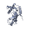
| Title | Catalase cryoEM structure from Micrococcus luteus at 1.9 Angstrom resolution. | |||||||||
 Map data Map data | ||||||||||
 Sample Sample |
| |||||||||
 Keywords Keywords | Catalase / NADPH / Protoporphyrin IX / OXIDOREDUCTASE | |||||||||
| Function / homology |  Function and homology information Function and homology informationcatalase / catalase activity / hydrogen peroxide catabolic process / response to hydrogen peroxide / heme binding / metal ion binding / cytoplasm Similarity search - Function | |||||||||
| Biological species |  Micrococcus luteus (bacteria) Micrococcus luteus (bacteria) | |||||||||
| Method | single particle reconstruction / cryo EM / Resolution: 1.9 Å | |||||||||
 Authors Authors | Li J / Henderson R / Russo CJ / Wilson H / Chen S | |||||||||
| Funding support |  United Kingdom, 1 items United Kingdom, 1 items
| |||||||||
 Citation Citation | Journal: To Be Published Title: Comparison of human and bacterial monofunctional catalase structures obtained by electron cryomicroscopy. Authors: Slowik D / Li J / Wilson H / Shtyrov A / Chen S / McMullan G / Russo CJ / Murshudov G / Henderson R | |||||||||
| History |
|
- Structure visualization
| Supplemental images |
|---|

- Downloads & links
-EMDB archive
| Map data | emd_55405.map.gz | 22.4 MB | EMDB map data format | |
|---|---|---|---|---|
| Header (meta data) | emd-55405-v30.xmlemd-55405.xml | 24 KB 24 KB | Display Display | EMDB header |
| FSC (resolution estimation) | emd_55405_fsc.xml | 14.1 KB | Display | FSC data file |
| Images |  emd_55405.png emd_55405.png | 203.9 KB | ||
| Masks | emd_55405_msk_1.map | 512 MB | Mask map | |
| Filedesc metadata | emd-55405.cif.gz | 7.2 KB | ||
| Others | emd_55405_half_map_1.map.gzemd_55405_half_map_2.map.gz | 398.1 MB 398.2 MB | ||
| Archive directory |  http://ftp.pdbj.org/pub/emdb/structures/EMD-55405ftp://ftp.pdbj.org/pub/emdb/structures/EMD-55405 http://ftp.pdbj.org/pub/emdb/structures/EMD-55405ftp://ftp.pdbj.org/pub/emdb/structures/EMD-55405 | HTTPS FTP |
-Related structure data
| Related structure data |  9t0mMC  9t0lC M: atomic model generated by this map C: citing same article ( |
|---|---|
| Similar structure data |

-Links
| EMDB pages | EMDB (EBI/PDBe) / EMDataResource |
|---|---|
| Related items in Molecule of the Month |

-Map
| File | Download / File: emd_55405.map.gz / Format: CCP4 / Size: 512 MB / Type: IMAGE STORED AS FLOATING POINT NUMBER (4 BYTES) | ||||||||||||||||||||||||||||||||||||
|---|---|---|---|---|---|---|---|---|---|---|---|---|---|---|---|---|---|---|---|---|---|---|---|---|---|---|---|---|---|---|---|---|---|---|---|---|---|
| Projections & slices | Image control
Images are generated by Spider. | ||||||||||||||||||||||||||||||||||||
| Voxel size | X=Y=Z: 0.8 Å | ||||||||||||||||||||||||||||||||||||
| Density |
| ||||||||||||||||||||||||||||||||||||
| Symmetry | Space group: 1 | ||||||||||||||||||||||||||||||||||||
| Details | EMDB XML:
|
 Z (Sec.)
 Y (Row.)
 X (Col.)


-Supplemental data
-Mask #1
| File | emd_55405_msk_1.map | ||||||||||||
|---|---|---|---|---|---|---|---|---|---|---|---|---|---|
| Projections & Slices |
| ||||||||||||
| Density Histograms |


-Half map: #2
| File | emd_55405_half_map_1.map | ||||||||||||
|---|---|---|---|---|---|---|---|---|---|---|---|---|---|
| Projections & Slices |
| ||||||||||||
| Density Histograms |


-Half map: #1
| File | emd_55405_half_map_2.map | ||||||||||||
|---|---|---|---|---|---|---|---|---|---|---|---|---|---|
| Projections & Slices |
| ||||||||||||
| Density Histograms |


- Sample components
-Entire : catalase with cofactor NADPH
| Entire | Name: catalase with cofactor NADPH |
|---|---|
| Components |
|
-Supramolecule #1: catalase with cofactor NADPH
| Supramolecule | Name: catalase with cofactor NADPH / type: organelle_or_cellular_component / ID: 1 / Parent: 0 / Macromolecule list: #1 |
|---|---|
| Source (natural) | Organism: Micrococcus luteus (bacteria) |
| Molecular weight | Theoretical: 220 KDa |
-Macromolecule #1: Catalase
| Macromolecule | Name: Catalase / type: protein_or_peptide / ID: 1 / Number of copies: 1 / Enantiomer: LEVO / EC number: catalase |
|---|---|
| Source (natural) | Organism: Micrococcus luteus (bacteria) |
| Molecular weight | Theoretical: 58.09875 KDa |
| Recombinant expression | Organism: |
| Sequence | String: HHHHHHHHSG EHQKTTPHAT GSTRQNGAPA VSDRQSLTVG SEGPIVLHDT HLLETHQHFN RMNIPERRPH AKGSGAFGEF EVTEDVSKY TKALVFQPGT KTETLLRFST VAGELGSPDT WRDVRGFALR FYTEEGNYDL VGNNTPIFFL RDPMKFTHFI R SQKRLPDS ...String: HHHHHHHHSG EHQKTTPHAT GSTRQNGAPA VSDRQSLTVG SEGPIVLHDT HLLETHQHFN RMNIPERRPH AKGSGAFGEF EVTEDVSKY TKALVFQPGT KTETLLRFST VAGELGSPDT WRDVRGFALR FYTEEGNYDL VGNNTPIFFL RDPMKFTHFI R SQKRLPDS GLRDATMQWD FWTNNPESAH QVTYLMGPRG LPRTWREMNG YGSHTYLWVN AQGEKHWVKY HFISQQGVHN LS NDEATKI AGENADFHRQ DLFESIAKGD HPKWDLYIQA IPYEEGKTYR FNPFDLTKTI SQKDYPRIKV GTLTLNRNPE NHF AQIESA AFSPSNTVPG IGLSPDRMLL GRAFAYHDAQ LYRVGAHVNQ LPVNRPKNAV HNYAFEGQMW YDHTGDRSTY VPNS NGDSW SDETGPVDDG WEADGTLTRE AQALRADDDD FGQAGTLVRE VFSDQERDDF VETVAGALKG VRQDVQARAF EYWKN VDAT IGQRIEDEVK RHEGDGIPGV EAGGEARM UniProtKB: Catalase |
-Macromolecule #2: PROTOPORPHYRIN IX CONTAINING FE
| Macromolecule | Name: PROTOPORPHYRIN IX CONTAINING FE / type: ligand / ID: 2 / Number of copies: 1 / Formula: HEM |
|---|---|
| Molecular weight | Theoretical: 616.487 Da |
| Chemical component information |  ChemComp-HEM: |
-Macromolecule #3: NADPH DIHYDRO-NICOTINAMIDE-ADENINE-DINUCLEOTIDE PHOSPHATE
| Macromolecule | Name: NADPH DIHYDRO-NICOTINAMIDE-ADENINE-DINUCLEOTIDE PHOSPHATE type: ligand / ID: 3 / Number of copies: 1 / Formula: NDP |
|---|---|
| Molecular weight | Theoretical: 745.421 Da |
| Chemical component information |  ChemComp-NDP: |
-Macromolecule #4: water
| Macromolecule | Name: water / type: ligand / ID: 4 / Number of copies: 422 / Formula: HOH |
|---|---|
| Molecular weight | Theoretical: 18.015 Da |
| Chemical component information |  ChemComp-HOH: |
-Experimental details
-Structure determination
| Method | cryo EM |
|---|---|
 Processing Processing | single particle reconstruction |
| Aggregation state | particle |
-Sample preparation
| Concentration | 25 mg/mL | |||||||||||||||
|---|---|---|---|---|---|---|---|---|---|---|---|---|---|---|---|---|
| Buffer | pH: 7.4 Component:
| |||||||||||||||
| Grid | Model: HexAuFoil / Material: GOLD / Mesh: 300 / Support film - Material: GOLD / Support film - topology: HOLEY / Support film - Film thickness: 45 / Pretreatment - Type: PLASMA CLEANING / Pretreatment - Time: 75 sec. / Pretreatment - Atmosphere: OTHER / Pretreatment - Pressure: 0.003 kPa | |||||||||||||||
| Vitrification | Cryogen name: ETHANE / Chamber humidity: 100 % / Chamber temperature: 277 K / Instrument: HOMEMADE PLUNGER |
- Electron microscopy
| Microscope | TFS KRIOS |
|---|---|
| Temperature | Min: 80.0 K / Max: 80.0 K |
| Alignment procedure | Coma free - Residual tilt: 0.1 mrad |
| Details | Only one optical group, with minimal beam tilt. |
| Image recording | Film or detector model: TFS FALCON 4i (4k x 4k) / Digitization - Dimensions - Width: 4096 pixel / Digitization - Dimensions - Height: 4096 pixel / Number grids imaged: 1 / Number real images: 27182 / Average exposure time: 3.0 sec. / Average electron dose: 50.4 e/Å2 Details: Images collected with AFIS with maximum image shift of 1.5 microns. |
| Electron beam | Acceleration voltage: 300 kV / Electron source:  FIELD EMISSION GUN FIELD EMISSION GUN |
| Electron optics | C2 aperture diameter: 50.0 µm / Calibrated defocus max: 18.1 µm / Calibrated defocus min: 11.4 µm / Calibrated magnification: 273437 / Illumination mode: FLOOD BEAM / Imaging mode: BRIGHT FIELD / Cs: 2.7 mm / Nominal defocus max: 18.1 µm / Nominal defocus min: 11.4 µm / Nominal magnification: 155000 |
| Sample stage | Cooling holder cryogen: NITROGEN |
| Experimental equipment |  Model: Titan Krios / Image courtesy: FEI Company |
+Image processing

-Atomic model buiding 1
| Initial model | PDB ID: Chain - Chain ID: A / Chain - Residue range: 7-505 / Chain - Source name: PDB / Chain - Initial model type: experimental model / Details: The initial model was PDB entry 1GWE. |
|---|---|
| Refinement | Space: REAL / Protocol: RIGID BODY FIT |
| Output model | PDB-9t0m: |