Movie
Movie Controller

[English] 日本語
 Yorodumi
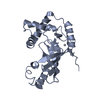
- PDB-9t0m: Catalase cryoEM structure from Micrococcus luteus at 1.9 Angstrom... -
+ Open data

- Basic information
| Entry | Database: PDB / ID: 9t0m | |||||||||||||||||||||||||||
|---|---|---|---|---|---|---|---|---|---|---|---|---|---|---|---|---|---|---|---|---|---|---|---|---|---|---|---|---|
| Title | Catalase cryoEM structure from Micrococcus luteus at 1.9 Angstrom resolution. | |||||||||||||||||||||||||||
 Components Components | Catalase | |||||||||||||||||||||||||||
 Keywords Keywords | OXIDOREDUCTASE / Catalase / NADPH / Protoporphyrin IX | |||||||||||||||||||||||||||
| Function / homology |  Function and homology information Function and homology informationcatalase / catalase activity / hydrogen peroxide catabolic process / response to hydrogen peroxide / heme binding / metal ion binding / cytoplasm Similarity search - Function | |||||||||||||||||||||||||||
| Biological species |  Micrococcus luteus (bacteria) Micrococcus luteus (bacteria) | |||||||||||||||||||||||||||
| Method | ELECTRON MICROSCOPY / single particle reconstruction / cryo EM / Resolution: 1.9 Å | |||||||||||||||||||||||||||
 Authors Authors | Li, J. / Henderson, R. / Russo, C.J. / Wilson, H. / Chen, S. | |||||||||||||||||||||||||||
| Funding support |  United Kingdom, 1items United Kingdom, 1items
| |||||||||||||||||||||||||||
 Citation Citation | Journal: To Be Published Title: Comparison of human and bacterial monofunctional catalase structures obtained by electron cryomicroscopy. Authors: Slowik, D. / Li, J. / Wilson, H. / Shtyrov, A. / Chen, S. / McMullan, G. / Russo, C.J. / Murshudov, G. / Henderson, R. | |||||||||||||||||||||||||||
| History |
|
- Structure visualization
| Structure viewer | Molecule: MolmilJmol/JSmol |
|---|
- Downloads & links
-Download
| PDBx/mmCIF format | 9t0m.cif.gz | 253.8 KB | Display | PDBx/mmCIF format |
|---|---|---|---|---|
| PDB format | pdb9t0m.ent.gz | 160.7 KB | Display | PDB format |
| PDBx/mmJSON format | 9t0m.json.gz | Tree view | PDBx/mmJSON format | |
| Others |  Other downloads Other downloads |
-Validation report
| Arichive directory | https://data.pdbj.org/pub/pdb/validation_reports/t0/9t0mftp://data.pdbj.org/pub/pdb/validation_reports/t0/9t0m | HTTPS FTP |
|---|
-Related structure data
| Related structure data |  55405MC  9t0lC M: map data used to model this data C: citing same article ( |
|---|---|
| Similar structure data |

-Links
 PDBj
PDBj

- Assembly
| Deposited unit | 
| ||||||||||||||||
|---|---|---|---|---|---|---|---|---|---|---|---|---|---|---|---|---|---|
| 1 | 
| ||||||||||||||||
| Noncrystallographic symmetry (NCS) | NCS oper:
|
-Components
| #1: Protein | Mass: 58098.750 Da / Num. of mol.: 1 Source method: isolated from a genetically manipulated source Source: (gene. exp.) Micrococcus luteus (bacteria) / Gene: katA / Plasmid: pUCIDT-Amp / Production host: |
|---|---|
| #2: Chemical | ChemComp-HEM /   Mass: 616.487 Da / Num. of mol.: 1 / Source method: obtained synthetically / Formula: C34H32FeN4O4 Mass: 616.487 Da / Num. of mol.: 1 / Source method: obtained synthetically / Formula: C34H32FeN4O4 |
| #3: Chemical | ChemComp-NDP /   Mass: 745.421 Da / Num. of mol.: 1 / Source method: obtained synthetically / Formula: C21H30N7O17P3 Mass: 745.421 Da / Num. of mol.: 1 / Source method: obtained synthetically / Formula: C21H30N7O17P3 |
| #4: Water | ChemComp-HOH /  Mass: 18.015 Da / Num. of mol.: 422 / Source method: isolated from a natural source / Formula: H2O Mass: 18.015 Da / Num. of mol.: 422 / Source method: isolated from a natural source / Formula: H2O |
| Has ligand of interest | N |
| Has protein modification | N |
-Experimental details
-Experiment
| Experiment | Method: ELECTRON MICROSCOPY |
|---|---|
| EM experiment | Aggregation state: PARTICLE / 3D reconstruction method: single particle reconstruction |
- Sample preparation
| Component | Name: catalase with cofactor NADPH / Type: ORGANELLE OR CELLULAR COMPONENT / Entity ID: #1 / Source: RECOMBINANT | |||||||||||||||||||||||||
|---|---|---|---|---|---|---|---|---|---|---|---|---|---|---|---|---|---|---|---|---|---|---|---|---|---|---|
| Molecular weight | Value: 0.22 MDa / Experimental value: NO | |||||||||||||||||||||||||
| Source (natural) | Organism: Micrococcus luteus (bacteria) | |||||||||||||||||||||||||
| Source (recombinant) | Organism: | |||||||||||||||||||||||||
| Buffer solution | pH: 7.4 | |||||||||||||||||||||||||
| Buffer component |
| |||||||||||||||||||||||||
| Specimen | Conc.: 25 mg/ml / Embedding applied: NO / Shadowing applied: NO / Staining applied: NO / Vitrification applied: YES | |||||||||||||||||||||||||
| Specimen support | Grid material: GOLD / Grid mesh size: 300 divisions/in. / Grid type: HexAuFoil | |||||||||||||||||||||||||
| Vitrification | Instrument: HOMEMADE PLUNGER / Cryogen name: ETHANE / Humidity: 100 % / Chamber temperature: 277 K |
- Electron microscopy imaging
| Experimental equipment |  Model: Titan Krios / Image courtesy: FEI Company |
|---|---|
| Microscopy | Model: TFS KRIOS / Details: Only one optical group, with minimal beam tilt. |
| Electron gun | Electron source:  FIELD EMISSION GUN / Accelerating voltage: 300 kV / Illumination mode: FLOOD BEAM FIELD EMISSION GUN / Accelerating voltage: 300 kV / Illumination mode: FLOOD BEAM |
| Electron lens | Mode: BRIGHT FIELD / Nominal magnification: 155000 X / Calibrated magnification: 273437 X / Nominal defocus max: 18100 nm / Nominal defocus min: 11400 nm / Calibrated defocus min: 11400 nm / Calibrated defocus max: 18100 nm / Cs: 2.7 mm / C2 aperture diameter: 50 µm / Alignment procedure: COMA FREE |
| Specimen holder | Cryogen: NITROGEN / Temperature (max): 80 K / Temperature (min): 80 K / Residual tilt: 0.1 mradians |
| Image recording | Average exposure time: 3 sec. / Electron dose: 50.4 e/Å2 / Film or detector model: TFS FALCON 4i (4k x 4k) / Num. of grids imaged: 1 / Num. of real images: 27182 Details: Images collected with AFIS with maximum image shift of 1.5 microns. |
| Image scans | Sampling size: 14 µm / Width: 4096 / Height: 4096 |
- Processing
| EM software |
| ||||||||||||||||||||||||||||||||||||||||||||||||||
|---|---|---|---|---|---|---|---|---|---|---|---|---|---|---|---|---|---|---|---|---|---|---|---|---|---|---|---|---|---|---|---|---|---|---|---|---|---|---|---|---|---|---|---|---|---|---|---|---|---|---|---|
| CTF correction | Type: PHASE FLIPPING AND AMPLITUDE CORRECTION | ||||||||||||||||||||||||||||||||||||||||||||||||||
| Particle selection | Num. of particles selected: 1046104 | ||||||||||||||||||||||||||||||||||||||||||||||||||
| Symmetry | Point symmetry: D2 (2x2 fold dihedral) | ||||||||||||||||||||||||||||||||||||||||||||||||||
| 3D reconstruction | Resolution: 1.9 Å / Resolution method: FSC 0.143 CUT-OFF / Num. of particles: 261172 / Algorithm: FOURIER SPACE / Details: Standard Relion gold standard. / Num. of class averages: 1 / Symmetry type: POINT | ||||||||||||||||||||||||||||||||||||||||||||||||||
| Atomic model building | Protocol: RIGID BODY FIT / Space: REAL | ||||||||||||||||||||||||||||||||||||||||||||||||||
| Atomic model building | PDB-ID: 1GZE Pdb chain-ID: A / Accession code: 1GZE / Chain residue range: 7-505 / Details: The initial model was PDB entry 1GWE. / Pdb chain residue range: 7-505 / Source name: PDB / Type: experimental model | ||||||||||||||||||||||||||||||||||||||||||||||||||
| Refine LS restraints |
|