[English] 日本語
Yorodumi
- EMDB-13256: Human erythrocyte catalase cryoEM -

+
Open data


ID or keywords:

Loading...

-
Basic information

Entry
Database: EMDB / ID: EMD-13256
TitleHuman erythrocyte catalase cryoEM
Map dataHuman erythrocyte catalase cryo-EM map from Chen et al., 2021, to be published.
Sample
  • Organelle or cellular component: Human erythrocyte catalase with cofactors Heme and NADPH
    • Protein or peptide: Catalase
  • Ligand: NADPH DIHYDRO-NICOTINAMIDE-ADENINE-DINUCLEOTIDE PHOSPHATE
  • Ligand: PROTOPORPHYRIN IX CONTAINING FE
  • Ligand: water
KeywordsHuman erythrocyte catalase cryoEM / OXIDOREDUCTASE
Function / homology
Function and homology information


response to phenylpropanoid / aminoacylase activity / catalase complex / hemoglobin metabolic process / response to inactivity / cellular detoxification of hydrogen peroxide / response to L-ascorbic acid / response to ozone / oxidoreductase activity, acting on peroxide as acceptor / response to light intensity ...response to phenylpropanoid / aminoacylase activity / catalase complex / hemoglobin metabolic process / response to inactivity / cellular detoxification of hydrogen peroxide / response to L-ascorbic acid / response to ozone / oxidoreductase activity, acting on peroxide as acceptor / response to light intensity / catalase / UV protection / response to fatty acid / response to vitamin A / catalase activity / peroxisomal membrane / ureteric bud development / triglyceride metabolic process / Detoxification of Reactive Oxygen Species / antioxidant activity / peroxisomal matrix / positive regulation of cell division / response to vitamin E / response to hyperoxia / FOXO-mediated transcription of oxidative stress, metabolic and neuronal genes / response to cadmium ion / aerobic respiration / cholesterol metabolic process / response to activity / response to reactive oxygen species / hydrogen peroxide catabolic process / Peroxisomal protein import / response to lead ion / response to insulin / response to hydrogen peroxide / cellular response to growth factor stimulus / osteoblast differentiation / peroxisome / response to estradiol / NADP binding / secretory granule lumen / response to ethanol / ficolin-1-rich granule lumen / positive regulation of phosphatidylinositol 3-kinase/protein kinase B signal transduction / response to hypoxia / response to xenobiotic stimulus / intracellular membrane-bounded organelle / focal adhesion / heme binding / Neutrophil degranulation / negative regulation of apoptotic process / enzyme binding / protein homodimerization activity / protein-containing complex / mitochondrion / extracellular exosome / extracellular region / identical protein binding / membrane / metal ion binding / cytoplasm / cytosol
Similarity search - Function
Catalase, clade 3 / Catalase, mono-functional, haem-containing, clades 1 and 3 / Catalase haem-binding site / Catalase proximal heme-ligand signature. / Catalase / Catalase active site / Catalase proximal active site signature. / Catalase immune-responsive domain / Catalase-related immune-responsive / Catalase core domain ...Catalase, clade 3 / Catalase, mono-functional, haem-containing, clades 1 and 3 / Catalase haem-binding site / Catalase proximal heme-ligand signature. / Catalase / Catalase active site / Catalase proximal active site signature. / Catalase immune-responsive domain / Catalase-related immune-responsive / Catalase core domain / Catalase, mono-functional, haem-containing / Catalase / catalase family profile. / Catalase superfamily
Similarity search - Domain/homology
Biological speciesHomo sapiens (human)
Methodsingle particle reconstruction / cryo EM / Resolution: 2.2 Å
AuthorsChen S / Li J
Funding support United Kingdom, 1 items
OrganizationGrant numberCountry
Medical Research Council (MRC, United Kingdom)MC_U105184322 United Kingdom
Citation
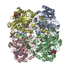
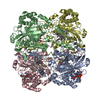
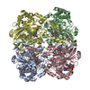
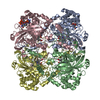
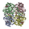
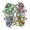
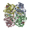
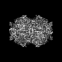
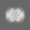
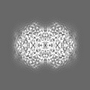
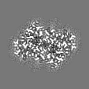
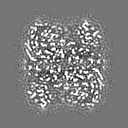
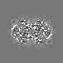
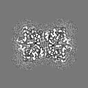
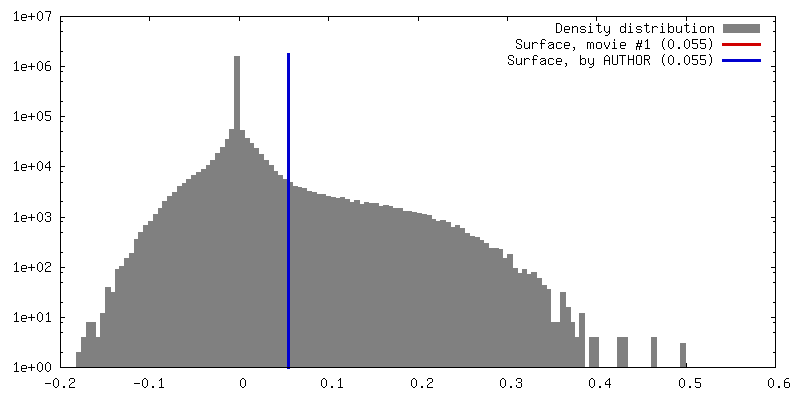
Journal: Microscopy (Oxf) / Year: 2022
Title: Interaction of human erythrocyte catalase with air-water interface in cryoEM.
Authors: Shaoxia Chen / Jade Li / Kutti R Vinothkumar / Richard Henderson /
Abstract: One of the key goals in single-particle cryo-microscopy is to obtain a uniform distribution of particle orientations, so that the three-dimensional structure has isotropic resolution in Fourier space. ...One of the key goals in single-particle cryo-microscopy is to obtain a uniform distribution of particle orientations, so that the three-dimensional structure has isotropic resolution in Fourier space. A common problem arises from the interaction of protein molecules with the air-water interface that exists on both surfaces of the thin film of liquid that is formed prior to plunge-freezing into liquid ethane. Some proteins and other macromolecular complexes are disrupted by interaction with the air-water interface. Other proteins or macromolecules either become concentrated through their interaction with the interface or are excluded because they bind strongly to some other part of the grid or the filter paper used in blotting. In this paper, the interaction of human erythrocyte catalase with the air-water interface is investigated and minimized by the addition of certain detergents. Detergents can form an amphipathic monolayer at the air-water interface that creates a barrier and leaves the molecules free to adopt a variety of orientations, thus facilitating the 3D structure determination. These results suggest that further characterization and development of detergents for cryo-microscopy plunge-freezing would be useful.
History
DepositionJul 23, 2021-
Header (metadata) releaseAug 25, 2021-
Map releaseAug 25, 2021-
UpdateJul 17, 2024-
Current statusJul 17, 2024Processing site: PDBe / Status: Released

-
Structure visualization
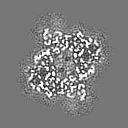
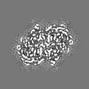
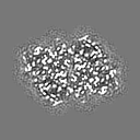
Movie
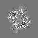
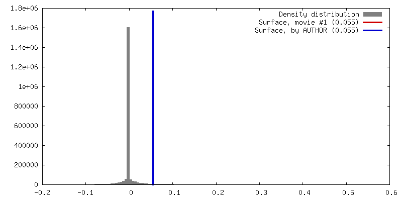
  • Surface view with section colored by density value
  • Surface level: 0.055
  • Imaged by UCSF Chimera
  • Download
  • Surface view colored by radius
  • Surface level: 0.055
  • Imaged by UCSF Chimera
  • Download
  • Surface view with fitted model
  • Atomic models: PDB-7p8w
  • Surface level: 0.055
  • Imaged by UCSF Chimera
  • Download
  • Simplified surface model + fitted atomic model
  • Atomic modelsPDB-7p8w
  • Imaged by Jmol
  • Download
Movie viewer
Structure viewerEM map:
SurfViewMolmilJmol/JSmol
Supplemental images

Downloads & links

-
Map

FileDownload / File: emd_13256.map.gz / Format: CCP4 / Size: 8 MB / Type: IMAGE STORED AS FLOATING POINT NUMBER (4 BYTES)
AnnotationHuman erythrocyte catalase cryo-EM map from Chen et al., 2021, to be published.
Voxel sizeX=Y=Z: 1.07031 Å
Density
Contour LevelBy AUTHOR: 0.055 / Movie #1: 0.055
Minimum - Maximum-0.17919458 - 0.5014312
Average (Standard dev.)0.004404877 (±0.030695038)
SymmetrySpace group: 1
Details

EMDB XML:

Map geometry
Axis orderXYZ
Origin000
Dimensions128128128
Spacing128128128
CellA=B=C: 137.0 Å
α=β=γ: 90.0 °

CCP4 map header:

modeImage stored as Reals
Å/pix. X/Y/Z1.07031251.07031251.0703125
M x/y/z128128128
origin x/y/z0.0000.0000.000
length x/y/z137.000137.000137.000
α/β/γ90.00090.00090.000
MAP C/R/S123
start NC/NR/NS000
NC/NR/NS128128128
D min/max/mean-0.1790.5010.004

-
Supplemental data

-
Mask #1

Fileemd_13256_msk_1.map
Projections & Slices
AxesZYX

Projections

Slices (1/2)
Density Histograms

-
Sample components

-
Entire : Human erythrocyte catalase with cofactors Heme and NADPH

EntireName: Human erythrocyte catalase with cofactors Heme and NADPH
Components
  • Organelle or cellular component: Human erythrocyte catalase with cofactors Heme and NADPH
    • Protein or peptide: Catalase
  • Ligand: NADPH DIHYDRO-NICOTINAMIDE-ADENINE-DINUCLEOTIDE PHOSPHATE
  • Ligand: PROTOPORPHYRIN IX CONTAINING FE
  • Ligand: water

-
Supramolecule #1: Human erythrocyte catalase with cofactors Heme and NADPH

SupramoleculeName: Human erythrocyte catalase with cofactors Heme and NADPH
type: organelle_or_cellular_component / ID: 1 / Parent: 0 / Macromolecule list: #1 / Details: Purified protein
Source (natural)Organism: Homo sapiens (human)
Molecular weightTheoretical: 240 KDa

-
Macromolecule #1: Catalase

MacromoleculeName: Catalase / type: protein_or_peptide / ID: 1 / Number of copies: 4 / Enantiomer: LEVO / EC number: catalase
Source (natural)Organism: Homo sapiens (human) / Cell: erythrocyte
Molecular weightTheoretical: 59.836996 KDa
Recombinant expressionOrganism: Homo sapiens (human)
SequenceString: MADSRDPASD QMQHWKEQRA AQKADVLTTG AGNPVGDKLN VITVGPRGPL LVQDVVFTDE MAHFDRERIP ERVVHAKGAG AFGYFEVTH DITKYSKAKV FEHIGKKTPI AVRFSTVAGE SGSADTVRDP RGFAVKFYTE DGNWDLVGNN TPIFFIRDPI L FPSFIHSQ ...String:
MADSRDPASD QMQHWKEQRA AQKADVLTTG AGNPVGDKLN VITVGPRGPL LVQDVVFTDE MAHFDRERIP ERVVHAKGAG AFGYFEVTH DITKYSKAKV FEHIGKKTPI AVRFSTVAGE SGSADTVRDP RGFAVKFYTE DGNWDLVGNN TPIFFIRDPI L FPSFIHSQ KRNPQTHLKD PDMVWDFWSL RPESLHQVSF LFSDRGIPDG HRHMNGYGSH TFKLVNANGE AVYCKFHYKT DQ GIKNLSV EDAARLSQED PDYGIRDLFN AIATGKYPSW TFYIQVMTFN QAETFPFNPF DLTKVWPHKD YPLIPVGKLV LNR NPVNYF AEVEQIAFDP SNMPPGIEAS PDKMLQGRLF AYPDTHRHRL GPNYLHIPVN CPYRARVANY QRDGPMCMQD NQGG APNYY PNSFGAPEQQ PSALEHSIQY SGEVRRFNTA NDDNVTQVRA FYVNVLNEEQ RKRLCENIAG HLKDAQIFIQ KKAVK NFTE VHPDYGSHIQ ALLDKYNAEK PKNAIHTFVQ SGSHLAAREK ANL

UniProtKB: Catalase

-
Macromolecule #2: NADPH DIHYDRO-NICOTINAMIDE-ADENINE-DINUCLEOTIDE PHOSPHATE

MacromoleculeName: NADPH DIHYDRO-NICOTINAMIDE-ADENINE-DINUCLEOTIDE PHOSPHATE
type: ligand / ID: 2 / Number of copies: 4 / Formula: NDP
Molecular weightTheoretical: 745.421 Da
Chemical component information

ChemComp-NDP:
NADPH DIHYDRO-NICOTINAMIDE-ADENINE-DINUCLEOTIDE PHOSPHATE

-
Macromolecule #3: PROTOPORPHYRIN IX CONTAINING FE

MacromoleculeName: PROTOPORPHYRIN IX CONTAINING FE / type: ligand / ID: 3 / Number of copies: 4 / Formula: HEM
Molecular weightTheoretical: 616.487 Da
Chemical component information

ChemComp-HEM:
PROTOPORPHYRIN IX CONTAINING FE

-
Macromolecule #4: water

MacromoleculeName: water / type: ligand / ID: 4 / Number of copies: 1110 / Formula: HOH
Molecular weightTheoretical: 18.015 Da
Chemical component information

ChemComp-HOH:
WATER

-
Experimental details

-
Structure determination

Methodcryo EM
Processingsingle particle reconstruction
Aggregation stateparticle

-
Sample preparation

Concentration40.0 mg/mL
BufferpH: 7.4
Component:
ConcentrationFormulaName
0.15 MNaClSodium chloride
10.0 mMC4H11NO3Tris Chloride
0.2 mMNADPHNADPH
GridModel: Quantifoil R1.2/1.3 / Material: COPPER / Mesh: 300 / Support film - Material: CARBON / Support film - topology: HOLEY ARRAY / Support film - Film thickness: 30 / Pretreatment - Type: GLOW DISCHARGE / Pretreatment - Time: 30 sec. / Pretreatment - Atmosphere: AIR / Pretreatment - Pressure: 0.015 kPa
VitrificationCryogen name: ETHANE / Chamber humidity: 100 % / Chamber temperature: 277 K / Instrument: HOMEMADE PLUNGER
Details: Controlled environment plunge-freezer (Bellare et al, 1988; Technion University).
DetailsThis sample was monodisperse.

-
Electron microscopy

MicroscopeFEI TITAN KRIOS
Alignment procedureComa free - Residual tilt: 0.25 mrad
Image recordingFilm or detector model: FEI FALCON III (4k x 4k) / Detector mode: COUNTING / Digitization - Dimensions - Width: 4096 pixel / Digitization - Dimensions - Height: 4096 pixel / Number grids imaged: 1 / Number real images: 356 / Average exposure time: 59.0 sec. / Average electron dose: 38.0 e/Å2
Electron beamAcceleration voltage: 300 kV / Electron source: FIELD EMISSION GUN
Electron opticsC2 aperture diameter: 50.0 µm / Illumination mode: FLOOD BEAM / Imaging mode: BRIGHT FIELD / Cs: 2.7 mm / Nominal magnification: 75000
Sample stageSpecimen holder model: FEI TITAN KRIOS AUTOGRID HOLDER / Cooling holder cryogen: NITROGEN
Experimental equipment
Model: Titan Krios / Image courtesy: FEI Company

+
Image processing

Particle selectionNumber selected: 150000 / Details: Autopicked using RELION Laplacian operator
Startup modelType of model: PDB ENTRY
PDB model - PDB ID:

Details: Resolution truncated to 60A.
Final reconstructionNumber classes used: 1 / Applied symmetry - Point group: D2 (2x2 fold dihedral) / Algorithm: FOURIER SPACE / Resolution.type: BY AUTHOR / Resolution: 2.2 Å / Resolution method: FSC 0.143 CUT-OFF / Software - Name: RELION (ver. 3.1) / Number images used: 119169
Initial angle assignmentType: MAXIMUM LIKELIHOOD / Software - Name: RELION (ver. 3.1)
Final angle assignmentType: MAXIMUM LIKELIHOOD / Software - Name: RELION (ver. 3.1)
Final 3D classificationNumber classes: 25 / Software - Name: RELION (ver. 3.1) / Details: 10 2D classes were selected.
FSC plot (resolution estimation)

-
Atomic model buiding 1

Initial model
PDB IDChain

residue_range: 24-502, source_name: PDB, initial_model_type: experimental model

residue_range: 5-23, source_name: PDB, initial_model_type: experimental model
RefinementSpace: RECIPROCAL / Protocol: OTHER / Overall B value: 40
Output model

PDB-7p8w:
Human erythrocyte catalase cryoEM

+
About Yorodumi

-
News

-
Feb 9, 2022. New format data for meta-information of EMDB entries

New format data for meta-information of EMDB entries

  • Version 3 of the EMDB header file is now the official format.
  • The previous official version 1.9 will be removed from the archive.

Related info.:EMDB header

External links:wwPDB to switch to version 3 of the EMDB data model

-
Aug 12, 2020. Covid-19 info

Covid-19 info

URL: https://pdbjlvh1.pdbj.org/emnavi/covid19.php

New page: Covid-19 featured information page in EM Navigator.

Related info.:Covid-19 info / Mar 5, 2020. Novel coronavirus structure data

+
Mar 5, 2020. Novel coronavirus structure data

Novel coronavirus structure data

Related info.:Yorodumi Speices / Aug 12, 2020. Covid-19 info

External links:COVID-19 featured content - PDBj / Molecule of the Month (242):Coronavirus Proteases

+
Jan 31, 2019. EMDB accession codes are about to change! (news from PDBe EMDB page)

EMDB accession codes are about to change! (news from PDBe EMDB page)

  • The allocation of 4 digits for EMDB accession codes will soon come to an end. Whilst these codes will remain in use, new EMDB accession codes will include an additional digit and will expand incrementally as the available range of codes is exhausted. The current 4-digit format prefixed with “EMD-” (i.e. EMD-XXXX) will advance to a 5-digit format (i.e. EMD-XXXXX), and so on. It is currently estimated that the 4-digit codes will be depleted around Spring 2019, at which point the 5-digit format will come into force.
  • The EM Navigator/Yorodumi systems omit the EMD- prefix.

Related info.:Q: What is EMD? / ID/Accession-code notation in Yorodumi/EM Navigator

External links:EMDB Accession Codes are Changing Soon! / Contact to PDBj

+
Jul 12, 2017. Major update of PDB

Major update of PDB

  • wwPDB released updated PDB data conforming to the new PDBx/mmCIF dictionary.
  • This is a major update changing the version number from 4 to 5, and with Remediation, in which all the entries are updated.
  • In this update, many items about electron microscopy experimental information are reorganized (e.g. em_software).
  • Now, EM Navigator and Yorodumi are based on the updated data.

External links:wwPDB Remediation / Enriched Model Files Conforming to OneDep Data Standards Now Available in the PDB FTP Archive

-
Yorodumi

Thousand views of thousand structures

  • Yorodumi is a browser for structure data from EMDB, PDB, SASBDB, etc.
  • This page is also the successor to EM Navigator detail page, and also detail information page/front-end page for Omokage search.
  • The word "yorodu" (or yorozu) is an old Japanese word meaning "ten thousand". "mi" (miru) is to see.

Related info.:EMDB / PDB / SASBDB / Comparison of 3 databanks / Yorodumi Search / Aug 31, 2016. New EM Navigator & Yorodumi / Yorodumi Papers / Jmol/JSmol / Function and homology information / Changes in new EM Navigator and Yorodumi

Read more