 ムービー
ムービー コントローラー


+ データを開く

- 基本情報
基本情報
| 登録情報 | データベース: EMDB / ID: EMD-5375 | |||||||||
|---|---|---|---|---|---|---|---|---|---|---|
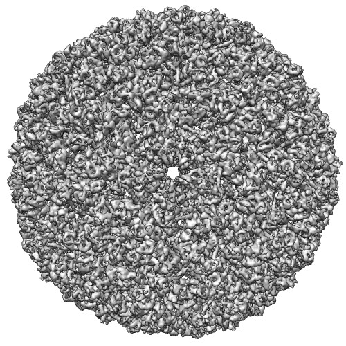
| タイトル | Direct electron detection yields cryo-EM reconstructions at resolutions beyond .75 Nyquist frequency | |||||||||
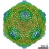
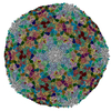
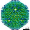
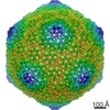
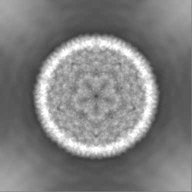
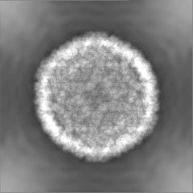
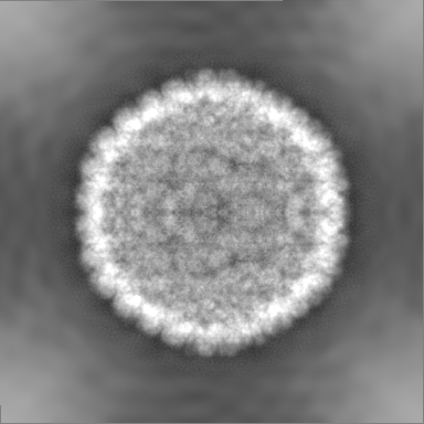
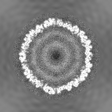
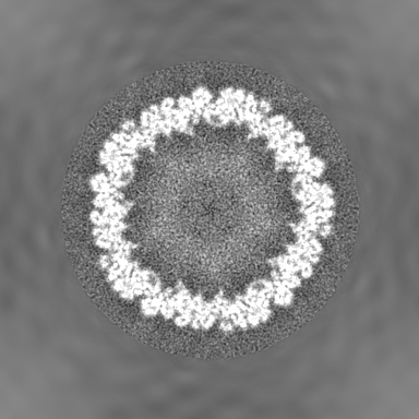
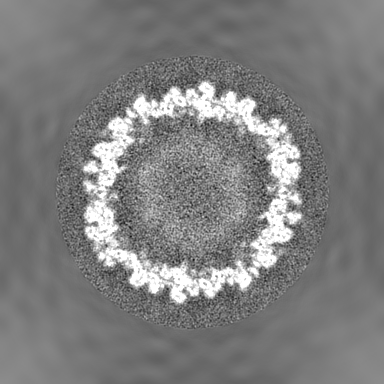
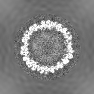
 マップデータ マップデータ | Icosahedral reconstruction of bacteriophage P22 | |||||||||
 試料 試料 |
| |||||||||
 キーワード キーワード | Cryo-EM / Electron cryo-microscopy / Direct detection device / Active pixel sensor / CMOS detector / Nyquist frequency | |||||||||
| 生物種 |  Enterobacteria phage P22 (ファージ) Enterobacteria phage P22 (ファージ) | |||||||||
| 手法 | 単粒子再構成法 / クライオ電子顕微鏡法 / 解像度: 8.5 Å | |||||||||
 データ登録者 データ登録者 | Bammes BE / Rochat RH / Jakana J / Chen D / Chiu W | |||||||||
 引用 引用 | ジャーナル: J Struct Biol / 年: 2012 タイトル: Direct electron detection yields cryo-EM reconstructions at resolutions beyond 3/4 Nyquist frequency. 著者: Benjamin E Bammes / Ryan H Rochat / Joanita Jakana / Dong-Hua Chen / Wah Chiu /  要旨: One limitation in electron cryo-microscopy (cryo-EM) is the inability to recover high-resolution signal from the image-recording media at the full-resolution limit of the transmission electron ...One limitation in electron cryo-microscopy (cryo-EM) is the inability to recover high-resolution signal from the image-recording media at the full-resolution limit of the transmission electron microscope. Direct electron detection using CMOS-based sensors for digitally recording images has the potential to alleviate this shortcoming. Here, we report a practical performance evaluation of a Direct Detection Device (DDD®) for biological cryo-EM at two different microscope voltages: 200 and 300 kV. Our DDD images of amorphous and graphitized carbon show strong per-pixel contrast with image resolution near the theoretical sampling limit of the data. Single-particle reconstructions of two frozen-hydrated bacteriophages, P22 and ε15, establish that the DDD is capable of recording usable signal for 3D reconstructions at about 4/5 of the Nyquist frequency, which is a vast improvement over the performance of conventional imaging media. We anticipate the unparalleled performance of this digital recording device will dramatically benefit cryo-EM for routine tomographic and single-particle structural determination of biological specimens. | |||||||||
| 履歴 |
|
- 構造の表示
構造の表示
| ムービー |
 ムービービューア ムービービューア |
|---|---|
| 構造ビューア | EMマップ: SurfViewMolmilJmol/JSmol |
| 添付画像 |
- ダウンロードとリンク
-EMDBアーカイブ
| マップデータ | emd_5375.map.gz | 196.4 MB | EMDBマップデータ形式 | |
|---|---|---|---|---|
| ヘッダ (付随情報) | emd-5375-v30.xmlemd-5375.xml | 9.9 KB 9.9 KB | 表示 表示 | EMDBヘッダ |
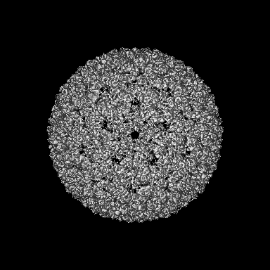
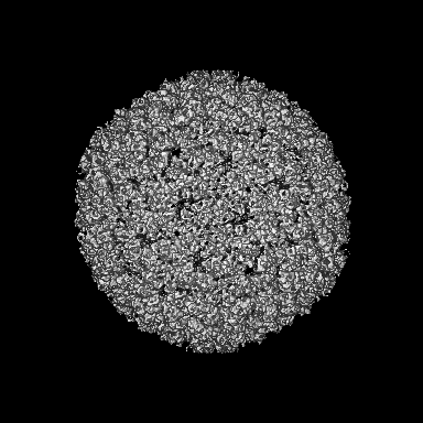
| 画像 |  emd_5375_1.jpg emd_5375_1.jpg | 116.4 KB | ||
| アーカイブディレクトリ |  http://ftp.pdbj.org/pub/emdb/structures/EMD-5375ftp://ftp.pdbj.org/pub/emdb/structures/EMD-5375 http://ftp.pdbj.org/pub/emdb/structures/EMD-5375ftp://ftp.pdbj.org/pub/emdb/structures/EMD-5375 | HTTPS FTP |
-検証レポート
| 文書・要旨 | emd_5375_validation.pdf.gz | 77.6 KB | 表示 | EMDB検証レポート |
|---|---|---|---|---|
| 文書・詳細版 | emd_5375_full_validation.pdf.gz | 76.7 KB | 表示 | |
| XML形式データ | emd_5375_validation.xml.gz | 494 B | 表示 | |
| アーカイブディレクトリ | https://ftp.pdbj.org/pub/emdb/validation_reports/EMD-5375ftp://ftp.pdbj.org/pub/emdb/validation_reports/EMD-5375 | HTTPS FTP |
-関連構造データ


-リンク
| EMDBのページ | EMDB (EBI/PDBe) / EMDataResource |
|---|
-マップ
| ファイル | ダウンロード / ファイル: emd_5375.map.gz / 形式: CCP4 / 大きさ: 210.9 MB / タイプ: IMAGE STORED AS FLOATING POINT NUMBER (4 BYTES) | ||||||||||||||||||||||||||||||||||||||||||||||||||||||||||||||||||||
|---|---|---|---|---|---|---|---|---|---|---|---|---|---|---|---|---|---|---|---|---|---|---|---|---|---|---|---|---|---|---|---|---|---|---|---|---|---|---|---|---|---|---|---|---|---|---|---|---|---|---|---|---|---|---|---|---|---|---|---|---|---|---|---|---|---|---|---|---|---|
| 注釈 | Icosahedral reconstruction of bacteriophage P22 | ||||||||||||||||||||||||||||||||||||||||||||||||||||||||||||||||||||
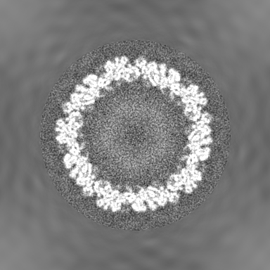
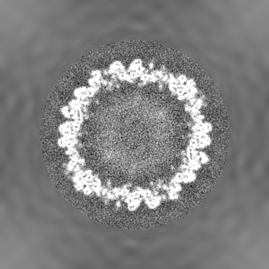
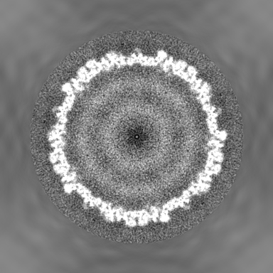
| 投影像・断面図 | 画像のコントロール
画像は Spider により作成 | ||||||||||||||||||||||||||||||||||||||||||||||||||||||||||||||||||||
| ボクセルのサイズ | X=Y=Z: 3.48 Å | ||||||||||||||||||||||||||||||||||||||||||||||||||||||||||||||||||||
| 密度 |
| ||||||||||||||||||||||||||||||||||||||||||||||||||||||||||||||||||||
| 対称性 | 空間群: 1 | ||||||||||||||||||||||||||||||||||||||||||||||||||||||||||||||||||||
| 詳細 | EMDB XML:
CCP4マップ ヘッダ情報:
| ||||||||||||||||||||||||||||||||||||||||||||||||||||||||||||||||||||
 Z (Sec.)
 Y (Row.)
 X (Col.)


-添付データ
- 試料の構成要素
-全体 : Bacteriophage P22 procapsid
| 全体 | 名称: Bacteriophage P22 procapsid |
|---|---|
| 要素 |
|
-超分子 #1000: Bacteriophage P22 procapsid
| 超分子 | 名称: Bacteriophage P22 procapsid / タイプ: sample / ID: 1000 / Number unique components: 1 |
|---|
-超分子 #1: Enterobacteria phage P22
| 超分子 | 名称: Enterobacteria phage P22 / タイプ: virus / ID: 1 / Name.synonym: P22 / NCBI-ID: 10754 / 生物種: Enterobacteria phage P22 / データベース: NCBI / ウイルスタイプ: VIRION / ウイルス・単離状態: STRAIN / ウイルス・エンベロープ: No / ウイルス・中空状態: Yes / Syn species name: P22 |
|---|---|
| 宿主 | 生物種:  Salmonella (サルモネラ菌) / 別称: BACTERIA(EUBACTERIA) Salmonella (サルモネラ菌) / 別称: BACTERIA(EUBACTERIA) |
| ウイルス殻 | Shell ID: 1 / 名称: Procapsid / 直径: 525 Å / T番号(三角分割数): 7 |
-実験情報
-構造解析
| 手法 | クライオ電子顕微鏡法 |
|---|---|
 解析 解析 | 単粒子再構成法 |
| 試料の集合状態 | particle |
-試料調製
| 濃度 | 1 mg/mL |
|---|---|
| 緩衝液 | pH: 7.6 / 詳細: 50 mM Tris pH 7.6, 25 mM NaCl, 2mM EDTA |
| グリッド | 詳細: 400 mesh copper grid |
| 凍結 | 凍結剤: ETHANE / チャンバー内湿度: 95 % / チャンバー内温度: 170 K / 装置: OTHER / 詳細: Vitrification instrument: Vitrobot / 手法: Blot for 2 seconds before plunging |
- 電子顕微鏡法
| 顕微鏡 | JEOL 2010F |
|---|---|
| 温度 | 最低: 100 K / 最高: 100 K / 平均: 100 K |
| アライメント法 | Legacy - 非点収差: objective lens astigmatism was corrected at 100,000 times magnification |
| 日付 | 2010年6月10日 |
| 撮影 | カテゴリ: CCD フィルム・検出器のモデル: DIRECT ELECTRON DE-12 (4k x 3k) デジタル化 - サンプリング間隔: 6 µm / 実像数: 200 / 平均電子線量: 17 e/Å2 詳細: Images were collected on a CMOS type detector DE-12 from Direct Electron |
| 電子線 | 加速電圧: 200 kV / 電子線源:  FIELD EMISSION GUN FIELD EMISSION GUN |
| 電子光学系 | 倍率(補正後): 17200 / 照射モード: FLOOD BEAM / 撮影モード: BRIGHT FIELD / Cs: 2.0 mm / 最大 デフォーカス(公称値): 5.0 µm / 最小 デフォーカス(公称値): 2.0 µm / 倍率(公称値): 15000 |
| 試料ステージ | 試料ホルダー: Side Entry / 試料ホルダーモデル: GATAN LIQUID NITROGEN |
-画像解析
| CTF補正 | 詳細: Each Micrograph |
|---|---|
| 最終 再構成 | アルゴリズム: OTHER / 解像度のタイプ: BY AUTHOR / 解像度: 8.5 Å / 解像度の算出法: FSC 0.5 CUT-OFF / ソフトウェア - 名称: MPSA 詳細: Resolution was calculated to be 8.5 Angstroms at 0.5 FSC and 7.3 Angstroms at 0.143 FSC as compared against the 3.8 Angstrom reconstruction of P22. 使用した粒子像数: 7500 |
