Netherlands Organisation for Scientific Research (NWO)
OCENW.XL21.XL21.048
Netherlands
National Institutes of Health/National Institute of General Medical Sciences (NIH/NIGMS)
1R01GM138854
United States
National Institutes of Health/National Institute of General Medical Sciences (NIH/NIGMS)
1R01GM141109
United States
National Institutes of Health/National Institute of General Medical Sciences (NIH/NIGMS)
1R01GM143183
United States
Citation
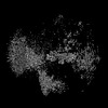
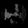
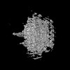
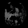
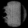
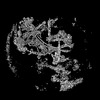
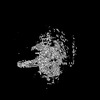
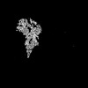
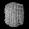
Journal: Nature / Year: 2025 Title: Structural diversity of axonemes across mammalian motile cilia. Authors: Miguel Ricardo Leung / Chen Sun / Jianwei Zeng / Jacob R Anderson / Qingwei Niu / Wei Huang / Willem E M Noteborn / Alan Brown / Tzviya Zeev-Ben-Mordehai / Rui Zhang / Abstract: Reproduction, development and homeostasis depend on motile cilia, whose rhythmic beating is powered by a microtubule-based molecular machine called the axoneme. Although an atomic model of the ...Reproduction, development and homeostasis depend on motile cilia, whose rhythmic beating is powered by a microtubule-based molecular machine called the axoneme. Although an atomic model of the axoneme is available for the alga Chlamydomonas reinhardtii, structures of mammalian axonemes are incomplete. Furthermore, we do not fully understand how molecular structures of axonemes vary across motile-ciliated cell types in the body. Here we use cryoelectron microscopy, cryoelectron tomography and proteomics to resolve the 96-nm modular repeat of axonemal doublet microtubules (DMTs) from both sperm flagella and epithelial cilia of the oviduct, brain ventricles and respiratory tract. We find that sperm DMTs are the most specialized, with epithelial cilia having only minor differences across tissues. We build a model of the mammalian sperm DMT, defining the positions and interactions of 181 proteins including 34 newly identified proteins. We elucidate the composition of radial spoke 3 and uncover binding sites of kinases associated with regeneration of ATP and regulation of ciliary motility. We discover a sperm-specific, axoneme-tethered T-complex protein ring complex (TRiC) chaperone that may contribute to construction or maintenance of the long flagella of mammalian sperm. We resolve axonemal dyneins in their prestroke states, illuminating conformational changes that occur during ciliary movement. Our results illustrate how elements of chemical and mechanical regulation are embedded within the axoneme, providing valuable resources for understanding the aetiology of ciliopathy and infertility, and exemplifying the discovery power of modern structural biology.
In the structure databanks used in Yorodumi, some data are registered as the other names, "COVID-19 virus" and "2019-nCoV". Here are the details of the virus and the list of structure data.
Jan 31, 2019. EMDB accession codes are about to change! (news from PDBe EMDB page)
EMDB accession codes are about to change! (news from PDBe EMDB page)
The allocation of 4 digits for EMDB accession codes will soon come to an end. Whilst these codes will remain in use, new EMDB accession codes will include an additional digit and will expand incrementally as the available range of codes is exhausted. The current 4-digit format prefixed with “EMD-” (i.e. EMD-XXXX) will advance to a 5-digit format (i.e. EMD-XXXXX), and so on. It is currently estimated that the 4-digit codes will be depleted around Spring 2019, at which point the 5-digit format will come into force.
The EM Navigator/Yorodumi systems omit the EMD- prefix.
Related info.:Q: What is EMD? / ID/Accession-code notation in Yorodumi/EM Navigator
Yorodumi is a browser for structure data from EMDB, PDB, SASBDB, etc.
This page is also the successor to EM Navigator detail page, and also detail information page/front-end page for Omokage search.
The word "yorodu" (or yorozu) is an old Japanese word meaning "ten thousand". "mi" (miru) is to see.
Related info.:EMDB / PDB / SASBDB / Comparison of 3 databanks / Yorodumi Search / Aug 31, 2016. New EM Navigator & Yorodumi / Yorodumi Papers / Jmol/JSmol / Function and homology information / Changes in new EM Navigator and Yorodumi
 Movie
Movie Controller

 Yorodumi
 Open data
 Basic information
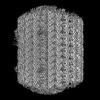
 Map data
 Sample
 Keywords
 Authors
 Netherlands,
  United States, 4 items
  Citation
 Structure visualization
 Downloads & links
 EMDB map data format
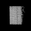
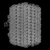
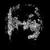
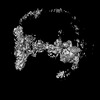
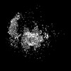
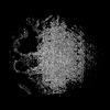
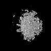
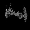
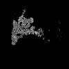
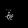
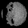
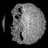
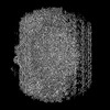
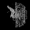
 emd_53135.png
 http://ftp.pdbj.org/pub/emdb/structures/EMD-53135


 Z (Sec.)
 Y (Row.)
 X (Col.)


 Sample components
 Processing
 Electron microscopy
 FIELD EMISSION GUN

