 Movie
Movie Controller

[English] 日本語
 Yorodumi
- EMDB-5125: PSRP1 is not a bona fide ribosomal protein, but a stress response... -
+ Open data

- Basic information
| Entry | Database: EMDB / ID: EMD-5125 | |||||||||
|---|---|---|---|---|---|---|---|---|---|---|
| Title | PSRP1 is not a bona fide ribosomal protein, but a stress response factor | |||||||||
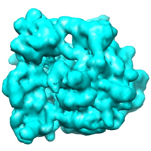
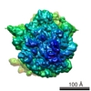
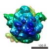
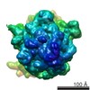
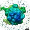
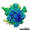
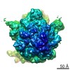
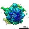
 Map data Map data | 70S E.coli Ribosome with PSRP1, in vitro | |||||||||
 Sample Sample |
| |||||||||
 Keywords Keywords | 70S / E.coli Ribosome / Cryo-EM PSRP1 / PSRP-1 / ribosomal protein / stress response factor. | |||||||||
| Biological species |   Spinacia oleracea (spinach) Spinacia oleracea (spinach) | |||||||||
| Method | single particle reconstruction / cryo EM / Resolution: 9.8 Å | |||||||||
 Authors Authors | Sharma MR / Donhofer A / Barat C / Datta P / Fucini P / Wilson DN / Agrawal RK | |||||||||
 Citation Citation | Journal: J Biol Chem / Year: 2010 Title: PSRP1 is not a ribosomal protein, but a ribosome-binding factor that is recycled by the ribosome-recycling factor (RRF) and elongation factor G (EF-G). Authors: Manjuli R Sharma / Alexandra Dönhöfer / Chandana Barat / Viter Marquez / Partha P Datta / Paola Fucini / Daniel N Wilson / Rajendra K Agrawal /   Abstract: Plastid-specific ribosomal proteins (PSRPs) have been proposed to play roles in the light-dependent regulation of chloroplast translation. Here we demonstrate that PSRP1 is not a bona fide ribosomal ...Plastid-specific ribosomal proteins (PSRPs) have been proposed to play roles in the light-dependent regulation of chloroplast translation. Here we demonstrate that PSRP1 is not a bona fide ribosomal protein, but rather a functional homologue of the Escherichia coli cold-shock protein pY. Three-dimensional Cryo-electron microscopic (Cryo-EM) reconstructions reveal that, like pY, PSRP1 binds within the intersubunit space of the 70S ribosome, at a site overlapping the positions of mRNA and A- and P-site tRNAs. PSRP1 induces conformational changes within ribosomal components that comprise several intersubunit bridges, including bridge B2a, thereby stabilizes the ribosome against dissociation. We find that the presence of PSRP1/pY lowers the binding of tRNA to the ribosome. Furthermore, similarly to tRNAs, PSRP1/pY is recycled from the ribosome by the concerted action of the ribosome-recycling factor (RRF) and elongation factor G (EF-G). These results suggest a novel function for EF-G and RRF in the post-stress return of PSRP1/pY-inactivated ribosomes to the actively translating pool. | |||||||||
| History |
|
- Structure visualization
| Movie |
 Movie viewer Movie viewer |
|---|---|
| Structure viewer | EM map: SurfViewMolmilJmol/JSmol |
| Supplemental images |
- Downloads & links
-EMDB archive
| Map data | emd_5125.map.gz | 7.8 MB | EMDB map data format | |
|---|---|---|---|---|
| Header (meta data) | emd-5125-v30.xmlemd-5125.xml | 10.1 KB 10.1 KB | Display Display | EMDB header |
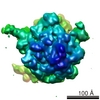
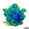
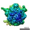
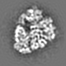
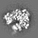
| Images |  emd_5125_1.jpg emd_5125_1.jpg | 51.7 KB | ||
| Archive directory |  http://ftp.pdbj.org/pub/emdb/structures/EMD-5125ftp://ftp.pdbj.org/pub/emdb/structures/EMD-5125 http://ftp.pdbj.org/pub/emdb/structures/EMD-5125ftp://ftp.pdbj.org/pub/emdb/structures/EMD-5125 | HTTPS FTP |
-Validation report
| Summary document | emd_5125_validation.pdf.gz | 78 KB | Display | EMDB validaton report |
|---|---|---|---|---|
| Full document | emd_5125_full_validation.pdf.gz | 77.1 KB | Display | |
| Data in XML | emd_5125_validation.xml.gz | 493 B | Display | |
| Arichive directory | https://ftp.pdbj.org/pub/emdb/validation_reports/EMD-5125ftp://ftp.pdbj.org/pub/emdb/validation_reports/EMD-5125 | HTTPS FTP |
-Related structure data

-Links
| EMDB pages | EMDB (EBI/PDBe) / EMDataResource |
|---|---|
| Related items in Molecule of the Month |

-Map
| File | Download / File: emd_5125.map.gz / Format: CCP4 / Size: 8.2 MB / Type: IMAGE STORED AS FLOATING POINT NUMBER (4 BYTES) | ||||||||||||||||||||||||||||||||||||||||||||||||||||||||||||||||||||
|---|---|---|---|---|---|---|---|---|---|---|---|---|---|---|---|---|---|---|---|---|---|---|---|---|---|---|---|---|---|---|---|---|---|---|---|---|---|---|---|---|---|---|---|---|---|---|---|---|---|---|---|---|---|---|---|---|---|---|---|---|---|---|---|---|---|---|---|---|---|
| Annotation | 70S E.coli Ribosome with PSRP1, in vitro | ||||||||||||||||||||||||||||||||||||||||||||||||||||||||||||||||||||
| Projections & slices | Image control
Images are generated by Spider. | ||||||||||||||||||||||||||||||||||||||||||||||||||||||||||||||||||||
| Voxel size | X=Y=Z: 2.76 Å | ||||||||||||||||||||||||||||||||||||||||||||||||||||||||||||||||||||
| Density |
| ||||||||||||||||||||||||||||||||||||||||||||||||||||||||||||||||||||
| Symmetry | Space group: 1 | ||||||||||||||||||||||||||||||||||||||||||||||||||||||||||||||||||||
| Details | EMDB XML:
CCP4 map header:
| ||||||||||||||||||||||||||||||||||||||||||||||||||||||||||||||||||||
 Z (Sec.)
 Y (Row.)
 X (Col.)


-Supplemental data
- Sample components
-Entire : 70S E.coli ribosome with PSRP1 in vitro
| Entire | Name: 70S E.coli ribosome with PSRP1 in vitro |
|---|---|
| Components |
|
-Supramolecule #1000: 70S E.coli ribosome with PSRP1 in vitro
| Supramolecule | Name: 70S E.coli ribosome with PSRP1 in vitro / type: sample / ID: 1000 / Number unique components: 2 |
|---|---|
| Molecular weight | Experimental: 2.8 MDa / Theoretical: 2.8 MDa |
-Supramolecule #1: 70S Ribosome
| Supramolecule | Name: 70S Ribosome / type: complex / ID: 1 / Recombinant expression: No / Database: NCBI / Ribosome-details: ribosome-prokaryote: LSU 50S, SSU 30S |
|---|---|
| Source (natural) | Organism: |
| Molecular weight | Experimental: 2.5 MDa / Theoretical: 2.5 MDa |
-Macromolecule #1: plastid specific ribosomal protein-1
| Macromolecule | Name: plastid specific ribosomal protein-1 / type: ligand / ID: 1 / Name.synonym: PSRP1 / Recombinant expression: No / Database: NCBI |
|---|---|
| Source (natural) | Organism: Spinacia oleracea (spinach) / synonym: Spinach / Organelle: Chloroplast |
| Molecular weight | Experimental: 270 KDa / Theoretical: 270 KDa |
-Experimental details
-Structure determination
| Method | cryo EM |
|---|---|
 Processing Processing | single particle reconstruction |
| Aggregation state | particle |
-Sample preparation
| Buffer | pH: 7.6 Details: 20mM Hepes-KOH, 8.2mM MgCl2, 80mM NH4Cl, 4mM beta-mercaptoethanol |
|---|---|
| Grid | Details: 300 mesh coper grid |
| Vitrification | Cryogen name: ETHANE / Chamber humidity: 80 % / Instrument: HOMEMADE PLUNGER / Details: Vitrification instrument: Plunger / Method: Blot for 3 seconds then plunge |
- Electron microscopy
| Microscope | FEI TECNAI F20 |
|---|---|
| Image recording | Category: FILM / Film or detector model: KODAK SO-163 FILM / Digitization - Scanner: ZEISS SCAI / Digitization - Sampling interval: 14 µm / Number real images: 53 / Average electron dose: 24 e/Å2 / Bits/pixel: 12 |
| Electron beam | Acceleration voltage: 200 kV / Electron source:  FIELD EMISSION GUN FIELD EMISSION GUN |
| Electron optics | Calibrated magnification: 50760 / Illumination mode: FLOOD BEAM / Imaging mode: BRIGHT FIELD / Nominal defocus max: 4.2 µm / Nominal defocus min: 1.2 µm / Nominal magnification: 50000 |
| Sample stage | Specimen holder: Eucentric / Specimen holder model: GATAN LIQUID NITROGEN |
| Experimental equipment |  Model: Tecnai F20 / Image courtesy: FEI Company |
-Image processing
| CTF correction | Details: Each Micrograph |
|---|---|
| Final reconstruction | Algorithm: OTHER / Resolution.type: BY AUTHOR / Resolution: 9.8 Å / Resolution method: FSC 0.5 CUT-OFF / Software - Name: Spider / Number images used: 32611 |
| Final two d classification | Number classes: 18 |
