 ムービー
ムービー コントローラー


+ データを開く

- 基本情報
基本情報
| 登録情報 |  | |||||||||
|---|---|---|---|---|---|---|---|---|---|---|
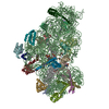
| タイトル | Structure of Kasugamycin-30S-IF1-IF3-mRNA-tRNA translation pre-initiation complex, open form | |||||||||
 マップデータ マップデータ | ||||||||||
 試料 試料 |
| |||||||||
 キーワード キーワード | Ribosome / Antibiotic / TRANSLATION | |||||||||
| 生物種 |  | |||||||||
| 手法 | 単粒子再構成法 / クライオ電子顕微鏡法 / 解像度: 2.9 Å | |||||||||
 データ登録者 データ登録者 | Safdari HA / Wilson DN | |||||||||
| 資金援助 |  ドイツ, 1件 ドイツ, 1件
| |||||||||
 引用 引用 | ジャーナル: Nat Commun / 年: 2025 タイトル: The translation inhibitors kasugamycin, edeine and GE81112 target distinct steps during 30S initiation complex formation. 著者: Haaris A Safdari / Martino Morici / Ana Sanchez-Castro / Andrea Dallapè / Helge Paternoga / Anna Maria Giuliodori / Attilio Fabbretti / Pohl Milón / Daniel N Wilson /   要旨: During bacterial translation initiation, the 30S ribosomal subunit, initiation factors, and initiator tRNA define the reading frame of the mRNA. This process is inhibited by kasugamycin, edeine and ...During bacterial translation initiation, the 30S ribosomal subunit, initiation factors, and initiator tRNA define the reading frame of the mRNA. This process is inhibited by kasugamycin, edeine and GE81112, however, their mechanisms of action have not been fully elucidated. Here we present cryo-electron microscopy structures of 30S initiation intermediate complexes formed in the presence of kasugamycin, edeine and GE81112 at resolutions of 2.0-2.9 Å. The structures reveal that all three antibiotics bind within the E-site of the 30S and preclude 30S initiation complex formation. While kasugamycin and edeine affect early steps of 30S pre-initiation complex formation, GE81112 stalls pre-initiation complex formation at a further step by allowing start codon recognition, but impeding IF3 departure. Collectively, our work highlights how chemically distinct compounds binding at a conserved site on the 30S can interfere with translation initiation in a unique manner. | |||||||||
| 履歴 |
|
- 構造の表示
構造の表示
| 添付画像 |
|---|

- ダウンロードとリンク
-EMDBアーカイブ
| マップデータ | emd_51214.map.gz | 193.5 MB |  EMDBマップデータ形式 EMDBマップデータ形式 | |
|---|---|---|---|---|
| ヘッダ (付随情報) | emd-51214-v30.xmlemd-51214.xml | 16.3 KB 16.3 KB | 表示 表示 | EMDBヘッダ |
| FSC (解像度算出) | emd_51214_fsc.xml | 14.2 KB | 表示 | FSCデータファイル |
| 画像 |  emd_51214.png emd_51214.png | 45.6 KB | ||
| マスクデータ | emd_51214_msk_1.map | 244.1 MB | マスクマップ | |
| Filedesc metadata | emd-51214.cif.gz | 4 KB | ||
| その他 | emd_51214_additional_1.map.gzemd_51214_half_map_1.map.gzemd_51214_half_map_2.map.gz | 22.5 MB 193.9 MB 193.9 MB | ||
| アーカイブディレクトリ |  http://ftp.pdbj.org/pub/emdb/structures/EMD-51214ftp://ftp.pdbj.org/pub/emdb/structures/EMD-51214 http://ftp.pdbj.org/pub/emdb/structures/EMD-51214ftp://ftp.pdbj.org/pub/emdb/structures/EMD-51214 | HTTPS FTP |
-検証レポート
| 文書・要旨 | emd_51214_validation.pdf.gz | 1 MB | 表示 | EMDB検証レポート |
|---|---|---|---|---|
| 文書・詳細版 | emd_51214_full_validation.pdf.gz | 1 MB | 表示 | |
| XML形式データ | emd_51214_validation.xml.gz | 21.3 KB | 表示 | |
| CIF形式データ | emd_51214_validation.cif.gz | 27.3 KB | 表示 | |
| アーカイブディレクトリ | https://ftp.pdbj.org/pub/emdb/validation_reports/EMD-51214ftp://ftp.pdbj.org/pub/emdb/validation_reports/EMD-51214 | HTTPS FTP |
-関連構造データ


-リンク
| EMDBのページ | EMDB (EBI/PDBe) / EMDataResource |
|---|
-マップ
| ファイル | ダウンロード / ファイル: emd_51214.map.gz / 形式: CCP4 / 大きさ: 244.1 MB / タイプ: IMAGE STORED AS FLOATING POINT NUMBER (4 BYTES) | ||||||||||||||||||||||||||||||||||||
|---|---|---|---|---|---|---|---|---|---|---|---|---|---|---|---|---|---|---|---|---|---|---|---|---|---|---|---|---|---|---|---|---|---|---|---|---|---|
| 投影像・断面図 | 画像のコントロール
画像は Spider により作成 | ||||||||||||||||||||||||||||||||||||
| ボクセルのサイズ | X=Y=Z: 0.832 Å | ||||||||||||||||||||||||||||||||||||
| 密度 |
| ||||||||||||||||||||||||||||||||||||
| 対称性 | 空間群: 1 | ||||||||||||||||||||||||||||||||||||
| 詳細 | EMDB XML:
|
 Z (Sec.)
 Y (Row.)
 X (Col.)


-添付データ
-マスク #1
| ファイル | emd_51214_msk_1.map | ||||||||||||
|---|---|---|---|---|---|---|---|---|---|---|---|---|---|
| 投影像・断面図 |
| ||||||||||||
| 密度ヒストグラム |


-追加マップ: #1
| ファイル | emd_51214_additional_1.map | ||||||||||||
|---|---|---|---|---|---|---|---|---|---|---|---|---|---|
| 投影像・断面図 |
| ||||||||||||
| 密度ヒストグラム |


-ハーフマップ: #1
| ファイル | emd_51214_half_map_1.map | ||||||||||||
|---|---|---|---|---|---|---|---|---|---|---|---|---|---|
| 投影像・断面図 |
| ||||||||||||
| 密度ヒストグラム |


-ハーフマップ: #2
| ファイル | emd_51214_half_map_2.map | ||||||||||||
|---|---|---|---|---|---|---|---|---|---|---|---|---|---|
| 投影像・断面図 |
| ||||||||||||
| 密度ヒストグラム |


- 試料の構成要素
-全体 : Structure of Kasugamycin-30S-IF1-IF3-mRNA-tRNA translation pre-in...
| 全体 | 名称: Structure of Kasugamycin-30S-IF1-IF3-mRNA-tRNA translation pre-initiation complex, open form |
|---|---|
| 要素 |
|
-超分子 #1: Structure of Kasugamycin-30S-IF1-IF3-mRNA-tRNA translation pre-in...
| 超分子 | 名称: Structure of Kasugamycin-30S-IF1-IF3-mRNA-tRNA translation pre-initiation complex, open form タイプ: complex / ID: 1 / 親要素: 0 / 含まれる分子: #2-#7, #9-#16, #1 |
|---|---|
| 由来(天然) | 生物種: |
| 分子量 | 理論値: 850 KDa |
-実験情報
-構造解析
| 手法 | クライオ電子顕微鏡法 |
|---|---|
 解析 解析 | 単粒子再構成法 |
| 試料の集合状態 | particle |
-試料調製
| 緩衝液 | pH: 7.5 |
|---|---|
| 凍結 | 凍結剤: ETHANE-PROPANE |
- 電子顕微鏡法
| 顕微鏡 | TFS KRIOS |
|---|---|
| 撮影 | フィルム・検出器のモデル: GATAN K3 BIOQUANTUM (6k x 4k) 平均電子線量: 1.14 e/Å2 |
| 電子線 | 加速電圧: 300 kV / 電子線源:  FIELD EMISSION GUN FIELD EMISSION GUN |
| 電子光学系 | 照射モード: FLOOD BEAM / 撮影モード: BRIGHT FIELD / 最大 デフォーカス(公称値): 1.1 µm / 最小 デフォーカス(公称値): 0.3 µm |
| 実験機器 |  モデル: Titan Krios / 画像提供: FEI Company |


