 Movie
Movie Controller


+ Open data

- Basic information
| Entry | Database: PDB / ID: 9fco | ||||||
|---|---|---|---|---|---|---|---|
| Title | Structure of E. coli 30S-IF1-IF3-mRNA-Kasugamycin complex | ||||||
 Components Components |
| ||||||
 Keywords Keywords | TRANSLATION / Ribosome / Antibiotic | ||||||
| Function / homology |  Function and homology information Function and homology informationribosome disassembly / ornithine decarboxylase inhibitor activity / misfolded RNA binding / Group I intron splicing / RNA folding / four-way junction DNA binding / regulation of mRNA stability / translation initiation factor activity / negative regulation of translational initiation / response to cold ...ribosome disassembly / ornithine decarboxylase inhibitor activity / misfolded RNA binding / Group I intron splicing / RNA folding / four-way junction DNA binding / regulation of mRNA stability / translation initiation factor activity / negative regulation of translational initiation / response to cold / mRNA regulatory element binding translation repressor activity / positive regulation of RNA splicing / transcription antitermination / DNA endonuclease activity / DNA-templated transcription termination / maintenance of translational fidelity / mRNA 5'-UTR binding / regulation of translation / ribosomal small subunit assembly / ribosome binding / ribosomal small subunit biogenesis / small ribosomal subunit / small ribosomal subunit rRNA binding / cytosolic small ribosomal subunit / cytoplasmic translation / tRNA binding / rRNA binding / structural constituent of ribosome / ribosome / translation / response to antibiotic / hydrolase activity / RNA binding / zinc ion binding / membrane / cytosol / cytoplasm Similarity search - Function | ||||||
| Biological species |  | ||||||
| Method | ELECTRON MICROSCOPY / single particle reconstruction / cryo EM / Resolution: 2.4 Å | ||||||
 Authors Authors | Safdari, H.A. / Wilson, D.N. | ||||||
| Funding support |  Germany, 1items Germany, 1items
| ||||||
 Citation Citation | Journal: Nat Commun / Year: 2025 Title: The translation inhibitors kasugamycin, edeine and GE81112 target distinct steps during 30S initiation complex formation. Authors: Haaris A Safdari / Martino Morici / Ana Sanchez-Castro / Andrea Dallapè / Helge Paternoga / Anna Maria Giuliodori / Attilio Fabbretti / Pohl Milón / Daniel N Wilson /   Abstract: During bacterial translation initiation, the 30S ribosomal subunit, initiation factors, and initiator tRNA define the reading frame of the mRNA. This process is inhibited by kasugamycin, edeine and ...During bacterial translation initiation, the 30S ribosomal subunit, initiation factors, and initiator tRNA define the reading frame of the mRNA. This process is inhibited by kasugamycin, edeine and GE81112, however, their mechanisms of action have not been fully elucidated. Here we present cryo-electron microscopy structures of 30S initiation intermediate complexes formed in the presence of kasugamycin, edeine and GE81112 at resolutions of 2.0-2.9 Å. The structures reveal that all three antibiotics bind within the E-site of the 30S and preclude 30S initiation complex formation. While kasugamycin and edeine affect early steps of 30S pre-initiation complex formation, GE81112 stalls pre-initiation complex formation at a further step by allowing start codon recognition, but impeding IF3 departure. Collectively, our work highlights how chemically distinct compounds binding at a conserved site on the 30S can interfere with translation initiation in a unique manner. | ||||||
| History |
|
- Structure visualization
| Structure viewer | Molecule: MolmilJmol/JSmol |
|---|
- Downloads & links
-Download
| PDBx/mmCIF format | 9fco.cif.gz | 954.2 KB | Display | PDBx/mmCIF format |
|---|---|---|---|---|
| PDB format | pdb9fco.ent.gz | 725.9 KB | Display | PDB format |
| PDBx/mmJSON format | 9fco.json.gz | Tree view | PDBx/mmJSON format | |
| Others |  Other downloads Other downloads |
-Validation report
| Arichive directory | https://data.pdbj.org/pub/pdb/validation_reports/fc/9fcoftp://data.pdbj.org/pub/pdb/validation_reports/fc/9fco | HTTPS FTP |
|---|
-Related structure data
| Related structure data |  50320MC  9fdaC  9fibC 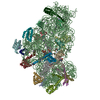 9g06C M: map data used to model this data C: citing same article ( |
|---|---|
| Similar structure data |


-Links
 PDBj
PDBj


- Assembly
| Deposited unit | 
|
|---|---|
| 1 |
|
-Components
-RNA chain , 2 types, 2 molecules BY
| #1: RNA chain | Mass: 339708.531 Da / Num. of mol.: 1 Source method: isolated from a genetically manipulated source Source: (gene. exp.) |
|---|---|
| #16: RNA chain | Mass: 13074.881 Da / Num. of mol.: 1 Source method: isolated from a genetically manipulated source Source: (gene. exp.) |
-Small ribosomal subunit protein ... , 12 types, 12 molecules DEFHKLOPQRTU
| #2: Protein | Mass: 23514.199 Da / Num. of mol.: 1 Source method: isolated from a genetically manipulated source Source: (gene. exp.) |
|---|---|
| #3: Protein | Mass: 17629.398 Da / Num. of mol.: 1 Source method: isolated from a genetically manipulated source Source: (gene. exp.) |
| #4: Protein | Mass: 15727.512 Da / Num. of mol.: 1 Source method: isolated from a genetically manipulated source Source: (gene. exp.) |
| #5: Protein | Mass: 14146.557 Da / Num. of mol.: 1 Source method: isolated from a genetically manipulated source Source: (gene. exp.) |
| #8: Protein | Mass: 15055.459 Da / Num. of mol.: 1 Source method: isolated from a genetically manipulated source Source: (gene. exp.) |
| #9: Protein | Mass: 13814.249 Da / Num. of mol.: 1 Source method: isolated from a genetically manipulated source Source: (gene. exp.) |
| #10: Protein | Mass: 10290.816 Da / Num. of mol.: 1 Source method: isolated from a genetically manipulated source Source: (gene. exp.) |
| #11: Protein | Mass: 9207.572 Da / Num. of mol.: 1 Source method: isolated from a genetically manipulated source Source: (gene. exp.) |
| #12: Protein | Mass: 9724.491 Da / Num. of mol.: 1 Source method: isolated from a genetically manipulated source Source: (gene. exp.) |
| #13: Protein | Mass: 9005.472 Da / Num. of mol.: 1 Source method: isolated from a genetically manipulated source Source: (gene. exp.) |
| #14: Protein | Mass: 9708.464 Da / Num. of mol.: 1 Source method: isolated from a genetically manipulated source Source: (gene. exp.) |
| #15: Protein | Mass: 8524.039 Da / Num. of mol.: 1 Source method: isolated from a genetically manipulated source Source: (gene. exp.) |
-Translation initiation factor IF- ... , 2 types, 2 molecules IJ
| #6: Protein | Mass: 8262.590 Da / Num. of mol.: 1 Source method: isolated from a genetically manipulated source Source: (gene. exp.) |
|---|---|
| #7: Protein | Mass: 20600.994 Da / Num. of mol.: 1 Source method: isolated from a genetically manipulated source Source: (gene. exp.) |
-Non-polymers , 4 types, 1928 molecules 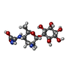


| #17: Chemical | ChemComp-KSG / ( Mass: 379.363 Da / Num. of mol.: 1 / Source method: obtained synthetically / Formula: C14H25N3O9 / Feature type: SUBJECT OF INVESTIGATION / Comment: antibiotic*YM Mass: 379.363 Da / Num. of mol.: 1 / Source method: obtained synthetically / Formula: C14H25N3O9 / Feature type: SUBJECT OF INVESTIGATION / Comment: antibiotic*YM | ||||
|---|---|---|---|---|---|
| #18: Chemical | ChemComp-MG /  Mass: 24.305 Da / Num. of mol.: 53 / Source method: obtained synthetically / Formula: Mg Mass: 24.305 Da / Num. of mol.: 53 / Source method: obtained synthetically / Formula: Mg#19: Chemical | ChemComp-K /  Mass: 39.098 Da / Num. of mol.: 25 / Source method: obtained synthetically / Formula: K Mass: 39.098 Da / Num. of mol.: 25 / Source method: obtained synthetically / Formula: K#20: Water | ChemComp-HOH / | Mass: 18.015 Da / Num. of mol.: 1849 / Source method: isolated from a natural source / Formula: H2O |
-Details
| Has ligand of interest | Y |
|---|---|
| Has protein modification | Y |
-Experimental details
-Experiment
| Experiment | Method: ELECTRON MICROSCOPY |
|---|---|
| EM experiment | Aggregation state: PARTICLE / 3D reconstruction method: single particle reconstruction |
- Sample preparation
| Component | Name: Structure of E.coli 30S-IF1-IF3-mRNA-Kasugamycin complex Type: RIBOSOME / Entity ID: #2-#7, #9-#16, #1 / Source: RECOMBINANT |
|---|---|
| Molecular weight | Experimental value: NO |
| Source (natural) | Organism: |
| Source (recombinant) | Organism: |
| Buffer solution | pH: 7.5 |
| Specimen | Embedding applied: NO / Shadowing applied: NO / Staining applied: NO / Vitrification applied: YES |
| Vitrification | Cryogen name: ETHANE-PROPANE |
- Electron microscopy imaging
| Experimental equipment |  Model: Titan Krios / Image courtesy: FEI Company |
|---|---|
| Microscopy | Model: TFS KRIOS |
| Electron gun | Electron source:  FIELD EMISSION GUN / Accelerating voltage: 300 kV / Illumination mode: FLOOD BEAM FIELD EMISSION GUN / Accelerating voltage: 300 kV / Illumination mode: FLOOD BEAM |
| Electron lens | Mode: BRIGHT FIELD / Nominal defocus max: 1100 nm / Nominal defocus min: 300 nm |
| Image recording | Electron dose: 1.14 e/Å2 / Film or detector model: GATAN K3 BIOQUANTUM (6k x 4k) |
- Processing
| EM software | Name: REFMAC / Version: 5.8.0415 / Category: model refinement |
|---|---|
| CTF correction | Type: PHASE FLIPPING AND AMPLITUDE CORRECTION |
| 3D reconstruction | Resolution: 2.4 Å / Resolution method: FSC 0.143 CUT-OFF / Num. of particles: 332988 / Symmetry type: POINT |
