[English] 日本語
 Yorodumi
- EMDB-51020: Structure of the Position 7 CMG-decorated gamma-Tubulin Ring Comp... -
+ Open data
- Basic information
| Entry |  | |||||||||
|---|---|---|---|---|---|---|---|---|---|---|
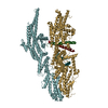
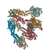
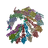
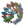
| Title | Structure of the Position 7 CMG-decorated gamma-Tubulin Ring Complex from Pig Brain | |||||||||
 Map data Map data | ||||||||||
 Sample Sample |
| |||||||||
 Keywords Keywords | Tubulin Complex / STRUCTURAL PROTEIN | |||||||||
| Function / homology |  Function and homology information Function and homology informationRecruitment of mitotic centrosome proteins and complexes / negative regulation of centriole replication / regulation of mitotic cell cycle spindle assembly checkpoint / microtubule organizing center organization / polar microtubule / gamma-tubulin complex / microtubule plus-end / microtubule nucleation / microtubule bundle formation / gamma-tubulin binding ...Recruitment of mitotic centrosome proteins and complexes / negative regulation of centriole replication / regulation of mitotic cell cycle spindle assembly checkpoint / microtubule organizing center organization / polar microtubule / gamma-tubulin complex / microtubule plus-end / microtubule nucleation / microtubule bundle formation / gamma-tubulin binding / centrosome cycle / regulation of neuron differentiation / Recruitment of NuMA to mitotic centrosomes / negative regulation of neuron differentiation / mitotic spindle pole / pericentriolar material / centriole replication / establishment of mitotic spindle orientation / spindle assembly / cytoplasmic microtubule organization / neurogenesis / positive regulation of microtubule polymerization / Loss of Nlp from mitotic centrosomes / Loss of proteins required for interphase microtubule organization from the centrosome / centriole / Recruitment of mitotic centrosome proteins and complexes / Recruitment of NuMA to mitotic centrosomes / Anchoring of the basal body to the plasma membrane / tubulin binding / AURKA Activation by TPX2 / meiotic cell cycle / chromosome segregation / brain development / microtubule cytoskeleton organization / spindle / neuron migration / spindle pole / cell junction / Regulation of PLK1 Activity at G2/M Transition / mitotic cell cycle / microtubule binding / microtubule / cytoskeleton / calmodulin binding / transcription cis-regulatory region binding / ciliary basal body / centrosome / protein kinase binding / positive regulation of DNA-templated transcription / protein-containing complex binding / perinuclear region of cytoplasm / Golgi apparatus / extracellular exosome / nucleoplasm / cytosol / cytoplasm Similarity search - Function | |||||||||
| Biological species |   Homo sapiens (human) Homo sapiens (human) | |||||||||
| Method | single particle reconstruction / cryo EM / Resolution: 4.3 Å | |||||||||
 Authors Authors | Munoz-Hernandez H / Krutyholowa R / Wieczorek M | |||||||||
| Funding support |  Switzerland, 2 items Switzerland, 2 items
| |||||||||
 Citation Citation |  Journal: Dev Cell / Year: 2024 Journal: Dev Cell / Year: 2024Title: Partial closure of the γ-tubulin ring complex by CDK5RAP2 activates microtubule nucleation. Authors: Yixin Xu / Hugo Muñoz-Hernández / Rościsław Krutyhołowa / Florina Marxer / Ferdane Cetin / Michal Wieczorek /  Abstract: Microtubule nucleation is templated by the γ-tubulin ring complex (γ-TuRC), but its structure deviates from the geometry of α-/β-tubulin in the microtubule, explaining the complex's poor ...Microtubule nucleation is templated by the γ-tubulin ring complex (γ-TuRC), but its structure deviates from the geometry of α-/β-tubulin in the microtubule, explaining the complex's poor nucleating activity. Several proteins may activate the γ-TuRC, but the mechanisms underlying activation are not known. Here, we determined the structure of the porcine γ-TuRC purified using CDK5RAP2's centrosomin motif 1 (CM1). We identified an unexpected conformation of the γ-TuRC bound to multiple protein modules containing MZT2, GCP2, and CDK5RAP2, resulting in a long-range constriction of the γ-tubulin ring that brings it in closer agreement with the 13-protofilament microtubule. Additional CDK5RAP2 promoted γ-TuRC decoration and stimulated the microtubule-nucleating activities of the porcine γ-TuRC and a reconstituted, CM1-free human complex in single-molecule assays. Our results provide a structural mechanism for the control of microtubule nucleation by CM1 proteins and identify conformational transitions in the γ-TuRC that prime it for microtubule nucleation. | |||||||||
| History |
|
- Structure visualization
| Supplemental images |
|---|
- Downloads & links
-EMDB archive
| Map data |  emd_51020.map.gz emd_51020.map.gz | 116.2 MB |  EMDB map data format EMDB map data format | |
|---|---|---|---|---|
| Header (meta data) |  emd-51020-v30.xml emd-51020-v30.xml emd-51020.xml emd-51020.xml | 24.2 KB 24.2 KB | Display Display |  EMDB header EMDB header |
| Images |  emd_51020.png emd_51020.png | 86.7 KB | ||
| Filedesc metadata |  emd-51020.cif.gz emd-51020.cif.gz | 8.7 KB | ||
| Others |  emd_51020_half_map_1.map.gz emd_51020_half_map_1.map.gz emd_51020_half_map_2.map.gz emd_51020_half_map_2.map.gz | 98.5 MB 98.5 MB | ||
| Archive directory |  http://ftp.pdbj.org/pub/emdb/structures/EMD-51020 http://ftp.pdbj.org/pub/emdb/structures/EMD-51020 ftp://ftp.pdbj.org/pub/emdb/structures/EMD-51020 ftp://ftp.pdbj.org/pub/emdb/structures/EMD-51020 | HTTPS FTP |
-Validation report
| Summary document |  emd_51020_validation.pdf.gz emd_51020_validation.pdf.gz | 978.7 KB | Display |  EMDB validaton report EMDB validaton report |
|---|---|---|---|---|
| Full document |  emd_51020_full_validation.pdf.gz emd_51020_full_validation.pdf.gz | 978.5 KB | Display | |
| Data in XML |  emd_51020_validation.xml.gz emd_51020_validation.xml.gz | 13.8 KB | Display | |
| Data in CIF |  emd_51020_validation.cif.gz emd_51020_validation.cif.gz | 16.4 KB | Display | |
| Arichive directory |  https://ftp.pdbj.org/pub/emdb/validation_reports/EMD-51020 https://ftp.pdbj.org/pub/emdb/validation_reports/EMD-51020 ftp://ftp.pdbj.org/pub/emdb/validation_reports/EMD-51020 ftp://ftp.pdbj.org/pub/emdb/validation_reports/EMD-51020 | HTTPS FTP |
-Related structure data
| Related structure data |  9g40MC  9g3xC  9g3yC  9g3zC M: atomic model generated by this map C: citing same article ( |
|---|---|
| Similar structure data | Similarity search - Function & homology  F&H Search F&H Search |
- Links
Links
| EMDB pages |  EMDB (EBI/PDBe) / EMDB (EBI/PDBe) /  EMDataResource EMDataResource |
|---|
- Map
Map
| File |  Download / File: emd_51020.map.gz / Format: CCP4 / Size: 125 MB / Type: IMAGE STORED AS FLOATING POINT NUMBER (4 BYTES) Download / File: emd_51020.map.gz / Format: CCP4 / Size: 125 MB / Type: IMAGE STORED AS FLOATING POINT NUMBER (4 BYTES) | ||||||||||||||||||||||||||||||||||||
|---|---|---|---|---|---|---|---|---|---|---|---|---|---|---|---|---|---|---|---|---|---|---|---|---|---|---|---|---|---|---|---|---|---|---|---|---|---|
| Projections & slices | Image control
Images are generated by Spider. | ||||||||||||||||||||||||||||||||||||
| Voxel size | X=Y=Z: 1.41333 Å | ||||||||||||||||||||||||||||||||||||
| Density |
| ||||||||||||||||||||||||||||||||||||
| Symmetry | Space group: 1 | ||||||||||||||||||||||||||||||||||||
| Details | EMDB XML:
|
-Supplemental data
-Half map: #2
| File | emd_51020_half_map_1.map | ||||||||||||
|---|---|---|---|---|---|---|---|---|---|---|---|---|---|
| Projections & Slices |
| ||||||||||||
| Density Histograms |
-Half map: #1
| File | emd_51020_half_map_2.map | ||||||||||||
|---|---|---|---|---|---|---|---|---|---|---|---|---|---|
| Projections & Slices |
| ||||||||||||
| Density Histograms |
- Sample components
-Entire : Gamma-Tubulin Ring Complex in native pig brain
| Entire | Name: Gamma-Tubulin Ring Complex in native pig brain |
|---|---|
| Components |
|
-Supramolecule #1: Gamma-Tubulin Ring Complex in native pig brain
| Supramolecule | Name: Gamma-Tubulin Ring Complex in native pig brain / type: complex / ID: 1 / Parent: 0 / Macromolecule list: all |
|---|---|
| Source (natural) | Organism:  |
-Macromolecule #1: Gamma-tubulin complex component 3
| Macromolecule | Name: Gamma-tubulin complex component 3 / type: protein_or_peptide / ID: 1 / Details: SsGCP3 / Number of copies: 1 / Enantiomer: LEVO |
|---|---|
| Source (natural) | Organism:  |
| Molecular weight | Theoretical: 103.172477 KDa |
| Sequence | String: MATPDQKSPN VLLQNLCCRI LGKSEADVAQ QFQYAVRVIG SNFAPTVERD EFLVAEKIKK ELTRQRREAD AALFSELHRK LHSQGVLKN KWSILYLLLS LSEDPRKQPS KVSGYAALFA QALPRDAHST PYYYARPQSL PLNYQERGAP SAQSAGSAGS S GVSSLGTY ...String: MATPDQKSPN VLLQNLCCRI LGKSEADVAQ QFQYAVRVIG SNFAPTVERD EFLVAEKIKK ELTRQRREAD AALFSELHRK LHSQGVLKN KWSILYLLLS LSEDPRKQPS KVSGYAALFA QALPRDAHST PYYYARPQSL PLNYQERGAP SAQSAGSAGS S GVSSLGTY ALNGPTPPPP PPALLPGQPL PAPGVGDGLR QQLGSRLAWT LTASQPSLPS TTSKAVPSSG SRGAARPRRE GD AAAGAVE VTEAALVRDI LYVFQGIDGK HVKMSNADNC YTVEGKANLS KSLRDTAVRL AELGWLHNKI RKYTDQRSLD RSF GLVGQS FCAALHQELR EYYRLLSVLH SQLQLEDDQG VNLGLESSLT LRRLLVWTYD PKMRLKTLAA LVDHCQGRKG GELA SAVHA YTKTGDPYAR SLVQHILSLV SHPVLSFLYR WIYDGELEDT YHEFFVASDP AVKADRLWHD KYALRKPMIP SFMTM DQCR KVLLIGKSIN FLHQVCHDQT PTTKMIAVTK SAESPQDAAD LFTDLENAFQ GKIDAAYFET SKYLLDVLNK KYSLLD HMQ AMRRYLLLGQ GDFIRHLMDL LKPELVRPAT TLYQHNLTGI LETAVRATNA QFDSPEILKR LDVRLLEVSP GDTGWDV FS LDYHVDGPIA TVFTRECMSH YLRAFNFLWR AKRVEYILTD IRKGHMCNAR LLRSMPEFSG VLHHCHILAS EMVHFIHQ M QYYVTFEVLE CSWDELWNRV QRAQDLDHII AAHEAFLGTV ISRCLLDSDS RALLNQLRAV FDQIIELQNT QDAIYRAAL EELQRRLQFE EKKKQREAEG QWGVSAAEEE QEKRRVQEFQ ESIPKMCSQL RILTHFYQGV VQQFLVSLTT SSDESLRFLS FRLDFNEHY RAREPRLRVS LGTRGRRSSH T UniProtKB: Gamma-tubulin complex component 3 |
-Macromolecule #2: Gamma-tubulin complex component
| Macromolecule | Name: Gamma-tubulin complex component / type: protein_or_peptide / ID: 2 / Number of copies: 1 / Enantiomer: LEVO |
|---|---|
| Source (natural) | Organism:  |
| Molecular weight | Theoretical: 102.609703 KDa |
| Sequence | String: MSEFRIHHDV NELLSLLRVH GGDGAEVYID LLQKNRTPYV TTTVSAHSAK VKIAEFSRTP EDFLKKYDEL KSKNTRNLDP LVYLLSKLM EDRETLQYLQ QNAKERAELA ASAAASSTAS FGASATASKI SMQELEELRK QLGSVATGPT WQQSLELTRK M LRDKQSKK ...String: MSEFRIHHDV NELLSLLRVH GGDGAEVYID LLQKNRTPYV TTTVSAHSAK VKIAEFSRTP EDFLKKYDEL KSKNTRNLDP LVYLLSKLM EDRETLQYLQ QNAKERAELA ASAAASSTAS FGASATASKI SMQELEELRK QLGSVATGPT WQQSLELTRK M LRDKQSKK NSGQRLPVLP AWVYERPALL GDFLPGTGGS ADTAVPIGSL PLASQEAAVV EDLLYVLVGV DGRYISAQPL TG RQGRTFL VDPNLDLSIR ELVSRILPVA ASYSTVTRFI EEKSSFEYGQ VNHALAAAMR TLVKEYLVLV TQLEQLQRQG LLS LQKLWF YIQPAMRSLD ILASLATSVD KGECIGGATL SLLHDRSFSY TGDSQAQELC LHLTKAASTP YFEILEKWIY RGII DDPYS EFMVEEHELR KEKIQEDYND KYWDQRYTVV QRQIPSFLQK MAGKVLSTGK YLNVVRECGH DVTCPVAKEV VYTLK ERAY VEQIEKAFSY ASKVLLDFLM GEKELLAHLR SIKRYFLMDQ GDFFVHFMDL TEEELKKPVD DITPTRLEAL LELALR MST ANTDPFKDDL KIDLMPHDLI TQLLRVLAIE TQQEKAMVHA DPTELTLSGL EAFSFDYVVT WPLSLIINRK ALTRYQM LF RHMFYCKHVE RQLCSVWISN KAAKRFSLHS AKWFAGAFTL RQRMLNFVQN IQSYMMFEVM EPTWHVLEQN LRSASNID D VLGHHASFLD NCLKDCMLTN PELLRVFSKL MSVCVMFTNC LQRFTQSMKL DSELGHPALE PGAMLGPPTE AERAEERAR KELARKCLAE HVDAPQLASS FEATITKFDK NFSAHLLDLL ARLSIYSTSD CEHGMASVIS RLDFNGFYTE RLERLSAERS QKAAPPVPG PRGPPALVPR VAVTAQ UniProtKB: Gamma-tubulin complex component |
-Macromolecule #3: Mitotic-spindle organizing protein 2A isoform X4
| Macromolecule | Name: Mitotic-spindle organizing protein 2A isoform X4 / type: protein_or_peptide / ID: 3 / Number of copies: 1 / Enantiomer: LEVO |
|---|---|
| Source (natural) | Organism:  |
| Molecular weight | Theoretical: 15.920321 KDa |
| Sequence | String: MAAPGAGPGP GAPPGLEAAL QKLALRRKKV LSAEETELFE LAQAAGGAMD PEVFKILVDL LKLNVAPLAV FQMLKSMCAG QRLASEPQD PVAVPLPTTS VPETRGRNRG SSALGGGPAL AERSGREGSS QRMPRQPSAT RLPKGGGPGK SPTRST UniProtKB: Mitotic-spindle organizing protein 2A isoform X4 |
-Macromolecule #4: CDK5 regulatory subunit-associated protein 2
| Macromolecule | Name: CDK5 regulatory subunit-associated protein 2 / type: protein_or_peptide / ID: 4 / Number of copies: 2 / Enantiomer: LEVO |
|---|---|
| Source (natural) | Organism:  Homo sapiens (human) Homo sapiens (human) |
| Molecular weight | Theoretical: 215.344219 KDa |
| Recombinant expression | Organism:  |
| Sequence | String: MMDLVLEEDV TVPGTLSGCS GLVPSVPDDL DGINPNAGLG NGLLPNVSEE TVSPTRARNM KDFENQITEL KKENFNLKLR IYFLEERMQ QEFHGPTEHI YKTNIELKVE VESLKRELQE REQLLIKASK AVESLAEAGG SEIQRVKEDA RKKVQQVEDL L TKRILLLE ...String: MMDLVLEEDV TVPGTLSGCS GLVPSVPDDL DGINPNAGLG NGLLPNVSEE TVSPTRARNM KDFENQITEL KKENFNLKLR IYFLEERMQ QEFHGPTEHI YKTNIELKVE VESLKRELQE REQLLIKASK AVESLAEAGG SEIQRVKEDA RKKVQQVEDL L TKRILLLE KDVTAAQAEL EKAFAGTETE KALRLRLESK LSEMKKMHEG DLAMALVLDE KDRLIEELKL SLKSKEALIQ CL KEEKSQM ACPDENVSSG ELRGLCAAPR EEKERETEAA QMEHQKERNS FEERIQALEE DLREKEREIA TEKKNSLKRD KAI QGLTMA LKSKEKKVEE LNSEIEKLSA AFAKAREALQ KAQTQEFQGS EDYETALSGK EALSAALRSQ NLTKSTENHR LRRS IKKIT QELSDLQQER ERLEKDLEEA HREKSKGDCT IRDLRNEVEK LRNEVNEREK AMENRYKSLL SESNKKLHNQ EQVIK HLTE STNQKDVLLQ KFNEKDLEVI QQNCYLMAAE DLELRSEGLI TEKCSSQQPP GSKTIFSKEK KQSSDYEELI QVLKKE QDI YTHLVKSLQE SDSINNLQAE LNKIFALRKQ LEQDVLSYQN LRKTLEEQIS EIRRREEESF SLYSDQTSYL SICLEEN NR FQVEHFSQEE LKKKVSDLIQ LVKELYTDNQ HLKKTIFDLS CMGFQGNGFP DRLASTEQTE LLASKEDEDT IKIGEDDE I NFLSDQHLQQ SNEIMKDLSK GGCKNGYLRH TESKISDCDG AHAPGCLEEG AFINLLAPLF NEKATLLLES RPDLLKVVR ELLLGQLFLT EQEVSGEHLD GKTEKTPKQK GELVHFVQTN SFSKPHDELK LSCEAQLVKA GEVPKVGLKD ASVQTVATEG DLLRFKHEA TREAWEEKPI NTALSAEHRP ENLHGVPGWQ AALLSLPGIT NREAKKSRLP ILIKPSRSLG NMYRLPATQE V VTQLQSQI LELQGELKEF KTCNKQLHQK LILAEAVMEG RPTPDKTLLN AQPPVGAAYQ DSPGEQKGIK TTSSVWRDKE MD SDQQRSY EIDSEICPPD DLASLPSCKE NPEDVLSPTS VATYLSSKSQ PSAKVSVMGT DQSESINTSN ETEYLKQKIH DLE TELEGY QNFIFQLQKH SQCSEAIITV LCGTEGAQDG LSKPKNGSDG EEMTFSSLHQ VRYVKHVKIL GPLAPEMIDS RVLE NLKQQ LEEQEYKLQK EQNLNMQLFS EIHNLQNKFR DLSPPRYDSL VQSQARELSL QRQQIKDGHG ICVISRQHMN TMIKA FEEL LQASDVDYCV AEGFQEQLNQ CAELLEKLEK LFLNGKSVGV EMNTQNELME RIEEDNLTYQ HLLPESPEPS ASHALS DYE TSEKSFFSRD QKQDNETEKT SVMVNSFSQD LLMEHIQEIR TLRKRLEESI KTNEKLRKQL ERQGSEFVQG STSIFAS GS ELHSSLTSEI HFLRKQNQAL NAMLIKGSRD KQKENDKLRE SLSRKTVSLE HLQREYASVK EENERLQKEG SEKERHNQ Q LIQEVRCSGQ ELSRVQEEVK LRQQLLSQND KLLQSLRVEL KAYEKLDEEH RRLREASGEG WKGQDPFRDL HSLLMEIQA LRLQLERSIE TSSTLQSRLK EQLARGAEKA QEGALTLAVQ AVSIPEVPLQ PDKHDGDKYP MESDNSFDLF DSSQAVTPKS VSETPPLSG NDTDSLSCDS GSSATSTPCV SRLVTGHHLW ASKNGRHVLG LIEDYEALLK QISQGQRLLA EMDIQTQEAP S STSQELGT KGPHPAPLSK FVSSVSTAKL TLEEAYRRLK LLWRVSLPED GQCPLHCEQI GEMKAEVTKL HKKLFEQEKK LQ NTMKLLQ LSKRQEKVIF DQLVVTHKIL RKARGNLELR PGGAHPGTCS PSRPGS UniProtKB: CDK5 regulatory subunit-associated protein 2 |
-Experimental details
-Structure determination
| Method | cryo EM |
|---|---|
 Processing Processing | single particle reconstruction |
| Aggregation state | particle |
- Sample preparation
| Buffer | pH: 7.5 Component:
Details: Listed in "g-TuRC cryo-EM sample preparation" section. | ||||||||||||||||
|---|---|---|---|---|---|---|---|---|---|---|---|---|---|---|---|---|---|
| Vitrification | Cryogen name: ETHANE-PROPANE / Chamber humidity: 100 % / Chamber temperature: 277.15 K / Instrument: FEI VITROBOT MARK III |
- Electron microscopy
| Microscope | TFS KRIOS |
|---|---|
| Image recording | Film or detector model: GATAN K3 BIOQUANTUM (6k x 4k) / Average electron dose: 55.0 e/Å2 |
| Electron beam | Acceleration voltage: 300 kV / Electron source:  FIELD EMISSION GUN FIELD EMISSION GUN |
| Electron optics | Illumination mode: SPOT SCAN / Imaging mode: BRIGHT FIELD / Nominal defocus max: 2.7 µm / Nominal defocus min: 0.9 µm |
| Experimental equipment |  Model: Titan Krios / Image courtesy: FEI Company |
 Movie
Movie Controller


 Z (Sec.)
 Y (Row.)
 X (Col.)


