[English] 日本語
 Yorodumi
- EMDB-51010: Structure of the Position 10 to 12 Open gamma-Tubulin Ring Comple... -
+ Open data
- Basic information
| Entry |  | |||||||||
|---|---|---|---|---|---|---|---|---|---|---|
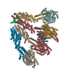
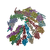
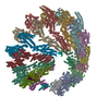
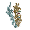
| Title | Structure of the Position 10 to 12 Open gamma-Tubulin Ring Complex from Pig Brain | |||||||||
 Map data Map data | ||||||||||
 Sample Sample |
| |||||||||
 Keywords Keywords | Tubulin Complex / STRUCTURAL PROTEIN | |||||||||
| Biological species |  | |||||||||
| Method | single particle reconstruction / cryo EM / Resolution: 4.9 Å | |||||||||
 Authors Authors | Munoz-Hernandez H / Wieczorek M | |||||||||
| Funding support |  Switzerland, 2 items Switzerland, 2 items
| |||||||||
 Citation Citation |  Journal: Dev Cell / Year: 2024 Journal: Dev Cell / Year: 2024Title: Partial closure of the γ-tubulin ring complex by CDK5RAP2 activates microtubule nucleation. Authors: Yixin Xu / Hugo Muñoz-Hernández / Rościsław Krutyhołowa / Florina Marxer / Ferdane Cetin / Michal Wieczorek /  Abstract: Microtubule nucleation is templated by the γ-tubulin ring complex (γ-TuRC), but its structure deviates from the geometry of α-/β-tubulin in the microtubule, explaining the complex's poor ...Microtubule nucleation is templated by the γ-tubulin ring complex (γ-TuRC), but its structure deviates from the geometry of α-/β-tubulin in the microtubule, explaining the complex's poor nucleating activity. Several proteins may activate the γ-TuRC, but the mechanisms underlying activation are not known. Here, we determined the structure of the porcine γ-TuRC purified using CDK5RAP2's centrosomin motif 1 (CM1). We identified an unexpected conformation of the γ-TuRC bound to multiple protein modules containing MZT2, GCP2, and CDK5RAP2, resulting in a long-range constriction of the γ-tubulin ring that brings it in closer agreement with the 13-protofilament microtubule. Additional CDK5RAP2 promoted γ-TuRC decoration and stimulated the microtubule-nucleating activities of the porcine γ-TuRC and a reconstituted, CM1-free human complex in single-molecule assays. Our results provide a structural mechanism for the control of microtubule nucleation by CM1 proteins and identify conformational transitions in the γ-TuRC that prime it for microtubule nucleation. | |||||||||
| History |
|
- Structure visualization
| Supplemental images |
|---|
- Downloads & links
-EMDB archive
| Map data |  emd_51010.map.gz emd_51010.map.gz | 107.2 MB |  EMDB map data format EMDB map data format | |
|---|---|---|---|---|
| Header (meta data) |  emd-51010-v30.xml emd-51010-v30.xml emd-51010.xml emd-51010.xml | 13.3 KB 13.3 KB | Display Display |  EMDB header EMDB header |
| FSC (resolution estimation) |  emd_51010_fsc.xml emd_51010_fsc.xml | 12.7 KB | Display |  FSC data file FSC data file |
| Images |  emd_51010.png emd_51010.png | 69.2 KB | ||
| Filedesc metadata |  emd-51010.cif.gz emd-51010.cif.gz | 4.6 KB | ||
| Others |  emd_51010_half_map_1.map.gz emd_51010_half_map_1.map.gz emd_51010_half_map_2.map.gz emd_51010_half_map_2.map.gz | 200.7 MB 200.7 MB | ||
| Archive directory |  http://ftp.pdbj.org/pub/emdb/structures/EMD-51010 http://ftp.pdbj.org/pub/emdb/structures/EMD-51010 ftp://ftp.pdbj.org/pub/emdb/structures/EMD-51010 ftp://ftp.pdbj.org/pub/emdb/structures/EMD-51010 | HTTPS FTP |
-Validation report
| Summary document |  emd_51010_validation.pdf.gz emd_51010_validation.pdf.gz | 1.1 MB | Display |  EMDB validaton report EMDB validaton report |
|---|---|---|---|---|
| Full document |  emd_51010_full_validation.pdf.gz emd_51010_full_validation.pdf.gz | 1.1 MB | Display | |
| Data in XML |  emd_51010_validation.xml.gz emd_51010_validation.xml.gz | 21.7 KB | Display | |
| Data in CIF |  emd_51010_validation.cif.gz emd_51010_validation.cif.gz | 28.4 KB | Display | |
| Arichive directory |  https://ftp.pdbj.org/pub/emdb/validation_reports/EMD-51010 https://ftp.pdbj.org/pub/emdb/validation_reports/EMD-51010 ftp://ftp.pdbj.org/pub/emdb/validation_reports/EMD-51010 ftp://ftp.pdbj.org/pub/emdb/validation_reports/EMD-51010 | HTTPS FTP |
-Related structure data
- Links
Links
| EMDB pages |  EMDB (EBI/PDBe) / EMDB (EBI/PDBe) /  EMDataResource EMDataResource |
|---|
- Map
Map
| File |  Download / File: emd_51010.map.gz / Format: CCP4 / Size: 216 MB / Type: IMAGE STORED AS FLOATING POINT NUMBER (4 BYTES) Download / File: emd_51010.map.gz / Format: CCP4 / Size: 216 MB / Type: IMAGE STORED AS FLOATING POINT NUMBER (4 BYTES) | ||||||||||||||||||||||||||||||||||||
|---|---|---|---|---|---|---|---|---|---|---|---|---|---|---|---|---|---|---|---|---|---|---|---|---|---|---|---|---|---|---|---|---|---|---|---|---|---|
| Projections & slices | Image control
Images are generated by Spider. | ||||||||||||||||||||||||||||||||||||
| Voxel size | X=Y=Z: 1.4133 Å | ||||||||||||||||||||||||||||||||||||
| Density |
| ||||||||||||||||||||||||||||||||||||
| Symmetry | Space group: 1 | ||||||||||||||||||||||||||||||||||||
| Details | EMDB XML:
|
-Supplemental data
-Half map: #1
| File | emd_51010_half_map_1.map | ||||||||||||
|---|---|---|---|---|---|---|---|---|---|---|---|---|---|
| Projections & Slices |
| ||||||||||||
| Density Histograms |
-Half map: #2
| File | emd_51010_half_map_2.map | ||||||||||||
|---|---|---|---|---|---|---|---|---|---|---|---|---|---|
| Projections & Slices |
| ||||||||||||
| Density Histograms |
- Sample components
-Entire : Gamma-Tubulin Ring Complex in native pig brain
| Entire | Name: Gamma-Tubulin Ring Complex in native pig brain |
|---|---|
| Components |
|
-Supramolecule #1: Gamma-Tubulin Ring Complex in native pig brain
| Supramolecule | Name: Gamma-Tubulin Ring Complex in native pig brain / type: complex / ID: 1 / Parent: 0 / Macromolecule list: all |
|---|---|
| Source (natural) | Organism:  |
-Macromolecule #1: Structure of the Position 10 to 12 Open gamma-Tubulin Ring Comple...
| Macromolecule | Name: Structure of the Position 10 to 12 Open gamma-Tubulin Ring Complex from Pig Brain type: protein_or_peptide / ID: 1 / Enantiomer: LEVO |
|---|---|
| Source (natural) | Organism:  |
| Sequence | String: MPREIITLQL GQCGNQIGFE FWKQLCAEHG ISPEGIVEEF ATEGTDRKDV FFYQADDEHY IPRAVLLDLE PRVIHSILNS PYAKLYNPEN IYLSEHGGGA GNNWASGFSQ GEKIHEDIFD IIDREADGSD SLEGFVLCHS IAGGTGSGLG SYLLERLNDR YPKKLVQTYS ...String: MPREIITLQL GQCGNQIGFE FWKQLCAEHG ISPEGIVEEF ATEGTDRKDV FFYQADDEHY IPRAVLLDLE PRVIHSILNS PYAKLYNPEN IYLSEHGGGA GNNWASGFSQ GEKIHEDIFD IIDREADGSD SLEGFVLCHS IAGGTGSGLG SYLLERLNDR YPKKLVQTYS VFPNQDEMSD VVVQPYNSLL TLKRLTQNAD CVVVLDNTAL NRIATDRLHI QNPSFSQINQ LVSTIMSAST TTLRYPGYMN NDLIGLIASL IPTPRLHFLM TGYTPLTTDQ SVASVRKTTV LDVMRRLLQP KNVMVSTGRD RQTNHCYIAI LNIIQGEVDP TQVHKSLQRI RERKLANFIP WGPASIQVAL SRKSPYLPSA HRVSGLMMAN HTSISSLFES SCQQYDKLRK REAFLEQFRK EDIFKENFDE LDRSREVVQE LIDEYHAATR PDYISWGTQE Q |
-Experimental details
-Structure determination
| Method | cryo EM |
|---|---|
 Processing Processing | single particle reconstruction |
| Aggregation state | particle |
- Sample preparation
| Buffer | pH: 7.5 |
|---|---|
| Grid | Model: Quantifoil R2/1 / Support film - Material: CARBON / Support film - topology: HOLEY |
| Vitrification | Cryogen name: ETHANE-PROPANE |
- Electron microscopy
| Microscope | TFS KRIOS |
|---|---|
| Image recording | Film or detector model: GATAN K3 BIOQUANTUM (6k x 4k) / Average electron dose: 55.0 e/Å2 |
| Electron beam | Acceleration voltage: 300 kV / Electron source:  FIELD EMISSION GUN FIELD EMISSION GUN |
| Electron optics | Illumination mode: SPOT SCAN / Imaging mode: BRIGHT FIELD / Nominal defocus max: 2.9 µm / Nominal defocus min: 0.9 µm |
| Experimental equipment |  Model: Titan Krios / Image courtesy: FEI Company |
+ Image processing
-Atomic model buiding 1
| Refinement | Space: REAL / Protocol: BACKBONE TRACE |
|---|
 Movie
Movie Controller


 Z (Sec.)
 Y (Row.)
 X (Col.)


