 Movie
Movie Controller

[English] 日本語
 Yorodumi
- EMDB-51019: Structure of the Open gamma-Tubulin Ring Complex from Pig Brain -
+ Open data

- Basic information
| Entry |  | |||||||||
|---|---|---|---|---|---|---|---|---|---|---|
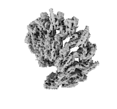
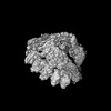
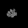
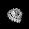
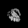
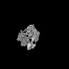
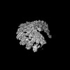
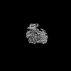
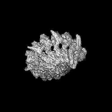
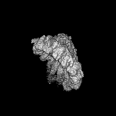
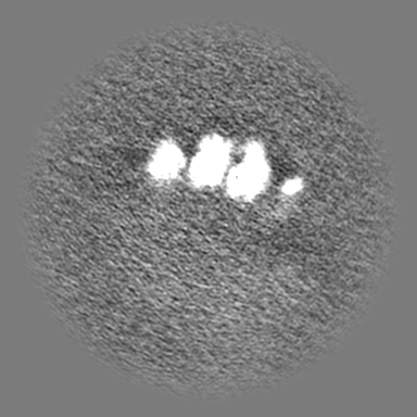
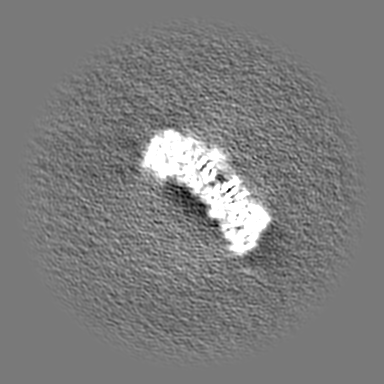
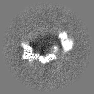
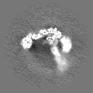
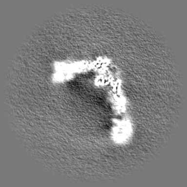
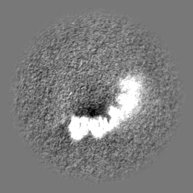
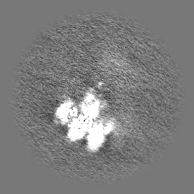
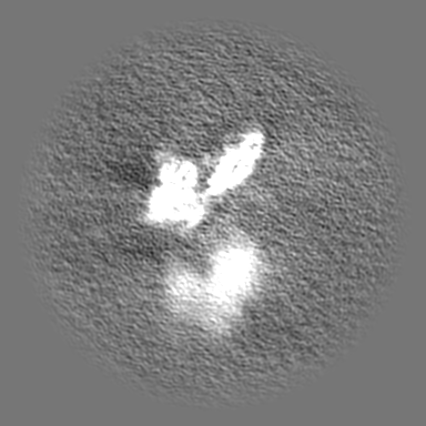
| Title | Structure of the Open gamma-Tubulin Ring Complex from Pig Brain | |||||||||
 Map data Map data | ||||||||||
 Sample Sample |
| |||||||||
 Keywords Keywords | Tubulin Complex / STRUCTURAL PROTEIN | |||||||||
| Function / homology |  Function and homology information Function and homology informationgamma-tubulin complex localization / microtubule nucleator activity / Recruitment of mitotic centrosome proteins and complexes / polar microtubule / gamma-tubulin complex / gamma-tubulin ring complex / meiotic spindle organization / microtubule nucleation / gamma-tubulin binding / Recruitment of NuMA to mitotic centrosomes ...gamma-tubulin complex localization / microtubule nucleator activity / Recruitment of mitotic centrosome proteins and complexes / polar microtubule / gamma-tubulin complex / gamma-tubulin ring complex / meiotic spindle organization / microtubule nucleation / gamma-tubulin binding / Recruitment of NuMA to mitotic centrosomes / pericentriolar material / mitotic sister chromatid segregation / cytoplasmic microtubule / spindle assembly / cytoplasmic microtubule organization / centriole / mitotic spindle organization / spindle microtubule / sperm principal piece / brain development / meiotic cell cycle / microtubule cytoskeleton organization / neuron migration / spindle / spindle pole / cell junction / mitotic cell cycle / sperm midpiece / microtubule binding / microtubule / calmodulin binding / ciliary basal body / centrosome / GTP binding / Golgi apparatus / nucleoplasm / nucleus / cytoplasm Similarity search - Function | |||||||||
| Biological species |   Homo sapiens (human) Homo sapiens (human) | |||||||||
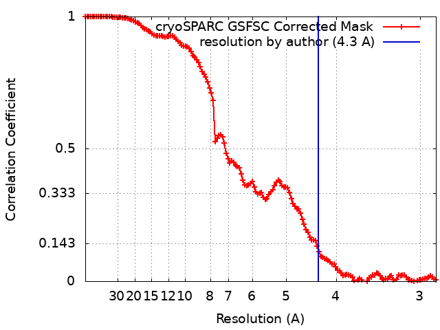
| Method | single particle reconstruction / cryo EM / Resolution: 4.3 Å | |||||||||
 Authors Authors | Munoz-Hernandez H / Wieczorek M | |||||||||
| Funding support |  Switzerland, 2 items Switzerland, 2 items
| |||||||||
 Citation Citation | Journal: Dev Cell / Year: 2024 Title: Partial closure of the γ-tubulin ring complex by CDK5RAP2 activates microtubule nucleation. Authors: Yixin Xu / Hugo Muñoz-Hernández / Rościsław Krutyhołowa / Florina Marxer / Ferdane Cetin / Michal Wieczorek / Abstract: Microtubule nucleation is templated by the γ-tubulin ring complex (γ-TuRC), but its structure deviates from the geometry of α-/β-tubulin in the microtubule, explaining the complex's poor ...Microtubule nucleation is templated by the γ-tubulin ring complex (γ-TuRC), but its structure deviates from the geometry of α-/β-tubulin in the microtubule, explaining the complex's poor nucleating activity. Several proteins may activate the γ-TuRC, but the mechanisms underlying activation are not known. Here, we determined the structure of the porcine γ-TuRC purified using CDK5RAP2's centrosomin motif 1 (CM1). We identified an unexpected conformation of the γ-TuRC bound to multiple protein modules containing MZT2, GCP2, and CDK5RAP2, resulting in a long-range constriction of the γ-tubulin ring that brings it in closer agreement with the 13-protofilament microtubule. Additional CDK5RAP2 promoted γ-TuRC decoration and stimulated the microtubule-nucleating activities of the porcine γ-TuRC and a reconstituted, CM1-free human complex in single-molecule assays. Our results provide a structural mechanism for the control of microtubule nucleation by CM1 proteins and identify conformational transitions in the γ-TuRC that prime it for microtubule nucleation. | |||||||||
| History |
|
- Structure visualization
| Supplemental images |
|---|
- Downloads & links
-EMDB archive
| Map data | emd_51019.map.gz | 108 MB | EMDB map data format | |
|---|---|---|---|---|
| Header (meta data) | emd-51019-v30.xmlemd-51019.xml | 29.3 KB 29.3 KB | Display Display | EMDB header |
| FSC (resolution estimation) | emd_51019_fsc.xml | 12.7 KB | Display | FSC data file |
| Images |  emd_51019.png emd_51019.png | 74.5 KB | ||
| Filedesc metadata | emd-51019.cif.gz | 10.5 KB | ||
| Others | emd_51019_half_map_1.map.gzemd_51019_half_map_2.map.gz | 200.4 MB 200.4 MB | ||
| Archive directory |  http://ftp.pdbj.org/pub/emdb/structures/EMD-51019ftp://ftp.pdbj.org/pub/emdb/structures/EMD-51019 http://ftp.pdbj.org/pub/emdb/structures/EMD-51019ftp://ftp.pdbj.org/pub/emdb/structures/EMD-51019 | HTTPS FTP |
-Related structure data
| Related structure data |  9g3zMC  9g3xC 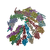 9g3yC 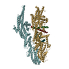 9g40C M: atomic model generated by this map C: citing same article ( |
|---|---|
| Similar structure data |


-Links
| EMDB pages | EMDB (EBI/PDBe) / EMDataResource |
|---|---|
| Related items in Molecule of the Month |


-Map
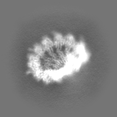
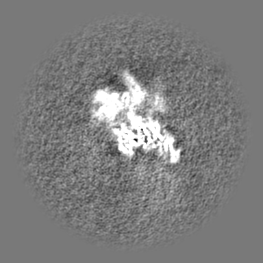
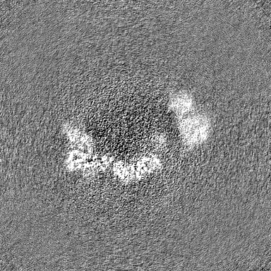
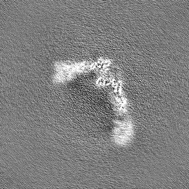
| File | Download / File: emd_51019.map.gz / Format: CCP4 / Size: 216 MB / Type: IMAGE STORED AS FLOATING POINT NUMBER (4 BYTES) | ||||||||||||||||||||||||||||||||||||
|---|---|---|---|---|---|---|---|---|---|---|---|---|---|---|---|---|---|---|---|---|---|---|---|---|---|---|---|---|---|---|---|---|---|---|---|---|---|
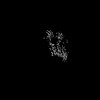
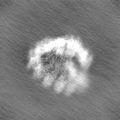
| Projections & slices | Image control
Images are generated by Spider. | ||||||||||||||||||||||||||||||||||||
| Voxel size | X=Y=Z: 1.41333 Å | ||||||||||||||||||||||||||||||||||||
| Density |
| ||||||||||||||||||||||||||||||||||||
| Symmetry | Space group: 1 | ||||||||||||||||||||||||||||||||||||
| Details | EMDB XML:
|
 Z (Sec.)
 Y (Row.)
 X (Col.)


-Supplemental data
-Half map: #2
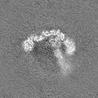
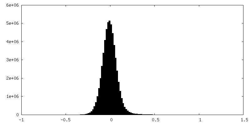
| File | emd_51019_half_map_1.map | ||||||||||||
|---|---|---|---|---|---|---|---|---|---|---|---|---|---|
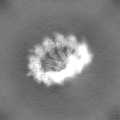
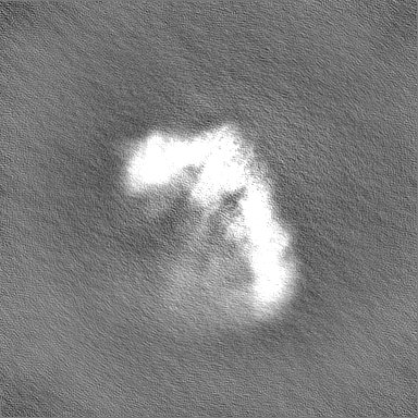
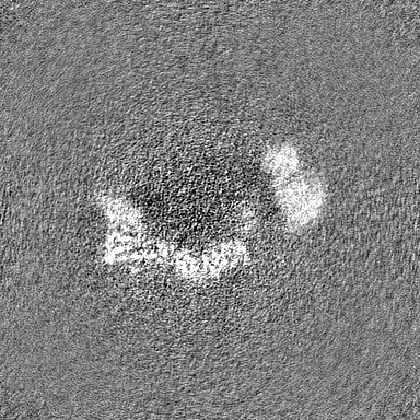
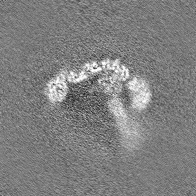
| Projections & Slices |
| ||||||||||||
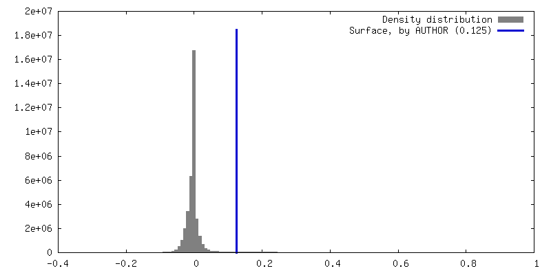
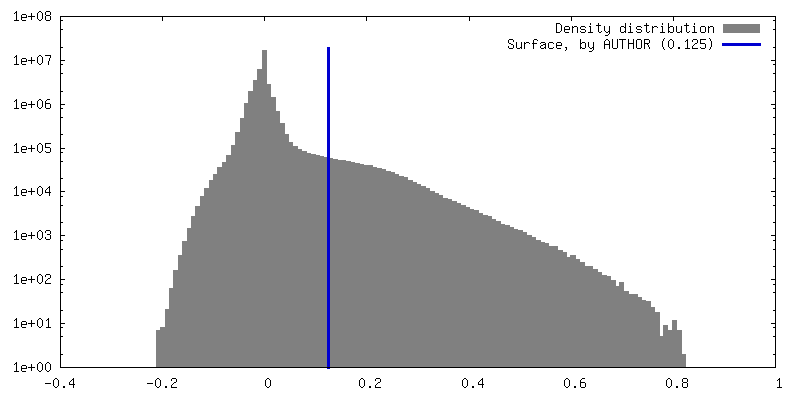
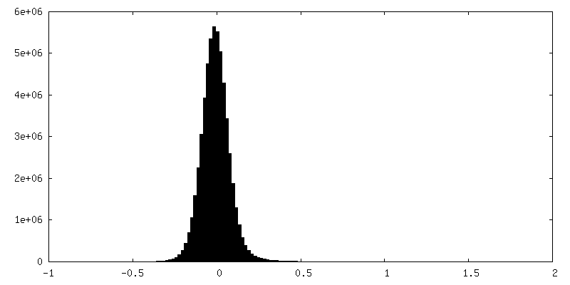
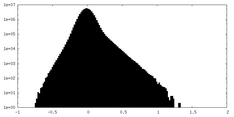
| Density Histograms |

-Half map: #1
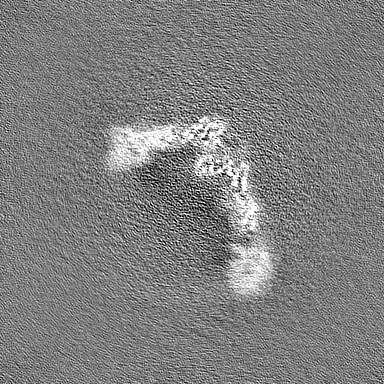
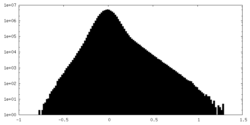
| File | emd_51019_half_map_2.map | ||||||||||||
|---|---|---|---|---|---|---|---|---|---|---|---|---|---|
| Projections & Slices |
| ||||||||||||
| Density Histograms |


- Sample components
+Entire : Gamma-Tubulin Ring Complex from native pig brain
+Supramolecule #1: Gamma-Tubulin Ring Complex from native pig brain
+Macromolecule #1: Mitotic spindle organizing protein 1
+Macromolecule #2: Mitotic-spindle organizing protein 2A isoform X4
+Macromolecule #3: Tubulin gamma chain
+Macromolecule #4: CDK5 regulatory subunit-associated protein 2

+Macromolecule #5: Gamma-tubulin complex component 3
+Macromolecule #6: Gamma-tubulin complex component
+Macromolecule #7: Tubulin gamma complex associated protein 6
+Macromolecule #8: Gamma-tubulin complex component
+Macromolecule #9: Gamma-tubulin complex component
-Experimental details
-Structure determination
| Method | cryo EM |
|---|---|
 Processing Processing | single particle reconstruction |
| Aggregation state | particle |
-Sample preparation
| Buffer | pH: 7.5 |
|---|---|
| Grid | Model: Quantifoil R2/1 / Material: COPPER / Support film - Material: CARBON / Support film - topology: HOLEY |
| Vitrification | Cryogen name: ETHANE-PROPANE |
- Electron microscopy
| Microscope | TFS KRIOS |
|---|---|
| Image recording | Film or detector model: GATAN K3 BIOQUANTUM (6k x 4k) / Average electron dose: 55.0 e/Å2 |
| Electron beam | Acceleration voltage: 300 kV / Electron source:  FIELD EMISSION GUN FIELD EMISSION GUN |
| Electron optics | Illumination mode: SPOT SCAN / Imaging mode: BRIGHT FIELD / Nominal defocus max: 2.9 µm / Nominal defocus min: 0.9 µm |
| Experimental equipment |  Model: Titan Krios / Image courtesy: FEI Company |
+Image processing
-Atomic model buiding 1
| Refinement | Space: REAL / Protocol: BACKBONE TRACE |
|---|---|
| Output model | PDB-9g3z: |
