Movie
Movie Controller

[English] 日本語
 Yorodumi
- EMDB-50619: Cryo-EM structure of the BcsE2F2 regulatory subcomplex from the E... -
+ Open data

- Basic information
| Entry | 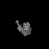 | |||||||||
|---|---|---|---|---|---|---|---|---|---|---|
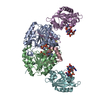
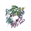
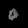
| Title | Cryo-EM structure of the BcsE2F2 regulatory subcomplex from the E. coli Bcs macrocomplex for cellulose secretion (local refinement) | |||||||||
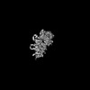
 Map data Map data | Deep EMhancer sharpened map from local refinement with mask around the BcsE2F2 complex from the c-di-GMP-saturated Bcs macrocomplex for E. coli cellulose secretion | |||||||||
 Sample Sample |
| |||||||||
 Keywords Keywords | Bacterial cellulose secretion / MEMBRANE PROTEIN | |||||||||
| Biological species |  | |||||||||
| Method | single particle reconstruction / cryo EM / Resolution: 2.85 Å | |||||||||
 Authors Authors | Anso I / Krasteva PV | |||||||||
| Funding support | European Union, 1 items
| |||||||||
 Citation Citation | Journal: Nat Commun / Year: 2024 Title: Structural basis for synthase activation and cellulose modification in the E. coli Type II Bcs secretion system. Authors: Itxaso Anso / Samira Zouhir / Thibault Géry Sana / Petya Violinova Krasteva /   Abstract: Bacterial cellulosic polymers constitute a prevalent class of biofilm matrix exopolysaccharides that are synthesized by several types of bacterial cellulose secretion (Bcs) systems, which include ...Bacterial cellulosic polymers constitute a prevalent class of biofilm matrix exopolysaccharides that are synthesized by several types of bacterial cellulose secretion (Bcs) systems, which include conserved cyclic diguanylate (c-di-GMP)-dependent cellulose synthase modules together with diverse accessory subunits. In E. coli, the biogenesis of phosphoethanolamine (pEtN)-modified cellulose relies on the BcsRQABEFG macrocomplex, encompassing inner-membrane and cytosolic subunits, and an outer membrane porin, BcsC. Here, we use cryogenic electron microscopy to shed light on the molecular mechanisms of BcsA-dependent recruitment and stabilization of a trimeric BcsG pEtN-transferase for polymer modification, and a dimeric BcsF-dependent recruitment of an otherwise cytosolic BcsERQ regulatory complex. We further demonstrate that BcsE, a secondary c-di-GMP sensor, can remain dinucleotide-bound and retain the essential-for-secretion BcsRQ partners onto the synthase even in the absence of direct c-di-GMP-synthase complexation, likely lowering the threshold for c-di-GMP-dependent synthase activation. Such activation-by-proxy mechanism could allow Bcs secretion system activity even in the absence of substantial intracellular c-di-GMP increase, and is reminiscent of other widespread synthase-dependent polysaccharide secretion systems where dinucleotide sensing and/or synthase stabilization are carried out by key co-polymerase subunits. | |||||||||
| History |
|
- Structure visualization
| Supplemental images |
|---|

- Downloads & links
-EMDB archive
| Map data | emd_50619.map.gz | 418 MB |  EMDB map data format EMDB map data format | |
|---|---|---|---|---|
| Header (meta data) | emd-50619-v30.xmlemd-50619.xml | 22.3 KB 22.3 KB | Display Display | EMDB header |
| FSC (resolution estimation) | emd_50619_fsc.xml | 16.5 KB | Display | FSC data file |
| Images |  emd_50619.png emd_50619.png | 48.8 KB | ||
| Filedesc metadata | emd-50619.cif.gz | 6.9 KB | ||
| Others | emd_50619_additional_1.map.gzemd_50619_half_map_1.map.gzemd_50619_half_map_2.map.gz | 226.2 MB 442.3 MB 442.3 MB | ||
| Archive directory |  http://ftp.pdbj.org/pub/emdb/structures/EMD-50619ftp://ftp.pdbj.org/pub/emdb/structures/EMD-50619 http://ftp.pdbj.org/pub/emdb/structures/EMD-50619ftp://ftp.pdbj.org/pub/emdb/structures/EMD-50619 | HTTPS FTP |
-Related structure data
| Related structure data | 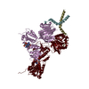 9fo7MC 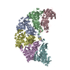 9fmtC 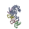 9fmvC 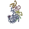 9fmzC 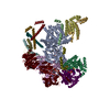 9fnnC 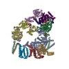 9fp0C 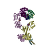 9fp2C C: citing same article ( M: atomic model generated by this map |
|---|
-Links
| EMDB pages | EMDB (EBI/PDBe) / EMDataResource |
|---|
-Map
| File | Download / File: emd_50619.map.gz / Format: CCP4 / Size: 476.8 MB / Type: IMAGE STORED AS FLOATING POINT NUMBER (4 BYTES) | ||||||||||||||||||||||||||||||||||||
|---|---|---|---|---|---|---|---|---|---|---|---|---|---|---|---|---|---|---|---|---|---|---|---|---|---|---|---|---|---|---|---|---|---|---|---|---|---|
| Annotation | Deep EMhancer sharpened map from local refinement with mask around the BcsE2F2 complex from the c-di-GMP-saturated Bcs macrocomplex for E. coli cellulose secretion | ||||||||||||||||||||||||||||||||||||
| Projections & slices | Image control
Images are generated by Spider. | ||||||||||||||||||||||||||||||||||||
| Voxel size | X=Y=Z: 0.839 Å | ||||||||||||||||||||||||||||||||||||
| Density |
| ||||||||||||||||||||||||||||||||||||
| Symmetry | Space group: 1 | ||||||||||||||||||||||||||||||||||||
| Details | EMDB XML:
|
 Z (Sec.)
 Y (Row.)
 X (Col.)


-Supplemental data
-Additional map: Unsharpened map from local refinement with mask around...
| File | emd_50619_additional_1.map | ||||||||||||
|---|---|---|---|---|---|---|---|---|---|---|---|---|---|
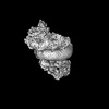
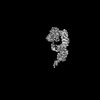
| Annotation | Unsharpened map from local refinement with mask around the BcsE2F2 complex from the c-di-GMP-saturated Bcs macrocomplex for E. coli cellulose secretion. | ||||||||||||
| Projections & Slices |
| ||||||||||||
| Density Histograms |


-Half map: Half-map from local refinement with mask around the...
| File | emd_50619_half_map_1.map | ||||||||||||
|---|---|---|---|---|---|---|---|---|---|---|---|---|---|
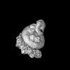
| Annotation | Half-map from local refinement with mask around the BcsE2F2 complex from the c-di-GMP-saturated Bcs macrocomplex for E. coli cellulose secretion. | ||||||||||||
| Projections & Slices |
| ||||||||||||
| Density Histograms |


-Half map: Half-map from local refinement with mask around the...
| File | emd_50619_half_map_2.map | ||||||||||||
|---|---|---|---|---|---|---|---|---|---|---|---|---|---|
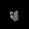
| Annotation | Half-map from local refinement with mask around the BcsE2F2 complex from the c-di-GMP-saturated Bcs macrocomplex for E. coli cellulose secretion. | ||||||||||||
| Projections & Slices |
| ||||||||||||
| Density Histograms |


- Sample components
-Entire : Locally refined BcsE2F2 regulatory subcomplex in saturating c-di-GMP
| Entire | Name: Locally refined BcsE2F2 regulatory subcomplex in saturating c-di-GMP |
|---|---|
| Components |
|
-Supramolecule #1: Locally refined BcsE2F2 regulatory subcomplex in saturating c-di-GMP
| Supramolecule | Name: Locally refined BcsE2F2 regulatory subcomplex in saturating c-di-GMP type: complex / ID: 1 / Parent: 0 / Macromolecule list: #1-#2 Details: Locally refined BcsE2F2 subcomplex in the context of the c-di-GMP-saturated Bcs macrocomplex for cellulose secretion in E. coli |
|---|---|
| Source (natural) | Organism: |
| Molecular weight | Theoretical: 990 KDa |
-Macromolecule #1: Cyclic di-GMP binding protein BcsE
| Macromolecule | Name: Cyclic di-GMP binding protein BcsE / type: protein_or_peptide / ID: 1 Details: Purified Bcs macrocomplex with stoichiometry BcsA-BcsB6-BcsR2-BcsQ2-BcsE2-BcsF2-BcsG3 Number of copies: 2 / Enantiomer: LEVO |
|---|---|
| Source (natural) | Organism: |
| Molecular weight | Theoretical: 60.967539 KDa |
| Recombinant expression | Organism: |
| Sequence | String: MASWSHPQFE KGSMRDIVDP VFSIGISSLW DELRHMPAGG VWWFNVDRHE DAISLANQTI ASQAETAHVA VISMDSDPAK IFQLDDSQG PEKIKLFSML NHEKGLYYLT RDLQCSIDPH NYLFILVCAN NAWQNIPAER LRSWLDKMNK WSRLNHCSLL V INPGNNND ...String: MASWSHPQFE KGSMRDIVDP VFSIGISSLW DELRHMPAGG VWWFNVDRHE DAISLANQTI ASQAETAHVA VISMDSDPAK IFQLDDSQG PEKIKLFSML NHEKGLYYLT RDLQCSIDPH NYLFILVCAN NAWQNIPAER LRSWLDKMNK WSRLNHCSLL V INPGNNND KQFSLLLEEY RSLFGLASLR FQGDQHLLDI AFWCNEKGVS ARQQLSVQQQ NGIWTLVQSE EAEIQPRSDE KR ILSNVAV LEGAPPLSEH WQLFNNNEVL FNEARTAQAA TVVFSLQQNA QIEPLARSIH TLRRQRGSAM KILVRENTAS LRA TDERLL LACGANMVIP WNAPLSRCLT MIESVQGQKF SRYVPEDITT LLSMTQPLKL RGFQKWDVFC NAVNNMMNNP LLPA HGKGV LVALRPVPGI RVEQALTLCR PNRTGDIMTI GGNRLVLFLS FCRINDLDTA LNHIFPLPTG DIFSNRMVWF EDDQI SAEL VQMRLLAPEQ WGMPLPLTQS SKPVINAEHD GRHWRRIPEP MRLLDDAVER SS |
-Macromolecule #2: Cellulose biosynthesis protein BcsF
| Macromolecule | Name: Cellulose biosynthesis protein BcsF / type: protein_or_peptide / ID: 2 Details: Purified Bcs macrocomplex with stoichiometry BcsA-BcsB6-BcsR2-BcsQ2-BcsE2-BcsF2-BcsG3 Number of copies: 2 / Enantiomer: LEVO |
|---|---|
| Source (natural) | Organism: |
| Molecular weight | Theoretical: 7.378975 KDa |
| Recombinant expression | Organism: |
| Sequence | String: MMTISDIIEI IVVCALIFFP LGYLARHSLR RIRDTLRLFF AKPRYVKPAG TLRRTEKARA TKK |
-Macromolecule #3: 9,9'-[(2R,3R,3aS,5S,7aR,9R,10R,10aS,12S,14aR)-3,5,10,12-tetrahydr...
| Macromolecule | Name: 9,9'-[(2R,3R,3aS,5S,7aR,9R,10R,10aS,12S,14aR)-3,5,10,12-tetrahydroxy-5,12-dioxidooctahydro-2H,7H-difuro[3,2-d:3',2'-j][1,3,7,9,2,8]tetraoxadiphosphacyclododecine-2,9-diyl]bis(2-amino-1,9-dihydro-6H-purin-6-one) type: ligand / ID: 3 / Number of copies: 4 / Formula: C2E |
|---|---|
| Molecular weight | Theoretical: 690.411 Da |
| Chemical component information | 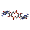 ChemComp-C2E: |
-Experimental details
-Structure determination
| Method | cryo EM |
|---|---|
 Processing Processing | single particle reconstruction |
| Aggregation state | particle |
-Sample preparation
| Concentration | 2 mg/mL |
|---|---|
| Buffer | pH: 8 Details: 120 mM NaCL 20 mM HEPES pH8 5 mM MgCl2 10 uM ApppCp 4 uM c-di-GMP 0.01% LM-NPG |
| Grid | Model: UltrAuFoil R1.2/1.3 / Material: GOLD / Mesh: 300 / Support film - Material: GOLD / Support film - topology: HOLEY |
| Vitrification | Cryogen name: ETHANE / Chamber humidity: 100 % / Chamber temperature: 277 K / Instrument: FEI VITROBOT MARK IV |
| Details | Purification from solubilized membrane fraction using an anti-FLAG M2 affinity resin |
- Electron microscopy
| Microscope | FEI TITAN KRIOS |
|---|---|
| Specialist optics | Energy filter - Name: GIF Quantum LS / Energy filter - Slit width: 20 eV |
| Image recording | Film or detector model: GATAN K3 (6k x 4k) / Number grids imaged: 2 / Number real images: 20022 / Average electron dose: 49.35 e/Å2 |
| Electron beam | Acceleration voltage: 300 kV / Electron source:  FIELD EMISSION GUN FIELD EMISSION GUN |
| Electron optics | Illumination mode: FLOOD BEAM / Imaging mode: BRIGHT FIELD / Cs: 2.7 mm / Nominal defocus max: 2.1 µm / Nominal defocus min: 0.3 µm |
| Experimental equipment |  Model: Titan Krios / Image courtesy: FEI Company |
+Image processing

-Atomic model buiding 1
| Initial model |
| ||||||||||||
|---|---|---|---|---|---|---|---|---|---|---|---|---|---|
| Refinement | Space: REAL / Protocol: FLEXIBLE FIT | ||||||||||||
| Output model | PDB-9fo7: |