 Movie
Movie Controller

[English] 日本語
 Yorodumi
- EMDB-50493: LSU structure derived from the monosome sample of the mitoribosom... -
+ Open data

- Basic information
| Entry |  | |||||||||
|---|---|---|---|---|---|---|---|---|---|---|
| Title | LSU structure derived from the monosome sample of the mitoribosome from T. gondii. | |||||||||
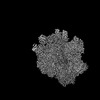
 Map data Map data | DeepEMhanced map of the LSU structure derived from the monosome sample of the mitoribosome from T. gondii. | |||||||||
 Sample Sample |
| |||||||||
 Keywords Keywords | Complex / translation / rRNA / RIBOSOME | |||||||||
| Biological species |  | |||||||||
| Method | single particle reconstruction / cryo EM / Resolution: 3.11 Å | |||||||||
 Authors Authors | Rocha REO / Barua S / Boissier F / Nguyen TT / Hashem Y | |||||||||
| Funding support | European Union, 1 items
| |||||||||
 Citation Citation | Journal: Nat Commun / Year: 2024 Title: Apicomplexan mitoribosome from highly fragmented rRNAs to a functional machine. Authors: Chaoyue Wang / Sari Kassem / Rafael Eduardo Oliveira Rocha / Pei Sun / Tan-Trung Nguyen / Joachim Kloehn / Xianyong Liu / Lorenzo Brusini / Alessandro Bonavoglia / Sramona Barua / Fanny ...Authors: Chaoyue Wang / Sari Kassem / Rafael Eduardo Oliveira Rocha / Pei Sun / Tan-Trung Nguyen / Joachim Kloehn / Xianyong Liu / Lorenzo Brusini / Alessandro Bonavoglia / Sramona Barua / Fanny Boissier / Mayara Lucia Del Cistia / Hongjuan Peng / Xinming Tang / Fujie Xie / Zixuan Wang / Oscar Vadas / Xun Suo / Yaser Hashem / Dominique Soldati-Favre / Yonggen Jia /    Abstract: The phylum Apicomplexa comprises eukaryotic parasites that cause fatal diseases affecting millions of people and animals worldwide. Their mitochondrial genomes have been significantly reduced, ...The phylum Apicomplexa comprises eukaryotic parasites that cause fatal diseases affecting millions of people and animals worldwide. Their mitochondrial genomes have been significantly reduced, leaving only three protein-coding genes and highly fragmented mitoribosomal rRNAs, raising challenging questions about mitoribosome composition, assembly and structure. Our study reveals how Toxoplasma gondii assembles over 40 mt-rRNA fragments using exclusively nuclear-encoded mitoribosomal proteins and three lineage-specific families of RNA-binding proteins. Among these are four proteins from the Apetala2/Ethylene Response Factor (AP2/ERF) family, originally known as transcription factors in plants and Apicomplexa, now repurposed as essential mitoribosome components. Cryo-EM analysis of the mitoribosome structure demonstrates how these AP2 proteins function as RNA binders to maintain mitoribosome integrity. The mitoribosome is also decorated with members of lineage-specific RNA-binding proteins belonging to RAP (RNA-binding domain abundant in Apicomplexa) proteins and HPR (heptatricopeptide repeat) families, highlighting the unique adaptations of these parasites. Solving the molecular puzzle of apicomplexan mitoribosome could inform the development of therapeutic strategies targeting organellar translation. | |||||||||
| History |
|
- Structure visualization
| Supplemental images |
|---|

- Downloads & links
-EMDB archive
| Map data | emd_50493.map.gz | 234.5 MB |  EMDB map data format EMDB map data format | |
|---|---|---|---|---|
| Header (meta data) | emd-50493-v30.xmlemd-50493.xml | 22.5 KB 22.5 KB | Display Display | EMDB header |
| FSC (resolution estimation) | emd_50493_fsc.xml | 13.8 KB | Display | FSC data file |
| Images |  emd_50493.png emd_50493.png | 136.2 KB | ||
| Masks | emd_50493_msk_1.map | 282.6 MB | Mask map | |
| Filedesc metadata | emd-50493.cif.gz | 4.9 KB | ||
| Others | emd_50493_additional_1.map.gzemd_50493_half_map_1.map.gzemd_50493_half_map_2.map.gz | 267 MB 261.9 MB 261.9 MB | ||
| Archive directory |  http://ftp.pdbj.org/pub/emdb/structures/EMD-50493ftp://ftp.pdbj.org/pub/emdb/structures/EMD-50493 http://ftp.pdbj.org/pub/emdb/structures/EMD-50493ftp://ftp.pdbj.org/pub/emdb/structures/EMD-50493 | HTTPS FTP |
-Validation report
| Summary document | emd_50493_validation.pdf.gz | 1 MB | Display | EMDB validaton report |
|---|---|---|---|---|
| Full document | emd_50493_full_validation.pdf.gz | 1 MB | Display | |
| Data in XML | emd_50493_validation.xml.gz | 22.7 KB | Display | |
| Data in CIF | emd_50493_validation.cif.gz | 30 KB | Display | |
| Arichive directory | https://ftp.pdbj.org/pub/emdb/validation_reports/EMD-50493ftp://ftp.pdbj.org/pub/emdb/validation_reports/EMD-50493 | HTTPS FTP |
-Related structure data


-Links
| EMDB pages | EMDB (EBI/PDBe) / EMDataResource |
|---|
-Map
| File | Download / File: emd_50493.map.gz / Format: CCP4 / Size: 282.6 MB / Type: IMAGE STORED AS FLOATING POINT NUMBER (4 BYTES) | ||||||||||||||||||||||||||||||||||||
|---|---|---|---|---|---|---|---|---|---|---|---|---|---|---|---|---|---|---|---|---|---|---|---|---|---|---|---|---|---|---|---|---|---|---|---|---|---|
| Annotation | DeepEMhanced map of the LSU structure derived from the monosome sample of the mitoribosome from T. gondii. | ||||||||||||||||||||||||||||||||||||
| Projections & slices | Image control
Images are generated by Spider. | ||||||||||||||||||||||||||||||||||||
| Voxel size | X=Y=Z: 1.5057 Å | ||||||||||||||||||||||||||||||||||||
| Density |
| ||||||||||||||||||||||||||||||||||||
| Symmetry | Space group: 1 | ||||||||||||||||||||||||||||||||||||
| Details | EMDB XML:
|
 Z (Sec.)
 Y (Row.)
 X (Col.)


-Supplemental data
-Mask #1
| File | emd_50493_msk_1.map | ||||||||||||
|---|---|---|---|---|---|---|---|---|---|---|---|---|---|
| Projections & Slices |
| ||||||||||||
| Density Histograms |


-Additional map: Sharpened map of the LSU structure derived from...
| File | emd_50493_additional_1.map | ||||||||||||
|---|---|---|---|---|---|---|---|---|---|---|---|---|---|
| Annotation | Sharpened map of the LSU structure derived from the monosome sample of the mitoribosome from T. gondii. | ||||||||||||
| Projections & Slices |
| ||||||||||||
| Density Histograms |


-Half map: Half map A of the LSU structure derived...
| File | emd_50493_half_map_1.map | ||||||||||||
|---|---|---|---|---|---|---|---|---|---|---|---|---|---|
| Annotation | Half map A of the LSU structure derived from the monosome sample of the mitoribosome from T. gondii. | ||||||||||||
| Projections & Slices |
| ||||||||||||
| Density Histograms |


-Half map: Half map B of the LSU structure derived...
| File | emd_50493_half_map_2.map | ||||||||||||
|---|---|---|---|---|---|---|---|---|---|---|---|---|---|
| Annotation | Half map B of the LSU structure derived from the monosome sample of the mitoribosome from T. gondii. | ||||||||||||
| Projections & Slices |
| ||||||||||||
| Density Histograms |


- Sample components
-Entire : LSU structure derived from the LSU sample of the mitoribosome fro...
| Entire | Name: LSU structure derived from the LSU sample of the mitoribosome from T. gondii. |
|---|---|
| Components |
|
-Supramolecule #1: LSU structure derived from the LSU sample of the mitoribosome fro...
| Supramolecule | Name: LSU structure derived from the LSU sample of the mitoribosome from T. gondii. type: complex / ID: 1 / Parent: 0 / Macromolecule list: #1-#11, #13-#22, #24-#25, #27 |
|---|---|
| Source (natural) | Organism: |
| Molecular weight | Theoretical: 4 MDa |
-Experimental details
-Structure determination
| Method | cryo EM |
|---|---|
 Processing Processing | single particle reconstruction |
| Aggregation state | particle |
-Sample preparation
| Buffer | pH: 7.5 |
|---|---|
| Grid | Model: Quantifoil R2/2 / Material: COPPER / Mesh: 300 / Support film - Material: CARBON / Support film - topology: HOLEY / Support film - Film thickness: 2 / Pretreatment - Type: GLOW DISCHARGE / Pretreatment - Time: 25 sec. / Pretreatment - Atmosphere: AIR / Pretreatment - Pressure: 2.6e-05 kPa |
| Vitrification | Cryogen name: ETHANE / Chamber humidity: 100 % / Chamber temperature: 277 K / Instrument: FEI VITROBOT MARK IV / Details: Blotting force 5, blotting time 2.5 seconds.. |
- Electron microscopy
| Microscope | FEI TALOS ARCTICA |
|---|---|
| Temperature | Min: 90.0 K / Max: 95.0 K |
| Image recording | Film or detector model: GATAN K2 SUMMIT (4k x 4k) / Detector mode: COUNTING / Digitization - Dimensions - Width: 4096 pixel / Digitization - Dimensions - Height: 4096 pixel / Digitization - Frames/image: 1-40 / Number grids imaged: 1 / Number real images: 9524 / Average exposure time: 6.0 sec. / Average electron dose: 50.0 e/Å2 |
| Electron beam | Acceleration voltage: 200 kV / Electron source:  FIELD EMISSION GUN FIELD EMISSION GUN |
| Electron optics | C2 aperture diameter: 30.0 µm / Illumination mode: FLOOD BEAM / Imaging mode: BRIGHT FIELD / Cs: 2.7 mm / Nominal defocus max: 2.5 µm / Nominal defocus min: 0.5 µm / Nominal magnification: 59000 |
| Sample stage | Specimen holder model: FEI TITAN KRIOS AUTOGRID HOLDER / Cooling holder cryogen: NITROGEN |
| Experimental equipment |  Model: Talos Arctica / Image courtesy: FEI Company |
+Image processing

-Atomic model buiding 1
| Refinement | Space: REAL / Protocol: AB INITIO MODEL |
|---|
