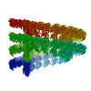
[日本語] English
- EMDB-5027: Kinesin13-Microtubule Ring Complex -

+
データを開く


IDまたはキーワード:

読み込み中...

-
基本情報

登録情報
データベース: EMDB / ID: EMD-5027
タイトルKinesin13-Microtubule Ring Complex
マップデータKinesin 13-Microtubule Ring Complex
試料
  • 試料: Kinesin13-Microtubule Ring Complex
  • タンパク質・ペプチド: KLP10a
  • タンパク質・ペプチド: alpha-tubulin
  • タンパク質・ペプチド: beta-tubulin
キーワードKinesin / Kinesin-13 / microtubule / tubulin / depolymerization / Kin-I / M-Kinesin / cytoskeleton
機能・相同性
機能・相同性情報


postsynaptic cytoskeleton organization / regulation of chromosome segregation / Amplification of signal from unattached kinetochores via a MAD2 inhibitory signal / establishment or maintenance of microtubule cytoskeleton polarity / Kinesins / Mitotic Prometaphase / EML4 and NUDC in mitotic spindle formation / Resolution of Sister Chromatid Cohesion / metaphase chromosome alignment / COPI-dependent Golgi-to-ER retrograde traffic ...postsynaptic cytoskeleton organization / regulation of chromosome segregation / Amplification of signal from unattached kinetochores via a MAD2 inhibitory signal / establishment or maintenance of microtubule cytoskeleton polarity / Kinesins / Mitotic Prometaphase / EML4 and NUDC in mitotic spindle formation / Resolution of Sister Chromatid Cohesion / metaphase chromosome alignment / COPI-dependent Golgi-to-ER retrograde traffic / RHO GTPases Activate Formins / Separation of Sister Chromatids / microtubule plus-end / attachment of mitotic spindle microtubules to kinetochore / MHC class II antigen presentation / microtubule plus-end binding / microtubule depolymerization / microtubule motor activity / motile cilium / tubulin complex / microtubule-based movement / mitotic metaphase chromosome alignment / regulation of neurotransmitter receptor localization to postsynaptic specialization membrane / chromosome, centromeric region / kinetochore / structural constituent of cytoskeleton / microtubule cytoskeleton organization / neuron migration / mitotic cell cycle / presynapse / 加水分解酵素; 酸無水物に作用; GTPに作用・細胞または細胞小器官の運動に関与 / microtubule / hydrolase activity / postsynapse / cell division / GTPase activity / GTP binding / glutamatergic synapse / ATP binding / metal ion binding / nucleus / cytoplasm
類似検索 - 分子機能
: / Kinesin-like protein KIF2A-like, N-terminal / Kinesin-like protein / Kinesin motor domain signature. / Kinesin motor domain, conserved site / Kinesin motor domain / Kinesin motor domain profile. / Kinesin motor, catalytic domain. ATPase. / Kinesin motor domain / Kinesin motor domain superfamily ...: / Kinesin-like protein KIF2A-like, N-terminal / Kinesin-like protein / Kinesin motor domain signature. / Kinesin motor domain, conserved site / Kinesin motor domain / Kinesin motor domain profile. / Kinesin motor, catalytic domain. ATPase. / Kinesin motor domain / Kinesin motor domain superfamily / Alpha tubulin / Alpha tubulin / Tubulin-beta mRNA autoregulation signal. / Beta tubulin, autoregulation binding site / Beta tubulin / Beta tubulin / Tubulin / Tubulin, C-terminal / Tubulin C-terminal domain / Tubulin, conserved site / Tubulin subunits alpha, beta, and gamma signature. / Tubulin/FtsZ family, C-terminal domain / Tubulin/FtsZ-like, C-terminal domain / Tubulin/FtsZ, C-terminal / Tubulin/FtsZ, 2-layer sandwich domain / Tubulin/FtsZ family, GTPase domain / Tubulin/FtsZ family, GTPase domain / Tubulin/FtsZ, GTPase domain / Tubulin/FtsZ, GTPase domain superfamily / P-loop containing nucleoside triphosphate hydrolase
類似検索 - ドメイン・相同性
Tubulin alpha-1A chain / Tubulin beta chain / Tubulin alpha-1D chain / Kinesin-like protein KIF2C
類似検索 - 構成要素
手法らせん対称体再構成法 / クライオ電子顕微鏡法 / 解像度: 28.0 Å
データ登録者Tan D / Rice WJ / Sosa H
引用ジャーナル: Structure / : 2008
タイトル: Structure of the kinesin13-microtubule ring complex.
著者: Dongyan Tan / William J Rice / Hernando Sosa /
要旨: To investigate the mechanism of kinesin13-induced microtubule depolymerization, we have calculated a three-dimensional (3D) map of the kinesin13-microtubule ring complex, using cryo-electron ...To investigate the mechanism of kinesin13-induced microtubule depolymerization, we have calculated a three-dimensional (3D) map of the kinesin13-microtubule ring complex, using cryo-electron microscopy (cryo-EM) and image analysis. An atomic model of the complex was produced by docking the crystal structures of tubulin and a kinesin13 motor domain (MD) into the 3D map. The model reveals a snapshot of the depolymerization mechanism by providing a 3D view of the complex formed between the kinesin13 MD and a curved tubulin protofilament (pf). It suggests that contacts mediated by kinesin13 class-specific residues in the putative microtubule-binding site stabilize intra-dimer tubulin curvature. In addition, a tubulin-binding site on the kinesin13 MD was identified. Mutations at this class-conserved site selectively disrupt the formation of microtubule-associated ring complexes.
履歴
登録2008年8月29日-
ヘッダ(付随情報) 公開2008年12月17日-
マップ公開2009年5月5日-
更新2014年4月2日-
現状2014年4月2日処理サイト: RCSB / 状態: 公開

-
構造の表示

ムービー
  • 表面図(断面を密度値に従い着色)
  • 表面レベル: 14
  • UCSF Chimeraによる作画
  • ダウンロード
  • 表面図(円筒半径に従い着色)
  • 表面レベル: 14
  • UCSF Chimeraによる作画
  • ダウンロード
  • あてはめたモデルとの重ね合わせ
  • 原子モデル: PDB-3edl
  • 表面レベル: 14
  • UCSF Chimeraによる作画
  • ダウンロード
  • 単純化した表面モデル + あてはめた原子モデル
  • 原子モデルPDB-3edl
  • Jmolによる作画
  • ダウンロード
ムービービューア
構造ビューアEMマップ:
SurfViewMolmilJmol/JSmol
添付画像

ダウンロードとリンク

-
マップ

ファイルダウンロード / ファイル: emd_5027.map.gz / 形式: CCP4 / 大きさ: 1.6 MB / タイプ: IMAGE STORED AS FLOATING POINT NUMBER (4 BYTES)
注釈Kinesin 13-Microtubule Ring Complex
投影像・断面図

画像のコントロール

大きさ
明度
コントラスト
その他
Z (Sec.)Y (Row.)X (Col.)
6 Å/pix.
x 41 pix.
= 246. Å
6 Å/pix.
x 101 pix.
= 606. Å
6 Å/pix.
x 101 pix.
= 606. Å

表面

投影像

断面 (1/3)

断面 (1/2)

断面 (2/3)

画像は Spider により作成

これらの図は立方格子座標系で作成されたものです

ボクセルのサイズX=Y=Z: 6 Å
密度
表面レベル登録者による: 11.1 / ムービー #1: 14
最小 - 最大-48.0 - 55.0
平均 (標準偏差)0.56261343 (±12.836534500000001)
対称性空間群: 1
詳細

EMDB XML:

マップ形状
Axis orderXYZ
Origin000
サイズ10110141
Spacing10110141
セルA: 606.0 Å / B: 606.0 Å / C: 246.0 Å
α=β=γ: 90.0 °

CCP4マップ ヘッダ情報:

modeImage stored as Reals
Å/pix. X/Y/Z666
M x/y/z10110141
origin x/y/z0.0000.0000.000
length x/y/z606.000606.000246.000
α/β/γ90.00090.00090.000
start NX/NY/NZ-127-127-127
NX/NY/NZ255255255
MAP C/R/S123
start NC/NR/NS000
NC/NR/NS10110141
D min/max/mean-48.00055.0000.563

-
添付データ

-
試料の構成要素

-
全体 : Kinesin13-Microtubule Ring Complex

全体名称: Kinesin13-Microtubule Ring Complex
要素
  • 試料: Kinesin13-Microtubule Ring Complex
  • タンパク質・ペプチド: KLP10a
  • タンパク質・ペプチド: alpha-tubulin
  • タンパク質・ペプチド: beta-tubulin

-
超分子 #1000: Kinesin13-Microtubule Ring Complex

超分子名称: Kinesin13-Microtubule Ring Complex / タイプ: sample / ID: 1000 / Number unique components: 3
分子量実験値: 250 KDa / 理論値: 250 KDa / 手法: SDS-PAGE

-
分子 #1: KLP10a

分子名称: KLP10a / タイプ: protein_or_peptide / ID: 1 / Name.synonym: Kinesin-13 / コピー数: 1 / 集合状態: monomer / 組換発現: Yes
由来(天然)細胞: BL21 / 細胞中の位置: cytosol
分子量実験値: 400 KDa / 理論値: 400 KDa
組換発現生物種: Escherichia coli (大腸菌) / 組換プラスミド: pRSET B

-
分子 #2: alpha-tubulin

分子名称: alpha-tubulin / タイプ: protein_or_peptide / ID: 2 / Name.synonym: tubulin / コピー数: 2 / 集合状態: Heterodimer / 組換発現: No / データベース: NCBI
由来(天然)組織: Bos taurus Brain
分子量実験値: 550 KDa
配列GO: tubulin complex / InterPro: Alpha tubulin

-
分子 #3: beta-tubulin

分子名称: beta-tubulin / タイプ: protein_or_peptide / ID: 3 / Name.synonym: tubulin / コピー数: 2 / 集合状態: Hterodimer / 組換発現: No / データベース: NCBI
由来(天然)組織: Brain
分子量実験値: 550 KDa
配列GO: tubulin complex / InterPro: Beta tubulin

-
実験情報

-
構造解析

手法クライオ電子顕微鏡法
解析らせん対称体再構成法
試料の集合状態filament

-
試料調製

濃度0.6 mg/mL
緩衝液pH: 6.8
詳細: 80 mM Pipes, pH 6.8, 2 mM MgCl2, 1 mM EGTA, 2 mM AMP-PNP, 0.02 mM taxol
グリッド詳細: Quantifoil grids
凍結凍結剤: ETHANE / チャンバー内湿度: 100 % / 装置: OTHER / 詳細: Vitrification instrument: VitroBot
手法: Blot for 2 seconds, Blot Offset (mm) -2, Plunge Time 1 sec, Wait Time 0 sec

-
電子顕微鏡法 #1

Microscopy ID1
顕微鏡FEI TECNAI 20
撮影カテゴリ: FILM / フィルム・検出器のモデル: KODAK SO-163 FILM / デジタル化 - スキャナー: OTHER / デジタル化 - サンプリング間隔: 12.5 µm / 実像数: 5 / 平均電子線量: 9 e/Å2 / ビット/ピクセル: 16
電子線加速電圧: 120 kV / 電子線源: LAB6
電子光学系照射モード: FLOOD BEAM / 撮影モード: BRIGHT FIELD / 最大 デフォーカス(公称値): 2.5 µm / 最小 デフォーカス(公称値): 1.5 µm / 倍率(公称値): 50000
試料ステージ試料ホルダー: Side entry liquid nitrogen-cooled cryo specimen holder
試料ホルダーモデル: GATAN LIQUID NITROGEN

-
電子顕微鏡法 #2

Microscopy ID2
顕微鏡FEI TECNAI F20
撮影カテゴリ: FILM / フィルム・検出器のモデル: KODAK SO-163 FILM / デジタル化 - スキャナー: OTHER / デジタル化 - サンプリング間隔: 12.5 µm / 実像数: 5 / 平均電子線量: 9 e/Å2 / ビット/ピクセル: 16
電子線加速電圧: 200 kV / 電子線源: FIELD EMISSION GUN
電子光学系照射モード: FLOOD BEAM / 撮影モード: BRIGHT FIELD / 倍率(公称値): 50000
試料ステージ試料ホルダー: Side entry liquid nitrogen-cooled cryo specimen holder
試料ホルダーモデル: GATAN LIQUID NITROGEN
実験機器
モデル: Tecnai F20 / 画像提供: FEI Company

-
画像解析

最終 再構成アルゴリズム: OTHER / 解像度のタイプ: BY AUTHOR / 解像度: 28.0 Å / 解像度の算出法: FSC 0.5 CUT-OFF / ソフトウェア - 名称: MRC, SUPRIM, Phoelix, Other
詳細: Final map is the averaged near and far layer line data from 5 filaments (10 datasets, 3296 asymmetric units)
CTF補正詳細: Each Particle

-
原子モデル構築 1

初期モデルPDB ID:

Chain - Chain ID: A
ソフトウェア名称: COLORES
詳細PDBEntryID_givenInChain. Protocol: Rigid-Body
精密化空間: REAL / プロトコル: RIGID BODY FIT
得られたモデル

PDB-3edl:
Kinesin13-Microtubule Ring complex

-
原子モデル構築 2

初期モデルPDB ID:
ソフトウェア名称: COLORES
詳細Protocol: Rigid Body
精密化空間: REAL / プロトコル: RIGID BODY FIT
得られたモデル

PDB-3edl:
Kinesin13-Microtubule Ring complex

-
原子モデル構築 3

初期モデルPDB ID:
ソフトウェア名称: COLORES
詳細Protocol: Rigid Body
精密化空間: REAL / プロトコル: RIGID BODY FIT
得られたモデル

PDB-3edl:
Kinesin13-Microtubule Ring complex

+
万見について

-
お知らせ

-
2022年2月9日: EMDBエントリの付随情報ファイルのフォーマットが新しくなりました

EMDBエントリの付随情報ファイルのフォーマットが新しくなりました

  • EMDBのヘッダファイルのバージョン3が、公式のフォーマットとなりました。
  • これまでは公式だったバージョン1.9は、アーカイブから削除されます。

関連情報:EMDBヘッダ

外部リンク:wwPDBはEMDBデータモデルのバージョン3へ移行します

-
2020年8月12日: 新型コロナ情報

新型コロナ情報

URL: https://pdbj.org/emnavi/covid19.php

新ページ: EM Navigatorに新型コロナウイルスの特設ページを開設しました。

関連情報:Covid-19情報 / 2020年3月5日: 新型コロナウイルスの構造データ

+
2020年3月5日: 新型コロナウイルスの構造データ

新型コロナウイルスの構造データ

関連情報:万見生物種 / 2020年8月12日: 新型コロナ情報

外部リンク:COVID-19特集ページ - PDBj / 今月の分子2020年2月:コロナウイルスプロテーアーゼ

+
2019年1月31日: EMDBのIDの桁数の変更

EMDBのIDの桁数の変更

  • EMDBエントリに付与されているアクセスコード(EMDB-ID)は4桁の数字(例、EMD-1234)でしたが、間もなく枯渇します。これまでの4桁のID番号は4桁のまま変更されませんが、4桁の数字を使い切った後に発行されるIDは5桁以上の数字(例、EMD-12345)になります。5桁のIDは2019年の春頃から発行される見通しです。
  • EM Navigator/万見では、接頭語「EMD-」は省略されています。

関連情報:Q: 「EMD」とは何ですか? / 万見/EM NavigatorにおけるID/アクセスコードの表記

外部リンク:EMDB Accession Codes are Changing Soon! / PDBjへお問い合わせ

+
2017年7月12日: PDB大規模アップデート

PDB大規模アップデート

  • 新バージョンのPDBx/mmCIF辞書形式に基づくデータがリリースされました。
  • 今回の更新はバージョン番号が4から5になる大規模なもので、全エントリデータの書き換えが行われる「Remediation」というアップデートに該当します。
  • このバージョンアップで、電子顕微鏡の実験手法に関する多くの項目の書式が改定されました(例:em_softwareなど)。
  • EM NavigatorとYorodumiでも、この改定に基づいた表示内容になります。

外部リンク:wwPDB Remediation / OneDepデータ基準に準拠した、より強化された内容のモデル構造ファイルが、PDBアーカイブで公開されました。

-
万見 (Yorodumi)

幾万の構造データを、幾万の視点から

  • 万見(Yorodumi)は、EMDB/PDB/SASBDBなどの構造データを閲覧するためのページです。
  • EM Navigatorの詳細ページの後継、Omokage検索のフロントエンドも兼ねています。

関連情報:EMDB / PDB / SASBDB / 3つのデータバンクの比較 / 万見検索 / 2016年8月31日: 新しいEM Navigatorと万見 / 万見文献 / Jmol/JSmol / 機能・相同性情報 / 新しいEM Navigatorと万見の変更点

他の情報も見る