 ムービー
ムービー コントローラー


+ データを開く

- 基本情報
基本情報
| 登録情報 | データベース: EMDB / ID: EMD-5014 | |||||||||
|---|---|---|---|---|---|---|---|---|---|---|
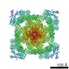
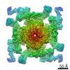
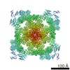
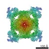
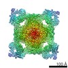
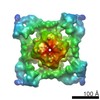
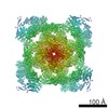
| タイトル | RyR1 in the closed state | |||||||||
 マップデータ マップデータ | 3D structure of RyR1 in the closed state. | |||||||||
 試料 試料 |
| |||||||||
 キーワード キーワード | ryanodine receptor / ion channel gating | |||||||||
| 生物種 |  | |||||||||
| 手法 | 単粒子再構成法 / クライオ電子顕微鏡法 / 解像度: 10.3 Å | |||||||||
 データ登録者 データ登録者 | Samso M / Wagenknecht T / Allen PD | |||||||||
 引用 引用 | ジャーナル: Nat Struct Mol Biol / 年: 2005 タイトル: Internal structure and visualization of transmembrane domains of the RyR1 calcium release channel by cryo-EM. 著者: Montserrat Samsó / Terence Wagenknecht / P D Allen /  要旨: RyR1 is an intracellular calcium channel with a central role in muscle contraction. We obtained a three-dimensional reconstruction of the RyR1 in the closed state at a nominal resolution of ...RyR1 is an intracellular calcium channel with a central role in muscle contraction. We obtained a three-dimensional reconstruction of the RyR1 in the closed state at a nominal resolution of approximately 10 A using cryo-EM. The cytoplasmic assembly consists of a series of interconnected tubular structures that merge into four columns that extend into the transmembrane assembly. The transmembrane assembly, which has at least six transmembrane alpha-helices per monomer, has four tilted rods that can be fitted with the inner helices of a closed K(+) channel atomic structure. The rods splay out at the lumenal side and converge into a dense ring at the cytoplasmic side. Another set of four rods emerges from this ring and shapes the inner part of the four columns. The resulting constricted axial structure provides direct continuity between cytoplasmic and transmembrane assemblies, and a possible mechanism for control of channel gating through conformational changes in the cytoplasmic assembly. | |||||||||
| 履歴 |
|
- 構造の表示
構造の表示
| ムービー |
 ムービービューア ムービービューア |
|---|---|
| 構造ビューア | EMマップ: SurfViewMolmilJmol/JSmol |
| 添付画像 |

- ダウンロードとリンク
-EMDBアーカイブ
| マップデータ | emd_5014.map.gz | 20.7 MB | EMDBマップデータ形式 | |
|---|---|---|---|---|
| ヘッダ (付随情報) | emd-5014-v30.xmlemd-5014.xml | 10.1 KB 10.1 KB | 表示 表示 | EMDBヘッダ |
| 画像 | 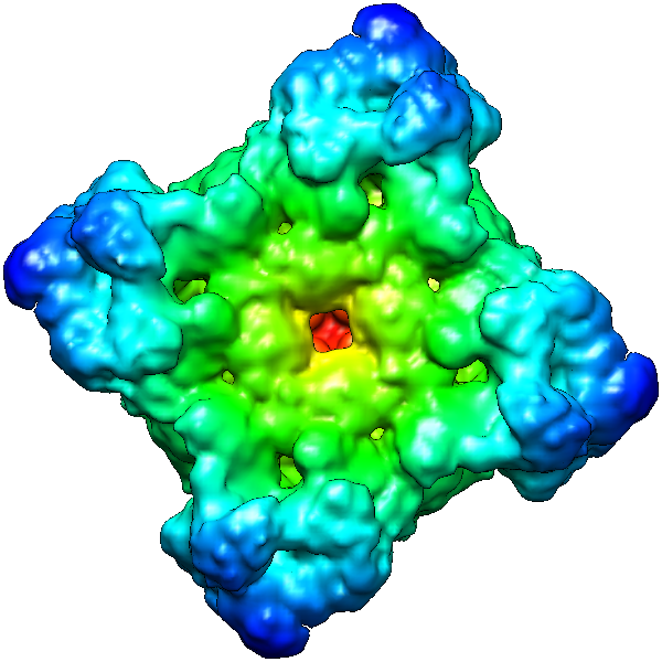 emd_5014_1.png emd_5014_1.png | 253.4 KB | ||
| アーカイブディレクトリ |  http://ftp.pdbj.org/pub/emdb/structures/EMD-5014ftp://ftp.pdbj.org/pub/emdb/structures/EMD-5014 http://ftp.pdbj.org/pub/emdb/structures/EMD-5014ftp://ftp.pdbj.org/pub/emdb/structures/EMD-5014 | HTTPS FTP |
-検証レポート
| 文書・要旨 | emd_5014_validation.pdf.gz | 77.4 KB | 表示 | EMDB検証レポート |
|---|---|---|---|---|
| 文書・詳細版 | emd_5014_full_validation.pdf.gz | 76.5 KB | 表示 | |
| XML形式データ | emd_5014_validation.xml.gz | 494 B | 表示 | |
| アーカイブディレクトリ | https://ftp.pdbj.org/pub/emdb/validation_reports/EMD-5014ftp://ftp.pdbj.org/pub/emdb/validation_reports/EMD-5014 | HTTPS FTP |
-関連構造データ
-リンク
| EMDBのページ | EMDB (EBI/PDBe) / EMDataResource |
|---|
-マップ
| ファイル | ダウンロード / ファイル: emd_5014.map.gz / 形式: CCP4 / 大きさ: 21.7 MB / タイプ: IMAGE STORED AS FLOATING POINT NUMBER (4 BYTES) | ||||||||||||||||||||||||||||||||||||||||||||||||||||||||||||||||||||
|---|---|---|---|---|---|---|---|---|---|---|---|---|---|---|---|---|---|---|---|---|---|---|---|---|---|---|---|---|---|---|---|---|---|---|---|---|---|---|---|---|---|---|---|---|---|---|---|---|---|---|---|---|---|---|---|---|---|---|---|---|---|---|---|---|---|---|---|---|---|
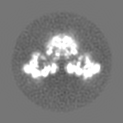
| 注釈 | 3D structure of RyR1 in the closed state. | ||||||||||||||||||||||||||||||||||||||||||||||||||||||||||||||||||||
| 投影像・断面図 | 画像のコントロール
画像は Spider により作成 | ||||||||||||||||||||||||||||||||||||||||||||||||||||||||||||||||||||
| ボクセルのサイズ | X=Y=Z: 2.8 Å | ||||||||||||||||||||||||||||||||||||||||||||||||||||||||||||||||||||
| 密度 |
| ||||||||||||||||||||||||||||||||||||||||||||||||||||||||||||||||||||
| 対称性 | 空間群: 1 | ||||||||||||||||||||||||||||||||||||||||||||||||||||||||||||||||||||
| 詳細 | EMDB XML:
CCP4マップ ヘッダ情報:
| ||||||||||||||||||||||||||||||||||||||||||||||||||||||||||||||||||||
 Z (Sec.)
 Y (Row.)
 X (Col.)


-添付データ
- 試料の構成要素
-全体 : ryanodine receptor 1
| 全体 | 名称: ryanodine receptor 1 |
|---|---|
| 要素 |
|
-超分子 #1000: ryanodine receptor 1
| 超分子 | 名称: ryanodine receptor 1 / タイプ: sample / ID: 1000 / 集合状態: homotetramer / Number unique components: 1 |
|---|---|
| 分子量 | 理論値: 565 KDa |
-超分子 #1: ryanodine receptor isoform 1
| 超分子 | 名称: ryanodine receptor isoform 1 / タイプ: organelle_or_cellular_component / ID: 1 / Name.synonym: RyR1 / コピー数: 4 / 集合状態: Tetramer / 組換発現: No / データベース: NCBI |
|---|---|
| Ref GO | 0: GO:0005219 |
| Ref INTERPRO | 0: IPR000699 |
| 由来(天然) | 生物種: |
| 分子量 | 実験値: 565 KDa / 理論値: 565 KDa |
-実験情報
-構造解析
| 手法 | クライオ電子顕微鏡法 |
|---|---|
 解析 解析 | 単粒子再構成法 |
| 試料の集合状態 | particle |
-試料調製
| 濃度 | 2.00 mg/mL |
|---|---|
| 緩衝液 | pH: 7.4 詳細: 20 mM Na-MOPS pH 7.4, 0.9 M NaCl, 0.5% (w/v) CHAPS, 2 mM DTT, 2 mM EGTA |
| グリッド | 詳細: 300 mesh holey copper grids |
| 凍結 | 凍結剤: ETHANE / 装置: HOMEMADE PLUNGER / 詳細: Vitrification instrument: two-side blotting plunger / 手法: Blot for 2 seconds before plunging |
- 電子顕微鏡法
| 顕微鏡 | FEI TECNAI F20 |
|---|---|
| 温度 | 平均: 87 K |
| アライメント法 | Legacy - 非点収差: objective lens astigmatism was corrected at 150,000 times magnification |
| 撮影 | カテゴリ: FILM / フィルム・検出器のモデル: KODAK SO-163 FILM / デジタル化 - スキャナー: ZEISS SCAI / デジタル化 - サンプリング間隔: 7.0 µm / 実像数: 818 / 平均電子線量: 10 e/Å2 / 詳細: after scanning pixels were averaged 2x2 / ビット/ピクセル: 8 |
| 電子線 | 加速電圧: 200 kV / 電子線源:  FIELD EMISSION GUN FIELD EMISSION GUN |
| 電子光学系 | 照射モード: FLOOD BEAM / 撮影モード: BRIGHT FIELD / Cs: 2.0 mm / 最大 デフォーカス(公称値): 4.0 µm / 最小 デフォーカス(公称値): 2.5 µm / 倍率(公称値): 50000 |
| 試料ステージ | 試料ホルダー: Side entry liquid nitrogen-cooled cryo specimen holder 試料ホルダーモデル: GATAN LIQUID NITROGEN |
| 実験機器 |  モデル: Tecnai F20 / 画像提供: FEI Company |
-画像解析
| CTF補正 | 詳細: Each particle |
|---|---|
| 最終 再構成 | アルゴリズム: OTHER / 解像度のタイプ: BY AUTHOR / 解像度: 10.3 Å / 解像度の算出法: OTHER / ソフトウェア - 名称: FREALIGN and SPIDER / 使用した粒子像数: 25722 |
-原子モデル構築 1
| 初期モデル | PDB ID: |
|---|---|
| 詳細 | The inner helices of KcsA (residues 72-119) were fitted by manual docking to the inner helices of RyR1 using program Chimera |
| 精密化 | 空間: REAL / プロトコル: RIGID BODY FIT |

