 Movie
Movie Controller


+ Open data

- Basic information
| Entry | Database: EMDB / ID: EMD-1274 | |||||||||
|---|---|---|---|---|---|---|---|---|---|---|
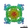
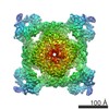
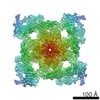
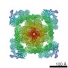
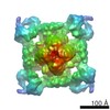
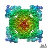
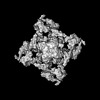
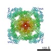
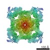
| Title | Structure of Ca2+ release channel at 14 A resolution. | |||||||||
 Map data Map data | The map is rendered from the cytoplasmic side (top view) along 4-fold axis of the protein | |||||||||
 Sample Sample |
| |||||||||
| Function / homology | ryanodine-sensitive calcium-release channel activity / RIH domain Function and homology information Function and homology information | |||||||||
| Biological species |  | |||||||||
| Method | single particle reconstruction / cryo EM / Resolution: 14.0 Å | |||||||||
 Authors Authors | Serysheva II / Hamilton SL / Chiu W / Ludtke SJ | |||||||||
 Citation Citation | Journal: J Mol Biol / Year: 2005 Title: Structure of Ca2+ release channel at 14 A resolution. Authors: Irina I Serysheva / Susan L Hamilton / Wah Chiu / Steven J Ludtke /  Abstract: The 14 A resolution structure of the 2.3 MDa Ca2+ release channel (also known as RyR1) was determined by electron cryomicroscopy and single particle reconstruction. This structure was produced using ...The 14 A resolution structure of the 2.3 MDa Ca2+ release channel (also known as RyR1) was determined by electron cryomicroscopy and single particle reconstruction. This structure was produced using collected data used for our previous published structures at 22-30 A resolution, but now taking advantage of recent algorithmic improvements in the EMAN software suite. This improved map clearly exhibits more structural detail and allows better defined docking of computationally predicted structural domain folds. Using sequence-based fold recognition, the N-terminal region of RyR1, residues 216-572, was predicted to have significant structural similarity with the IP3-binding core region of the type 1 IP3R. This putative structure was computationally localized to the clamp-shaped region of RyR1, which has been implicated to have a regulatory role in the channel activity. | |||||||||
| History |
|
- Structure visualization
| Movie |
Movie viewer |
|---|---|
| Structure viewer | EM map: SurfViewMolmilJmol/JSmol |
| Supplemental images |
 UCSF Chimera
- Downloads & links
-EMDB archive
| Map data | emd_1274.map.gz | 873.7 KB | EMDB map data format | |
|---|---|---|---|---|
| Header (meta data) | emd-1274-v30.xmlemd-1274.xml | 9.5 KB 9.5 KB | Display Display | EMDB header |
| Images |  1274.gif 1274.gif | 84.6 KB | ||
| Archive directory |  http://ftp.pdbj.org/pub/emdb/structures/EMD-1274ftp://ftp.pdbj.org/pub/emdb/structures/EMD-1274 http://ftp.pdbj.org/pub/emdb/structures/EMD-1274ftp://ftp.pdbj.org/pub/emdb/structures/EMD-1274 | HTTPS FTP |
-Related structure data
| Similar structure data |
|---|

-Links
| EMDB pages | EMDB (EBI/PDBe) / EMDataResource |
|---|
-Map
| File | Download / File: emd_1274.map.gz / Format: CCP4 / Size: 3.7 MB / Type: IMAGE STORED AS FLOATING POINT NUMBER (4 BYTES) | ||||||||||||||||||||||||||||||||||||||||||||||||||||||||||||||||||||
|---|---|---|---|---|---|---|---|---|---|---|---|---|---|---|---|---|---|---|---|---|---|---|---|---|---|---|---|---|---|---|---|---|---|---|---|---|---|---|---|---|---|---|---|---|---|---|---|---|---|---|---|---|---|---|---|---|---|---|---|---|---|---|---|---|---|---|---|---|---|
| Annotation | The map is rendered from the cytoplasmic side (top view) along 4-fold axis of the protein | ||||||||||||||||||||||||||||||||||||||||||||||||||||||||||||||||||||
| Projections & slices | Image control
Images are generated by Spider. | ||||||||||||||||||||||||||||||||||||||||||||||||||||||||||||||||||||
| Voxel size | X=Y=Z: 4.667 Å | ||||||||||||||||||||||||||||||||||||||||||||||||||||||||||||||||||||
| Density |
| ||||||||||||||||||||||||||||||||||||||||||||||||||||||||||||||||||||
| Symmetry | Space group: 1 | ||||||||||||||||||||||||||||||||||||||||||||||||||||||||||||||||||||
| Details | EMDB XML:
CCP4 map header:
| ||||||||||||||||||||||||||||||||||||||||||||||||||||||||||||||||||||
 Z (Sec.)
 Y (Row.)
 X (Col.)


-Supplemental data
- Sample components
-Entire : ryanodine receptor 1
| Entire | Name: ryanodine receptor 1 |
|---|---|
| Components |
|
-Supramolecule #1000: ryanodine receptor 1
| Supramolecule | Name: ryanodine receptor 1 / type: sample / ID: 1000 Details: The sample was purified after solubiliztion with detergent from rabbit skeletal muscle Oligomeric state: Homotetramer / Number unique components: 1 |
|---|---|
| Molecular weight | Theoretical: 565 KDa |
-Macromolecule #1: Ryanodine receptor type 1
| Macromolecule | Name: Ryanodine receptor type 1 / type: protein_or_peptide / ID: 1 / Name.synonym: Skeletal muscle calcium release channel / Details: detergent solubilized membrane protein / Number of copies: 4 / Oligomeric state: Homotetramer / Recombinant expression: No |
|---|---|
| Source (natural) | Organism: |
| Molecular weight | Experimental: 565 KDa |
| Sequence | GO: ryanodine-sensitive calcium-release channel activity / InterPro: RIH domain |
-Experimental details
-Structure determination
| Method | cryo EM |
|---|---|
 Processing Processing | single particle reconstruction |
| Aggregation state | particle |
-Sample preparation
| Concentration | 0.2 mg/mL |
|---|---|
| Buffer | pH: 7.4 Details: 300 mM KCl, 1 mM DTT, 0.4 % CHAPS, 5% sucrose, 1mM EGTA, 20 mM Mops |
| Grid | Details: thin carbon film support by holey carbon 400 mesh Cu grid |
| Vitrification | Cryogen name: ETHANE / Chamber temperature: 101 K / Instrument: HOMEMADE PLUNGER / Details: Vitrification instrument: Manual Plunger / Method: Blot for about 3 second before plunging |
- Electron microscopy
| Microscope | JEOL 1200EX |
|---|---|
| Temperature | Min: 109 K / Max: 110 K / Average: 109 K |
| Alignment procedure | Legacy - Astigmatism: objective lens astigmatism was corrected at 300,000X |
| Details | Electron Microscope JEM2010F |
| Image recording | Category: FILM / Film or detector model: KODAK SO-163 FILM / Digitization - Scanner: ZEISS SCAI / Digitization - Sampling interval: 4.667 µm / Number real images: 52 / Average electron dose: 7 e/Å2 |
| Electron beam | Acceleration voltage: 100 kV / Electron source: TUNGSTEN HAIRPIN |
| Electron optics | Illumination mode: FLOOD BEAM / Imaging mode: BRIGHT FIELD / Cs: 5.6 mm / Nominal defocus max: 4.1 µm / Nominal defocus min: 0.9 µm / Nominal magnification: 40000 |
| Sample stage | Specimen holder: Side entry / Specimen holder model: GATAN LIQUID NITROGEN |
-Image processing
| CTF correction | Details: CTF correction of each micrograph |
|---|---|
| Final reconstruction | Applied symmetry - Point group: C4 (4 fold cyclic) / Algorithm: OTHER / Resolution.type: BY AUTHOR / Resolution: 14.0 Å / Resolution method: FSC 0.5 CUT-OFF / Software - Name: EMAN / Number images used: 22000 |
