 Movie
Movie Controller


+ Open data

- Basic information
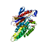
| Entry | Database: EMDB / ID: EMD-5011 | |||||||||
|---|---|---|---|---|---|---|---|---|---|---|
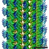
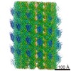
| Title | Crosslinked kinesin head-tail complex bound to the microtubule | |||||||||
 Map data Map data | null | |||||||||
 Sample Sample |
| |||||||||
 Keywords Keywords | kinesin / microtubule / tail domain / cargo / switch I / regulation | |||||||||
| Function / homology | kinesin complex / Kinesin motor domain / tubulin complex / Alpha tubulin / Beta tubulin Function and homology information Function and homology information | |||||||||
| Method | helical reconstruction / cryo EM / Resolution: 8.0 Å | |||||||||
 Authors Authors | Dietrich KA / Sindelar CV / Brewer PD / Downing KH / Cremo CR / Rice SE | |||||||||
 Citation Citation | Journal: Proc Natl Acad Sci U S A / Year: 2008 Title: The kinesin-1 motor protein is regulated by a direct interaction of its head and tail. Authors: Kristen A Dietrich / Charles V Sindelar / Paul D Brewer / Kenneth H Downing / Christine R Cremo / Sarah E Rice /  Abstract: Kinesin-1 is a molecular motor protein that transports cargo along microtubules. Inside cells, the vast majority of kinesin-1 is regulated to conserve ATP and to ensure its proper intracellular ...Kinesin-1 is a molecular motor protein that transports cargo along microtubules. Inside cells, the vast majority of kinesin-1 is regulated to conserve ATP and to ensure its proper intracellular distribution and coordination with other molecular motors. Regulated kinesin-1 folds in half at a hinge in its coiled-coil stalk. Interactions between coiled-coil regions near the enzymatically active heads at the N terminus and the regulatory tails at the C terminus bring these globular elements in proximity and stabilize the folded conformation. However, it has remained a mystery how kinesin-1's microtubule-stimulated ATPase activity is regulated in this folded conformation. Here, we present evidence for a direct interaction between the kinesin-1 head and tail. We photochemically cross-linked heads and tails and produced an 8-A cryoEM reconstruction of the cross-linked head-tail complex on microtubules. These data demonstrate that a conserved essential regulatory element in the kinesin-1 tail interacts directly and specifically with the enzymatically critical Switch I region of the head. This interaction suggests a mechanism for tail-mediated regulation of the ATPase activity of kinesin-1. In our structure, the tail makes simultaneous contacts with the kinesin-1 head and the microtubule, suggesting the tail may both regulate kinesin-1 in solution and hold it in a paused state with high ADP affinity on microtubules. The interaction of the Switch I region of the kinesin-1 head with the tail is strikingly similar to the interactions of small GTPases with their regulators, indicating that other kinesin motors may share similar regulatory mechanisms. | |||||||||
| History |
|
- Structure visualization
| Movie |
Movie viewer |
|---|---|
| Structure viewer | EM map: SurfViewMolmilJmol/JSmol |
| Supplemental images |

- Downloads & links
-EMDB archive
| Map data | emd_5011.map.gz | 15.5 MB | EMDB map data format | |
|---|---|---|---|---|
| Header (meta data) | emd-5011-v30.xmlemd-5011.xml | 14.1 KB 14.1 KB | Display Display | EMDB header |
| Images | emd_5011_1.tif | 732.7 KB | ||
| Archive directory |  http://ftp.pdbj.org/pub/emdb/structures/EMD-5011ftp://ftp.pdbj.org/pub/emdb/structures/EMD-5011 http://ftp.pdbj.org/pub/emdb/structures/EMD-5011ftp://ftp.pdbj.org/pub/emdb/structures/EMD-5011 | HTTPS FTP |
-Validation report
| Summary document | emd_5011_validation.pdf.gz | 78.6 KB | Display | EMDB validaton report |
|---|---|---|---|---|
| Full document | emd_5011_full_validation.pdf.gz | 77.8 KB | Display | |
| Data in XML | emd_5011_validation.xml.gz | 492 B | Display | |
| Arichive directory | https://ftp.pdbj.org/pub/emdb/validation_reports/EMD-5011ftp://ftp.pdbj.org/pub/emdb/validation_reports/EMD-5011 | HTTPS FTP |
-Related structure data
| Similar structure data |
|---|


-Links
| EMDB pages | EMDB (EBI/PDBe) / EMDataResource |
|---|
-Map
| File | Download / File: emd_5011.map.gz / Format: CCP4 / Size: 21.7 MB / Type: IMAGE STORED AS FLOATING POINT NUMBER (4 BYTES) | ||||||||||||||||||||||||||||||||||||||||||||||||||||||||||||||||||||
|---|---|---|---|---|---|---|---|---|---|---|---|---|---|---|---|---|---|---|---|---|---|---|---|---|---|---|---|---|---|---|---|---|---|---|---|---|---|---|---|---|---|---|---|---|---|---|---|---|---|---|---|---|---|---|---|---|---|---|---|---|---|---|---|---|---|---|---|---|---|
| Annotation | null | ||||||||||||||||||||||||||||||||||||||||||||||||||||||||||||||||||||
| Projections & slices | Image control
Images are generated by Spider. | ||||||||||||||||||||||||||||||||||||||||||||||||||||||||||||||||||||
| Voxel size | X=Y=Z: 2 Å | ||||||||||||||||||||||||||||||||||||||||||||||||||||||||||||||||||||
| Density |
| ||||||||||||||||||||||||||||||||||||||||||||||||||||||||||||||||||||
| Symmetry | Space group: 1 | ||||||||||||||||||||||||||||||||||||||||||||||||||||||||||||||||||||
| Details | EMDB XML:
CCP4 map header:
| ||||||||||||||||||||||||||||||||||||||||||||||||||||||||||||||||||||
 Z (Sec.)
 Y (Row.)
 X (Col.)


-Supplemental data
- Sample components
-Entire : Monomeric kinesin-1 head domain crosslinked in trans to dimerized...
| Entire | Name: Monomeric kinesin-1 head domain crosslinked in trans to dimerized kinesin-1 tail domain, bound to the microtubule |
|---|---|
| Components |
|
-Supramolecule #1000: Monomeric kinesin-1 head domain crosslinked in trans to dimerized...
| Supramolecule | Name: Monomeric kinesin-1 head domain crosslinked in trans to dimerized kinesin-1 tail domain, bound to the microtubule type: sample / ID: 1000 Oligomeric state: One head-tail complex binds to one tubulin alpha-beta dimer Number unique components: 3 |
|---|---|
| Molecular weight | Theoretical: 148 KDa |
-Macromolecule #1: microtubule
| Macromolecule | Name: microtubule / type: protein_or_peptide / ID: 1 / Name.synonym: microtubule Details: 13-protofilament microtubules with an asymmetric seam were selected for image processing Oligomeric state: quasi-helical assembly / Recombinant expression: No / Database: NCBI |
|---|---|
| Source (natural) | Tissue: brain / Cell: cow / Location in cell: cytoplasm |
| Molecular weight | Theoretical: 800 KDa |
| Sequence | GO: tubulin complex / InterPro: Alpha tubulin, Beta tubulin |
-Macromolecule #2: kinesin tail domain
| Macromolecule | Name: kinesin tail domain / type: protein_or_peptide / ID: 2 / Name.synonym: kinesin / Details: residues 823-944 of kinesin-1 / Oligomeric state: Dimer / Recombinant expression: Yes |
|---|---|
| Source (natural) | Cell: BL21 / Location in cell: cytoplasm |
| Molecular weight | Theoretical: 13 KDa |
| Recombinant expression | Organism:  |
| Sequence | GO: kinesin complex / InterPro: Kinesin motor domain |
-Macromolecule #3: human kinesin monomeric construct cys-lite K349
| Macromolecule | Name: human kinesin monomeric construct cys-lite K349 / type: protein_or_peptide / ID: 3 / Name.synonym: kinesin / Oligomeric state: monomer / Recombinant expression: Yes |
|---|---|
| Source (natural) | Cell: BL21 / Location in cell: cytoplasm |
| Molecular weight | Theoretical: 50 MDa |
| Recombinant expression | Organism: |
| Sequence | GO: kinesin complex / InterPro: Kinesin motor domain |
-Experimental details
-Structure determination
| Method | cryo EM |
|---|---|
 Processing Processing | helical reconstruction |
| Aggregation state | filament |
-Sample preparation
| Concentration | 1 mg/mL |
|---|---|
| Buffer | pH: 6.8 Details: 2.5 mM PIPES, 5 mM NaCl, 2 mM MgCl2, 1 mM EGTA, 5 mM Imidazole, 5 mM BME, and 40 uM ADP |
| Grid | Details: 300 mesh copper grid with homemade holey carbon |
| Vitrification | Cryogen name: ETHANE / Chamber temperature: 103 K / Instrument: HOMEMADE PLUNGER Details: Vitrification instrument: homemade. Vitrification carried out under ambient conditions Method: excess solution wicked away from grid before blotting and plunging immediately. Less than 0.5 seconds elapsed between blotting and plunging. |
- Electron microscopy
| Microscope | JEOL 4000EX |
|---|---|
| Temperature | Min: 98 K / Max: 108 K / Average: 100 K |
| Alignment procedure | Legacy - Astigmatism: objective lens astigmatism was approximately corrected at 400,000 times magnification |
| Date | Sep 1, 2007 |
| Image recording | Category: FILM / Film or detector model: KODAK SO-163 FILM / Digitization - Scanner: NIKON SUPER COOLSCAN 9000 / Digitization - Sampling interval: 6.3 µm / Number real images: 347 / Average electron dose: 16 e/Å2 / Bits/pixel: 16 |
| Electron beam | Acceleration voltage: 400 kV / Electron source: LAB6 |
| Electron optics | Illumination mode: FLOOD BEAM / Imaging mode: BRIGHT FIELD / Cs: 4.1 mm / Nominal defocus max: 1.8 µm / Nominal defocus min: 0.7 µm / Nominal magnification: 60000 |
| Sample stage | Specimen holder: Eucentric / Specimen holder model: GATAN LIQUID NITROGEN |
-Image processing
| Final reconstruction | Algorithm: OTHER / Resolution.type: BY AUTHOR / Resolution: 8.0 Å / Resolution method: OTHER / Software - Name: SPIDER Details: image defocus and astigmatism were determined by ctffind3 |
|---|---|
| CTF correction | Details: Integrated with fourier inversion reconstruction in the manner of FREALIGN by a customized c program |
| Final angle assignment | Details: SPIDER |
-Atomic model buiding 1
| Initial model | PDB ID: |
|---|---|
| Software | Name:  UCSF Chimera UCSF Chimera |
| Details | Protocol: Rigid Body. the menu option Fit Model in Map from UCSF Chimera was used to generate the fit |
| Refinement | Space: REAL / Protocol: RIGID BODY FIT / Target criteria: real-space density |
-Atomic model buiding 2
| Initial model | PDB ID: |
|---|---|
| Software | Name: UCSF Chimera |
| Details | Protocol: Rigid Body. the menu option Fit Model in Map from UCSF Chimera was used to generate the fit |
| Refinement | Space: REAL / Protocol: RIGID BODY FIT / Target criteria: real-space density |

