ムービー
ムービー コントローラー


+ データを開く

- 基本情報
基本情報
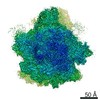
| 登録情報 | データベース: EMDB / ID: EMD-4935 | |||||||||
|---|---|---|---|---|---|---|---|---|---|---|
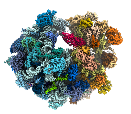
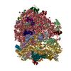
| タイトル | Evolutionary compaction and adaptation visualized by the structure of the dormant microsporidian ribosome | |||||||||
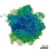
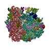
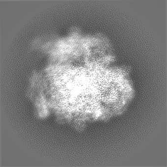
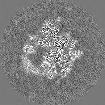
 マップデータ マップデータ | Overall map, state1 | |||||||||
 試料 試料 |
| |||||||||
 キーワード キーワード | Microsporidia / Ribosome / Intracellular Parasite | |||||||||
| 生物種 |  Vairimorpha necatrix (菌類) Vairimorpha necatrix (菌類) | |||||||||
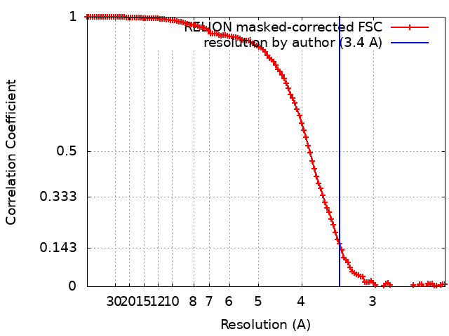
| 手法 | 単粒子再構成法 / クライオ電子顕微鏡法 / 解像度: 3.4 Å | |||||||||
 データ登録者 データ登録者 | Barandun J / Hunziker M | |||||||||
| 資金援助 |  米国, 米国,  スイス, 2件 スイス, 2件
| |||||||||
 引用 引用 | ジャーナル: Nat Microbiol / 年: 2019 タイトル: Evolutionary compaction and adaptation visualized by the structure of the dormant microsporidian ribosome. 著者: Jonas Barandun / Mirjam Hunziker / Charles R Vossbrinck / Sebastian Klinge /  要旨: Microsporidia are eukaryotic parasites that infect essentially all animal species, including many of agricultural importance, and are significant opportunistic parasites of humans. They are ...Microsporidia are eukaryotic parasites that infect essentially all animal species, including many of agricultural importance, and are significant opportunistic parasites of humans. They are characterized by having a specialized infection apparatus, an obligate intracellular lifestyle, rudimentary mitochondria and the smallest known eukaryotic genomes. Extreme genome compaction led to minimal gene sizes affecting even conserved ancient complexes such as the ribosome. In the present study, the cryo-electron microscopy structure of the ribosome from the microsporidium Vairimorpha necatrix is presented, which illustrates how genome compaction has resulted in the smallest known eukaryotic cytoplasmic ribosome. Selection pressure led to the loss of two ribosomal proteins and removal of essentially all eukaryote-specific ribosomal RNA (rRNA) expansion segments, reducing the rRNA to a functionally conserved core. The structure highlights how one microsporidia-specific and several repurposed existing ribosomal proteins compensate for the extensive rRNA reduction. The microsporidian ribosome is kept in an inactive state by two previously uncharacterized dormancy factors that specifically target the functionally important E-site, P-site and polypeptide exit tunnel. The present study illustrates the distinct effects of evolutionary pressure on RNA and protein-coding genes, provides a mechanism for ribosome inhibition and can serve as a structural basis for the development of inhibitors against microsporidian parasites. | |||||||||
| 履歴 |
|
- 構造の表示
構造の表示
| ムービー |
 ムービービューア ムービービューア |
|---|---|
| 構造ビューア | EMマップ: SurfViewMolmilJmol/JSmol |
| 添付画像 |
- ダウンロードとリンク
-EMDBアーカイブ
| マップデータ | emd_4935.map.gz | 137.2 MB | EMDBマップデータ形式 | |
|---|---|---|---|---|
| ヘッダ (付随情報) | emd-4935-v30.xmlemd-4935.xml | 89.2 KB 89.2 KB | 表示 表示 | EMDBヘッダ |
| FSC (解像度算出) | emd_4935_fsc.xml | 12.1 KB | 表示 | FSCデータファイル |
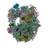
| 画像 |  emd_4935.png emd_4935.png | 270.4 KB | ||
| Filedesc metadata | emd-4935.cif.gz | 17.6 KB | ||
| その他 | emd_4935_additional.map.gz | 137 MB | ||
| アーカイブディレクトリ |  http://ftp.pdbj.org/pub/emdb/structures/EMD-4935ftp://ftp.pdbj.org/pub/emdb/structures/EMD-4935 http://ftp.pdbj.org/pub/emdb/structures/EMD-4935ftp://ftp.pdbj.org/pub/emdb/structures/EMD-4935 | HTTPS FTP |
-検証レポート
| 文書・要旨 | emd_4935_validation.pdf.gz | 673.8 KB | 表示 | EMDB検証レポート |
|---|---|---|---|---|
| 文書・詳細版 | emd_4935_full_validation.pdf.gz | 673.4 KB | 表示 | |
| XML形式データ | emd_4935_validation.xml.gz | 12.6 KB | 表示 | |
| CIF形式データ | emd_4935_validation.cif.gz | 17 KB | 表示 | |
| アーカイブディレクトリ | https://ftp.pdbj.org/pub/emdb/validation_reports/EMD-4935ftp://ftp.pdbj.org/pub/emdb/validation_reports/EMD-4935 | HTTPS FTP |
-関連構造データ
| 関連構造データ | 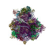 6rm3MC 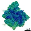 4931C 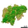 4932C  4933C 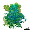 4934C C: 同じ文献を引用 ( M: このマップから作成された原子モデル |
|---|---|
| 類似構造データ | |
| 電子顕微鏡画像生データ | EMPIAR-11075 (タイトル: Single-particle cryo-EM dataset of the Vairimorpha necatrix ribosome Data size: 1.0 TB Data #1: Unaligned multi-frame micrographs of the Vairimorpha necatrix ribosome [micrographs - multiframe]) |


-リンク
| EMDBのページ | EMDB (EBI/PDBe) / EMDataResource |
|---|---|
| 「今月の分子」の関連する項目 |

-マップ
| ファイル | ダウンロード / ファイル: emd_4935.map.gz / 形式: CCP4 / 大きさ: 149.9 MB / タイプ: IMAGE STORED AS FLOATING POINT NUMBER (4 BYTES) | ||||||||||||||||||||||||||||||||||||||||||||||||||||||||||||||||||||
|---|---|---|---|---|---|---|---|---|---|---|---|---|---|---|---|---|---|---|---|---|---|---|---|---|---|---|---|---|---|---|---|---|---|---|---|---|---|---|---|---|---|---|---|---|---|---|---|---|---|---|---|---|---|---|---|---|---|---|---|---|---|---|---|---|---|---|---|---|---|
| 注釈 | Overall map, state1 | ||||||||||||||||||||||||||||||||||||||||||||||||||||||||||||||||||||
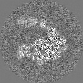
| 投影像・断面図 | 画像のコントロール
画像は Spider により作成 | ||||||||||||||||||||||||||||||||||||||||||||||||||||||||||||||||||||
| ボクセルのサイズ | X=Y=Z: 1.2 Å | ||||||||||||||||||||||||||||||||||||||||||||||||||||||||||||||||||||
| 密度 |
| ||||||||||||||||||||||||||||||||||||||||||||||||||||||||||||||||||||
| 対称性 | 空間群: 1 | ||||||||||||||||||||||||||||||||||||||||||||||||||||||||||||||||||||
| 詳細 | EMDB XML:
CCP4マップ ヘッダ情報:
| ||||||||||||||||||||||||||||||||||||||||||||||||||||||||||||||||||||
 Z (Sec.)
 Y (Row.)
 X (Col.)


-添付データ
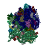
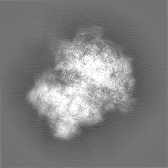
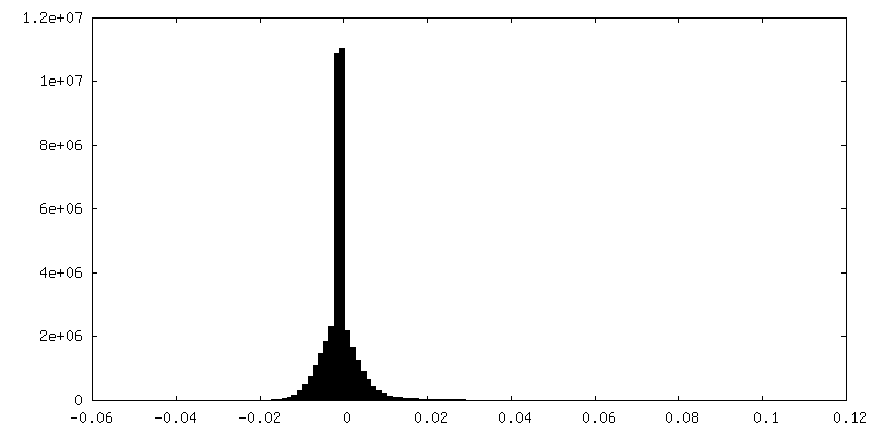
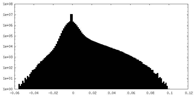
-追加マップ: Additional map, state2
| ファイル | emd_4935_additional.map | ||||||||||||
|---|---|---|---|---|---|---|---|---|---|---|---|---|---|
| 注釈 | Additional map, state2 | ||||||||||||
| 投影像・断面図 |
| ||||||||||||
| 密度ヒストグラム |

- 試料の構成要素
+全体 : Microsporidian Ribosome
+超分子 #1: Microsporidian Ribosome
+分子 #1: 16S rRNA
+分子 #2: 5S rRNA
+分子 #78: 23S rRNA
+分子 #3: uL2
+分子 #4: uS2
+分子 #5: uL15
+分子 #6: eS26
+分子 #7: uL3
+分子 #8: eS1
+分子 #9: eL29
+分子 #10: eS27
+分子 #11: uL4
+分子 #12: uS5
+分子 #13: eL30
+分子 #14: eS28
+分子 #15: uL18
+分子 #16: uS3
+分子 #17: eL31
+分子 #18: uS14
+分子 #19: eL6
+分子 #20: eS4
+分子 #21: eL32
+分子 #22: eS30
+分子 #23: uL30
+分子 #24: uS7
+分子 #25: eL33
+分子 #26: eS31
+分子 #27: eL8
+分子 #28: eS6
+分子 #29: eL34
+分子 #30: RACK1
+分子 #31: uL6
+分子 #32: eS7
+分子 #33: uL29
+分子 #34: uL16
+分子 #35: eS8
+分子 #36: eL36
+分子 #37: uL5
+分子 #38: uS4
+分子 #39: eL37
+分子 #40: eL13
+分子 #41: uS17
+分子 #42: eL39
+分子 #43: eL14
+分子 #44: eL40
+分子 #45: eS12
+分子 #46: eL15
+分子 #47: uS15
+分子 #48: MDF2
+分子 #49: MDF1
+分子 #50: uL13
+分子 #51: uS11
+分子 #52: eL42
+分子 #53: uL22
+分子 #54: uS19
+分子 #55: eL43
+分子 #56: eL18
+分子 #57: uS9
+分子 #58: eL19
+分子 #59: eS17
+分子 #60: eL20
+分子 #61: uS13
+分子 #62: eL21
+分子 #63: eS19
+分子 #64: eL22
+分子 #65: uS10
+分子 #66: uL14
+分子 #67: eS21
+分子 #68: eL24
+分子 #69: uS8
+分子 #70: uL23
+分子 #71: uS12
+分子 #72: msL1
+分子 #73: uL24
+分子 #74: eS24
+分子 #75: eL27
+分子 #76: eS25
+分子 #77: eS10
+分子 #79: MAGNESIUM ION
+分子 #80: ZINC ION
-実験情報
-構造解析
| 手法 | クライオ電子顕微鏡法 |
|---|---|
 解析 解析 | 単粒子再構成法 |
| 試料の集合状態 | particle |
-試料調製
| 濃度 | 11 mg/mL | ||||||||||||
|---|---|---|---|---|---|---|---|---|---|---|---|---|---|
| 緩衝液 | pH: 7.5 構成要素:
| ||||||||||||
| グリッド | 材質: COPPER / メッシュ: 400 / 支持フィルム - 材質: CARBON / 支持フィルム - トポロジー: CONTINUOUS / 支持フィルム - Film thickness: 2 / 前処理 - タイプ: GLOW DISCHARGE / 前処理 - 時間: 30 sec. / 前処理 - 雰囲気: AIR / 詳細: 50 mA | ||||||||||||
| 凍結 | 凍結剤: ETHANE / チャンバー内湿度: 100 % / チャンバー内温度: 295 K / 装置: FEI VITROBOT MARK IV |
- 電子顕微鏡法
| 顕微鏡 | FEI TALOS ARCTICA |
|---|---|
| 撮影 | フィルム・検出器のモデル: GATAN K2 SUMMIT (4k x 4k) 検出モード: SUPER-RESOLUTION / 撮影したグリッド数: 2 / 実像数: 3284 / 平均露光時間: 0.25 sec. / 平均電子線量: 5.55 e/Å2 |
| 電子線 | 加速電圧: 200 kV / 電子線源:  FIELD EMISSION GUN FIELD EMISSION GUN |
| 電子光学系 | 最小 デフォーカス(補正後): 4.0 µm / 照射モード: FLOOD BEAM / 撮影モード: BRIGHT FIELD / 最小 デフォーカス(公称値): 0.5 µm |
| 試料ステージ | 試料ホルダーモデル: FEI TITAN KRIOS AUTOGRID HOLDER ホルダー冷却材: NITROGEN |
| 実験機器 |  モデル: Talos Arctica / 画像提供: FEI Company |