 Movie
Movie Controller

[English] 日本語
 Yorodumi
- EMDB-48829: Structure of a native Drosophila melanogaster Nucleosome Elongati... -
+ Open data

- Basic information
| Entry |  | ||||||||||||
|---|---|---|---|---|---|---|---|---|---|---|---|---|---|
| Title | Structure of a native Drosophila melanogaster Nucleosome Elongation Complex (Pol II EC-nucleosome). Focused refinement of Pol II | ||||||||||||
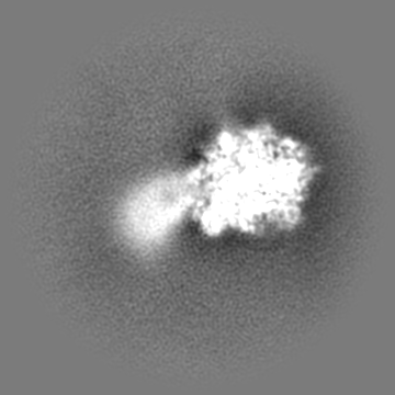
 Map data Map data | Locally refined, unsharpened Pol II from the Nucleosome Elongation Complex, resolved to 4.3 A | ||||||||||||
 Sample Sample |
| ||||||||||||
 Keywords Keywords | polymerase / Pol II / transcription / mRNA | ||||||||||||
| Biological species |  | ||||||||||||
| Method | single particle reconstruction / cryo EM / Resolution: 4.31 Å | ||||||||||||
 Authors Authors | Venette-Smith NL / Vishwakarma RK / Dollinger R / Schultz J / Venkatakrishnan V / Babitzke P / Anand G / Gilmour DS / Armache J-P / Murakami K | ||||||||||||
| Funding support |  United States, 3 items United States, 3 items
| ||||||||||||
 Citation Citation | Journal: To Be Published Title: Structural Characterization of Native RNA Polymerase II Transcription Complexes in Drosophila melanogaster Authors: Venette-Smith NL / Vishwakarma RK / Dollinger R / Schultz J / Venkatakrishnan V / Babitzke P / Anand G / Gilmour DS / Armache J-P / Murakami K | ||||||||||||
| History |
|
- Structure visualization
| Supplemental images |
|---|

- Downloads & links
-EMDB archive
| Map data | emd_48829.map.gz | 88.7 MB |  EMDB map data format EMDB map data format | |
|---|---|---|---|---|
| Header (meta data) | emd-48829-v30.xmlemd-48829.xml | 26 KB 26 KB | Display Display | EMDB header |
| FSC (resolution estimation) | emd_48829_fsc.xml | 11.9 KB | Display | FSC data file |
| Images |  emd_48829.png emd_48829.png | 28.5 KB | ||
| Filedesc metadata | emd-48829.cif.gz | 5.8 KB | ||
| Others | emd_48829_additional_1.map.gzemd_48829_half_map_1.map.gzemd_48829_half_map_2.map.gz | 168.2 MB 165.3 MB 165.3 MB | ||
| Archive directory |  http://ftp.pdbj.org/pub/emdb/structures/EMD-48829ftp://ftp.pdbj.org/pub/emdb/structures/EMD-48829 http://ftp.pdbj.org/pub/emdb/structures/EMD-48829ftp://ftp.pdbj.org/pub/emdb/structures/EMD-48829 | HTTPS FTP |
-Related structure data


-Links
| EMDB pages | EMDB (EBI/PDBe) / EMDataResource |
|---|
-Map
| File | Download / File: emd_48829.map.gz / Format: CCP4 / Size: 178 MB / Type: IMAGE STORED AS FLOATING POINT NUMBER (4 BYTES) | ||||||||||||||||||||||||||||||||||||
|---|---|---|---|---|---|---|---|---|---|---|---|---|---|---|---|---|---|---|---|---|---|---|---|---|---|---|---|---|---|---|---|---|---|---|---|---|---|
| Annotation | Locally refined, unsharpened Pol II from the Nucleosome Elongation Complex, resolved to 4.3 A | ||||||||||||||||||||||||||||||||||||
| Projections & slices | Image control
Images are generated by Spider. | ||||||||||||||||||||||||||||||||||||
| Voxel size | X=Y=Z: 1.1538 Å | ||||||||||||||||||||||||||||||||||||
| Density |
| ||||||||||||||||||||||||||||||||||||
| Symmetry | Space group: 1 | ||||||||||||||||||||||||||||||||||||
| Details | EMDB XML:
|
 Z (Sec.)
 Y (Row.)
 X (Col.)


-Supplemental data
-Additional map: Locally refined, sharpened Pol II from the Nucleosome...
| File | emd_48829_additional_1.map | ||||||||||||
|---|---|---|---|---|---|---|---|---|---|---|---|---|---|
| Annotation | Locally refined, sharpened Pol II from the Nucleosome Elongation Complex, resolved to 4.3 A (bfactor = -84.7) | ||||||||||||
| Projections & Slices |
| ||||||||||||
| Density Histograms |


-Half map: Half map 1 from cryoSPARC
| File | emd_48829_half_map_1.map | ||||||||||||
|---|---|---|---|---|---|---|---|---|---|---|---|---|---|
| Annotation | Half map 1 from cryoSPARC | ||||||||||||
| Projections & Slices |
| ||||||||||||
| Density Histograms |


-Half map: Half map 2 from cryoSPARC
| File | emd_48829_half_map_2.map | ||||||||||||
|---|---|---|---|---|---|---|---|---|---|---|---|---|---|
| Annotation | Half map 2 from cryoSPARC | ||||||||||||
| Projections & Slices |
| ||||||||||||
| Density Histograms |


- Sample components
-Entire : Native purified Nucleosome Elongation Complex from Drosophila mel...
| Entire | Name: Native purified Nucleosome Elongation Complex from Drosophila melanogaster |
|---|---|
| Components |
|
-Supramolecule #1: Native purified Nucleosome Elongation Complex from Drosophila mel...
| Supramolecule | Name: Native purified Nucleosome Elongation Complex from Drosophila melanogaster type: complex / ID: 1 / Parent: 0 / Macromolecule list: #1-#15 Details: An overall structure of a native purified Nucleosome Elongation Complex (Pol II EC - nucleosome), where the Pol II and nucleosome are both at a lower resolution due to their motion in respect to each other |
|---|---|
| Source (natural) | Organism: |
| Molecular weight | Theoretical: 0.750 kDa/nm |
-Experimental details
-Structure determination
| Method | cryo EM |
|---|---|
 Processing Processing | single particle reconstruction |
| Aggregation state | particle |
-Sample preparation
| Concentration | 1.0 mg/mL | ||||||||||||||||||
|---|---|---|---|---|---|---|---|---|---|---|---|---|---|---|---|---|---|---|---|
| Buffer | pH: 7.5 Component:
Details: 10 mM HEPES-HCl (pH = 7.5), 150 mM NaCl, 5% glycerol, 1 mM EDTA, 350 ug/mL 3x FLAG peptide, 1/1000th protease inhibitor | ||||||||||||||||||
| Grid | Model: Quantifoil / Material: GOLD / Mesh: 200 / Support film - Material: GOLD / Support film - topology: HOLEY / Pretreatment - Type: GLOW DISCHARGE / Pretreatment - Time: 10 sec. | ||||||||||||||||||
| Vitrification | Cryogen name: ETHANE / Chamber humidity: 100 % / Chamber temperature: 4 K / Instrument: FEI VITROBOT MARK IV | ||||||||||||||||||
| Details | double FLAG-tagged Pol II subunit Rpb1 was used for purification of Pol II complexes |
- Electron microscopy
| Microscope | TFS TALOS |
|---|---|
| Image recording | Film or detector model: FEI FALCON IV (4k x 4k) / Digitization - Dimensions - Width: 4096 pixel / Digitization - Dimensions - Height: 4096 pixel / Number grids imaged: 4 / Number real images: 31103 / Average electron dose: 50.65 e/Å2 Details: Although the data was collected, motion-corrected and CTF estimated using the pixel size of 0.944, all the subsequent data processing was performed using pixel size of 1.1538. This was ...Details: Although the data was collected, motion-corrected and CTF estimated using the pixel size of 0.944, all the subsequent data processing was performed using pixel size of 1.1538. This was achieved by extracting particles in box size 440x440 and Fourier-cropping it to box size 360x360 |
| Electron beam | Acceleration voltage: 200 kV / Electron source:  FIELD EMISSION GUN FIELD EMISSION GUN |
| Electron optics | Illumination mode: FLOOD BEAM / Imaging mode: BRIGHT FIELD / Nominal defocus max: 2.5 µm / Nominal defocus min: 1.0 µm |
| Sample stage | Specimen holder model: FEI TITAN KRIOS AUTOGRID HOLDER / Cooling holder cryogen: NITROGEN |
+Image processing

-Atomic model buiding 1
| Refinement | Overall B value: 50 |
|---|
