 Movie
Movie Controller

[English] 日本語
 Yorodumi
- EMDB-48619: Structure of a native Drosophila melanogaster octameric nucleosome -
+ Open data

- Basic information
| Entry |  | ||||||||||||
|---|---|---|---|---|---|---|---|---|---|---|---|---|---|
| Title | Structure of a native Drosophila melanogaster octameric nucleosome | ||||||||||||
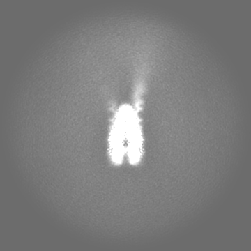
 Map data Map data | Refined, unsharpened Coulomb potential density map. Particles extracted in 440x440 Fourier cropped to 360x360. Map refined in cryoSPARC | ||||||||||||
 Sample Sample |
| ||||||||||||
 Keywords Keywords | Nucleosome / histones / histone / chromatin / DNA / GENE REGULATION | ||||||||||||
| Function / homology |  Function and homology information Function and homology informationHDMs demethylate histones / PKMTs methylate histone lysines / Interleukin-7 signaling / Chromatin modifying enzymes / : / SUMOylation of chromatin organization proteins / Metalloprotease DUBs / E3 ubiquitin ligases ubiquitinate target proteins / Factors involved in megakaryocyte development and platelet production / RCAF complex ...HDMs demethylate histones / PKMTs methylate histone lysines / Interleukin-7 signaling / Chromatin modifying enzymes / : / SUMOylation of chromatin organization proteins / Metalloprotease DUBs / E3 ubiquitin ligases ubiquitinate target proteins / Factors involved in megakaryocyte development and platelet production / RCAF complex / RMTs methylate histone arginines / Recruitment and ATM-mediated phosphorylation of repair and signaling proteins at DNA double strand breaks / SIRT1 negatively regulates rRNA expression / NoRC negatively regulates rRNA expression / Activated PKN1 stimulates transcription of AR (androgen receptor) regulated genes KLK2 and KLK3 / polytene chromosome band / PRC2 methylates histones and DNA / HDACs deacetylate histones / Ub-specific processing proteases / Negative Regulation of CDH1 Gene Transcription / Formation of the beta-catenin:TCF transactivating complex / MLL4 and MLL3 complexes regulate expression of PPARG target genes in adipogenesis and hepatic steatosis / RNA Polymerase I Promoter Escape / Regulation of endogenous retroelements by KRAB-ZFP proteins / larval somatic muscle development / RUNX1 regulates genes involved in megakaryocyte differentiation and platelet function / Senescence-Associated Secretory Phenotype (SASP) / Transcriptional regulation by small RNAs / Estrogen-dependent gene expression / HATs acetylate histones / Assembly of the ORC complex at the origin of replication / Oxidative Stress Induced Senescence / UCH proteinases / polytene chromosome / nucleosomal DNA binding / nuclear chromosome / structural constituent of chromatin / nucleosome / nucleosome assembly / heterochromatin formation / chromosome / chromatin organization / protein heterodimerization activity / chromatin / protein-containing complex binding / DNA binding / nucleus Similarity search - Function | ||||||||||||
| Biological species |  | ||||||||||||
| Method | single particle reconstruction / cryo EM / Resolution: 3.29 Å | ||||||||||||
 Authors Authors | Venette-Smith NL / Vishwakarma RK / Dollinger R / Schultz J / Venkatakrishnan V / Babitzke P / Anand G / Gilmour DS / Armache J-P / Murakami K | ||||||||||||
| Funding support |  United States, 3 items United States, 3 items
| ||||||||||||
 Citation Citation | Journal: To Be Published Title: Structural Characterization of Native RNA Polymerase II Transcription Complexes in Drosophila melanogaster Authors: Venette-Smith NL / Vishwakarma RK / Dollinger R / Schultz J / Venkatakrishnan V / Babitzke P / Anand G / Gilmour DS / Armache J-P / Murakami K | ||||||||||||
| History |
|
- Structure visualization
| Supplemental images |
|---|

- Downloads & links
-EMDB archive
| Map data | emd_48619.map.gz | 49.5 MB | EMDB map data format | |
|---|---|---|---|---|
| Header (meta data) | emd-48619-v30.xmlemd-48619.xml | 31.3 KB 31.3 KB | Display Display | EMDB header |
| FSC (resolution estimation) | emd_48619_fsc.xml | 11.8 KB | Display | FSC data file |
| Images |  emd_48619.png emd_48619.png | 19.9 KB | ||
| Filedesc metadata | emd-48619.cif.gz | 8 KB | ||
| Others | emd_48619_additional_1.map.gzemd_48619_half_map_1.map.gzemd_48619_half_map_2.map.gz | 166.8 MB 165 MB 165 MB | ||
| Archive directory |  http://ftp.pdbj.org/pub/emdb/structures/EMD-48619ftp://ftp.pdbj.org/pub/emdb/structures/EMD-48619 http://ftp.pdbj.org/pub/emdb/structures/EMD-48619ftp://ftp.pdbj.org/pub/emdb/structures/EMD-48619 | HTTPS FTP |
-Related structure data
| Related structure data |  9mu4MC  9mu5C  9mu6C  9mu7C  9mu8C  9mu9C M: atomic model generated by this map C: citing same article ( |
|---|---|
| Similar structure data |


-Links
| EMDB pages | EMDB (EBI/PDBe) / EMDataResource |
|---|---|
| Related items in Molecule of the Month |


-Map
| File | Download / File: emd_48619.map.gz / Format: CCP4 / Size: 178 MB / Type: IMAGE STORED AS FLOATING POINT NUMBER (4 BYTES) | ||||||||||||||||||||||||||||||||||||
|---|---|---|---|---|---|---|---|---|---|---|---|---|---|---|---|---|---|---|---|---|---|---|---|---|---|---|---|---|---|---|---|---|---|---|---|---|---|
| Annotation | Refined, unsharpened Coulomb potential density map. Particles extracted in 440x440 Fourier cropped to 360x360. Map refined in cryoSPARC | ||||||||||||||||||||||||||||||||||||
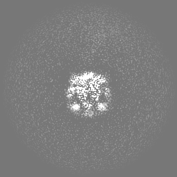
| Projections & slices | Image control
Images are generated by Spider. | ||||||||||||||||||||||||||||||||||||
| Voxel size | X=Y=Z: 1.1538 Å | ||||||||||||||||||||||||||||||||||||
| Density |
| ||||||||||||||||||||||||||||||||||||
| Symmetry | Space group: 1 | ||||||||||||||||||||||||||||||||||||
| Details | EMDB XML:
|
 Z (Sec.)
 Y (Row.)
 X (Col.)


-Supplemental data
-Additional map: Refined, auto-sharpened Coulomb potential density map with Bfactor...
| File | emd_48619_additional_1.map | ||||||||||||
|---|---|---|---|---|---|---|---|---|---|---|---|---|---|
| Annotation | Refined, auto-sharpened Coulomb potential density map with Bfactor = -50 | ||||||||||||
| Projections & Slices |
| ||||||||||||
| Density Histograms |


-Half map: Half map 1 from cryoSPARC
| File | emd_48619_half_map_1.map | ||||||||||||
|---|---|---|---|---|---|---|---|---|---|---|---|---|---|
| Annotation | Half map 1 from cryoSPARC | ||||||||||||
| Projections & Slices |
| ||||||||||||
| Density Histograms |


-Half map: Half map 2 from cryoSPARC
| File | emd_48619_half_map_2.map | ||||||||||||
|---|---|---|---|---|---|---|---|---|---|---|---|---|---|
| Annotation | Half map 2 from cryoSPARC | ||||||||||||
| Projections & Slices |
| ||||||||||||
| Density Histograms |


- Sample components
-Entire : Natively purified octameric nucleosome from Drosophila melanogaster
| Entire | Name: Natively purified octameric nucleosome from Drosophila melanogaster |
|---|---|
| Components |
|
-Supramolecule #1: Natively purified octameric nucleosome from Drosophila melanogaster
| Supramolecule | Name: Natively purified octameric nucleosome from Drosophila melanogaster type: complex / ID: 1 / Parent: 0 / Macromolecule list: all Details: This entry represents a native octameric nucleosome (i.e. containing two H2A/H2B dimers and two H3/H4 dimers), purified from Drosophila melanogaster embryos |
|---|---|
| Source (natural) | Organism: |
| Molecular weight | Theoretical: 240 kDa/nm |
-Macromolecule #1: Histone H2A
| Macromolecule | Name: Histone H2A / type: protein_or_peptide / ID: 1 / Number of copies: 2 / Enantiomer: LEVO |
|---|---|
| Source (natural) | Organism: |
| Molecular weight | Theoretical: 11.552494 KDa |
| Sequence | String: AKSRSNRAGL QFPVGRIHRL LRKGNYAERV GAGAPVYLAA VMEYLAAEVL ELAGNAARDN KKTRIIPRHL QLAIRNDEEL NKLLSGVTI AQGGVLPNIQ AVLLPKK UniProtKB: Histone H2A |
-Macromolecule #2: Histone H2B
| Macromolecule | Name: Histone H2B / type: protein_or_peptide / ID: 2 / Number of copies: 2 / Enantiomer: LEVO |
|---|---|
| Source (natural) | Organism: |
| Molecular weight | Theoretical: 10.979741 KDa |
| Sequence | String: KKRKRKESYA IYIYKVLKQV HPDTGISSKA MSIMNSFVND IFERIAAEAS RLAHYNKRST ITSREIQTAV RLLLPGELAK HAVSEGTKA VTKYTSSK UniProtKB: Histone H2B |
-Macromolecule #3: Histone H3
| Macromolecule | Name: Histone H3 / type: protein_or_peptide / ID: 3 / Number of copies: 2 / Enantiomer: LEVO |
|---|---|
| Source (natural) | Organism: |
| Molecular weight | Theoretical: 11.74677 KDa |
| Sequence | String: KKPHRYRPGT VALREIRRYQ KSTELLIRKL PFQRLVREIA QDFKTDLRFQ SSAVMALQEA SEAYLVGLFE DTNLCAIHAK RVTIMPKDI QLARRIRGER A UniProtKB: Histone H3 |
-Macromolecule #4: Histone H4
| Macromolecule | Name: Histone H4 / type: protein_or_peptide / ID: 4 / Number of copies: 2 / Enantiomer: LEVO |
|---|---|
| Source (natural) | Organism: |
| Molecular weight | Theoretical: 9.279875 KDa |
| Sequence | String: VLRDNIQGIT KPAIRRLARR GGVKRISGLI YEETRGVLKV FLENVIRDAV TYTEHAKRKT VTAMDVVYAL KRQGRTLYGF GG UniProtKB: Histone H4 |
-Macromolecule #5: DNA (164-MER)
| Macromolecule | Name: DNA (164-MER) / type: dna / ID: 5 / Number of copies: 1 / Classification: DNA |
|---|---|
| Source (natural) | Organism: |
| Molecular weight | Theoretical: 50.381172 KDa |
| Sequence | String: (DT)(DA)(DT)(DA)(DT)(DA)(DT)(DA)(DT)(DA) (DT)(DA)(DT)(DA)(DT)(DA)(DT)(DC)(DA)(DG) (DA)(DA)(DT)(DC)(DC)(DC)(DG)(DG)(DT) (DG)(DC)(DC)(DG)(DA)(DG)(DG)(DC)(DC)(DG) (DC) (DT)(DC)(DA)(DA)(DT)(DT) ...String: (DT)(DA)(DT)(DA)(DT)(DA)(DT)(DA)(DT)(DA) (DT)(DA)(DT)(DA)(DT)(DA)(DT)(DC)(DA)(DG) (DA)(DA)(DT)(DC)(DC)(DC)(DG)(DG)(DT) (DG)(DC)(DC)(DG)(DA)(DG)(DG)(DC)(DC)(DG) (DC) (DT)(DC)(DA)(DA)(DT)(DT)(DG)(DG) (DT)(DC)(DG)(DT)(DA)(DG)(DA)(DC)(DA)(DG) (DC)(DT) (DC)(DT)(DA)(DG)(DC)(DA)(DC) (DC)(DG)(DC)(DT)(DT)(DA)(DA)(DA)(DC)(DG) (DC)(DA)(DC) (DG)(DT)(DA)(DC)(DG)(DC) (DG)(DC)(DT)(DG)(DT)(DC)(DC)(DC)(DC)(DC) (DG)(DC)(DG)(DT) (DT)(DT)(DT)(DA)(DA) (DC)(DC)(DG)(DC)(DC)(DA)(DA)(DG)(DG)(DG) (DG)(DA)(DT)(DT)(DA) (DC)(DT)(DC)(DC) (DC)(DT)(DA)(DG)(DT)(DC)(DT)(DC)(DC)(DA) (DG)(DG)(DC)(DA)(DC)(DG) (DT)(DG)(DT) (DC)(DA)(DG)(DA)(DT)(DA)(DT)(DA)(DT)(DA) (DC)(DA)(DT)(DC)(DG)(DA)(DT) (DA)(DT) (DA)(DT) |
-Macromolecule #6: DNA (164-MER)
| Macromolecule | Name: DNA (164-MER) / type: dna / ID: 6 / Number of copies: 1 / Classification: DNA |
|---|---|
| Source (natural) | Organism: |
| Molecular weight | Theoretical: 50.861461 KDa |
| Sequence | String: (DA)(DT)(DA)(DT)(DA)(DT)(DC)(DG)(DA)(DT) (DG)(DT)(DA)(DT)(DA)(DT)(DA)(DT)(DC)(DT) (DG)(DA)(DC)(DA)(DC)(DG)(DT)(DG)(DC) (DC)(DT)(DG)(DG)(DA)(DG)(DA)(DC)(DT)(DA) (DG) (DG)(DG)(DA)(DG)(DT)(DA) ...String: (DA)(DT)(DA)(DT)(DA)(DT)(DC)(DG)(DA)(DT) (DG)(DT)(DA)(DT)(DA)(DT)(DA)(DT)(DC)(DT) (DG)(DA)(DC)(DA)(DC)(DG)(DT)(DG)(DC) (DC)(DT)(DG)(DG)(DA)(DG)(DA)(DC)(DT)(DA) (DG) (DG)(DG)(DA)(DG)(DT)(DA)(DA)(DT) (DC)(DC)(DC)(DC)(DT)(DT)(DG)(DG)(DC)(DG) (DG)(DT) (DT)(DA)(DA)(DA)(DA)(DC)(DG) (DC)(DG)(DG)(DG)(DG)(DG)(DA)(DC)(DA)(DG) (DC)(DG)(DC) (DG)(DT)(DA)(DC)(DG)(DT) (DG)(DC)(DG)(DT)(DT)(DT)(DA)(DA)(DG)(DC) (DG)(DG)(DT)(DG) (DC)(DT)(DA)(DG)(DA) (DG)(DC)(DT)(DG)(DT)(DC)(DT)(DA)(DC)(DG) (DA)(DC)(DC)(DA)(DA) (DT)(DT)(DG)(DA) (DG)(DC)(DG)(DG)(DC)(DC)(DT)(DC)(DG)(DG) (DC)(DA)(DC)(DC)(DG)(DG) (DG)(DA)(DT) (DT)(DC)(DT)(DG)(DA)(DT)(DA)(DT)(DA)(DT) (DA)(DT)(DA)(DT)(DA)(DT)(DA) (DT)(DA) (DT)(DA) |
-Experimental details
-Structure determination
| Method | cryo EM |
|---|---|
 Processing Processing | single particle reconstruction |
| Aggregation state | particle |
-Sample preparation
| Concentration | 1.0 mg/mL | ||||||||||||||||||
|---|---|---|---|---|---|---|---|---|---|---|---|---|---|---|---|---|---|---|---|
| Buffer | pH: 7.5 Component:
Details: 10 mM HEPES-HCl (pH = 7.5), 150 mM NaCl, 5% glycerol, 1 mM EDTA, 350 ug/mL 3x FLAG peptide, 1/1000th protease inhibitor | ||||||||||||||||||
| Grid | Model: Quantifoil / Material: GOLD / Mesh: 200 / Support film - Material: GOLD / Support film - topology: HOLEY / Pretreatment - Type: GLOW DISCHARGE / Pretreatment - Time: 10 sec. | ||||||||||||||||||
| Vitrification | Cryogen name: ETHANE / Chamber humidity: 100 % / Chamber temperature: 4 K / Instrument: FEI VITROBOT MARK IV | ||||||||||||||||||
| Details | double FLAG-tagged Pol II subunit Rpb1 was used for purification of Pol II complexes. We aimed to purify Pol II in tandem with nucleosomes. The free nucleosomes were co-purified with this sample |
- Electron microscopy
| Microscope | TFS TALOS |
|---|---|
| Image recording | Film or detector model: FEI FALCON IV (4k x 4k) / Digitization - Dimensions - Width: 4096 pixel / Digitization - Dimensions - Height: 4096 pixel / Number grids imaged: 4 / Number real images: 31103 / Average electron dose: 50.65 e/Å2 Details: Although the data was collected, motion-corrected and CTF estimated using the pixel size of 0.944, all the subsequent data processing was performed using pixel size of 1.1538. This was ...Details: Although the data was collected, motion-corrected and CTF estimated using the pixel size of 0.944, all the subsequent data processing was performed using pixel size of 1.1538. This was achieved by extracting particles in box size 440x440 and Fourier-cropping it to box size 360x360 |
| Electron beam | Acceleration voltage: 200 kV / Electron source:  FIELD EMISSION GUN FIELD EMISSION GUN |
| Electron optics | Illumination mode: FLOOD BEAM / Imaging mode: BRIGHT FIELD / Nominal defocus max: 2.5 µm / Nominal defocus min: 1.0 µm |
| Sample stage | Specimen holder model: FEI TITAN KRIOS AUTOGRID HOLDER / Cooling holder cryogen: NITROGEN |
+Image processing

-Atomic model buiding 1
| Initial model |
| ||||||||||||||||||||
|---|---|---|---|---|---|---|---|---|---|---|---|---|---|---|---|---|---|---|---|---|---|
| Details | The model was based on PDB: 3LZ0. First, 3LZ0 was rigid-body fit in the cryo-EM Coulomb potential density map using UCSF ChimeraX. Next, AlphaFold2 models for individual Drosophila melanogaster histones were aligned to their 3LZ0 counterparts, and further optimized in the density using rigid-body fit. Then, in Coot, the models were Real-space refined in conjunction with DNA 601 Widom sequence. The DNA sequence was extended, to fit into the density. For the final model optimization, phenix.real_space_refine was used | ||||||||||||||||||||
| Refinement | Protocol: AB INITIO MODEL / Overall B value: 50 | ||||||||||||||||||||
| Output model | PDB-9mu4: |


