 ムービー
ムービー コントローラー


+ データを開く

- 基本情報
基本情報
| 登録情報 |  | ||||||||||||||||||
|---|---|---|---|---|---|---|---|---|---|---|---|---|---|---|---|---|---|---|---|
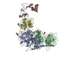
| タイトル | Cryo-EM structure of factor Va bound to activated protein C | ||||||||||||||||||
 マップデータ マップデータ | |||||||||||||||||||
 試料 試料 |
| ||||||||||||||||||
 キーワード キーワード | Coagulation / Activated Factor V / Activated Protein C / BLOOD CLOTTING | ||||||||||||||||||
| 機能・相同性 |  機能・相同性情報 機能・相同性情報activated protein C (thrombin-activated peptidase) / positive regulation of establishment of endothelial barrier / negative regulation of coagulation / response to vitamin K / platelet alpha granule / Cargo concentration in the ER / COPII-coated ER to Golgi transport vesicle / COPII-mediated vesicle transport / blood circulation / negative regulation of blood coagulation ...activated protein C (thrombin-activated peptidase) / positive regulation of establishment of endothelial barrier / negative regulation of coagulation / response to vitamin K / platelet alpha granule / Cargo concentration in the ER / COPII-coated ER to Golgi transport vesicle / COPII-mediated vesicle transport / blood circulation / negative regulation of blood coagulation / Transport of gamma-carboxylated protein precursors from the endoplasmic reticulum to the Golgi apparatus / Gamma-carboxylation of protein precursors / Common Pathway of Fibrin Clot Formation / Removal of aminoterminal propeptides from gamma-carboxylated proteins / Intrinsic Pathway of Fibrin Clot Formation / endoplasmic reticulum-Golgi intermediate compartment membrane / platelet alpha granule lumen / Cell surface interactions at the vascular wall / Post-translational protein phosphorylation / Golgi lumen / negative regulation of inflammatory response / Regulation of Insulin-like Growth Factor (IGF) transport and uptake by Insulin-like Growth Factor Binding Proteins (IGFBPs) / blood coagulation / Platelet degranulation / extracellular vesicle / endoplasmic reticulum lumen / copper ion binding / serine-type endopeptidase activity / calcium ion binding / negative regulation of apoptotic process / endoplasmic reticulum / Golgi apparatus / proteolysis / extracellular space / extracellular region / membrane / plasma membrane 類似検索 - 分子機能 | ||||||||||||||||||
| 生物種 |  Homo sapiens (ヒト) Homo sapiens (ヒト) | ||||||||||||||||||
| 手法 | 単粒子再構成法 / クライオ電子顕微鏡法 / 解像度: 3.0 Å | ||||||||||||||||||
 データ登録者 データ登録者 | Mohammed BM / Basore K / Di Cera E | ||||||||||||||||||
| 資金援助 |  米国, 5件 米国, 5件
| ||||||||||||||||||
 引用 引用 | ジャーナル: Blood / 年: 2025 タイトル: Cryo-EM structure of coagulation factor Va bound to activated protein C. 著者: Bassem M Mohammed / Katherine Basore / Enrico Di Cera / 要旨: Coagulation factor Va (FVa) is the cofactor component of the prothrombinase complex required for rapid generation of thrombin from prothrombin in the penultimate step of the coagulation cascade. In ...Coagulation factor Va (FVa) is the cofactor component of the prothrombinase complex required for rapid generation of thrombin from prothrombin in the penultimate step of the coagulation cascade. In addition, FVa is a target for proteolytic inactivation by activated protein C (APC). Like other protein-protein interactions in the coagulation cascade, the FVa-APC interaction has long posed a challenge to structural biology and its molecular underpinnings remain unknown. A recent cryogenic electron microscopy (cryo-EM) structure of FVa has revealed the arrangement of its A1-A2-A3-C1-C2 domains and the environment of the sites of APC cleavage at R306 and R506. Here, we report the cryo-EM structure of the FVa-APC complex at 3.15 Å resolution in which the protease domain of APC engages R506 in the A2 domain of FVa through electrostatic interactions between positively charged residues in the 30-loop and 70-loop of APC and an electronegative surface of FVa. The auxiliary γ-carboxyglutamic acid and epidermal growth factor domains of APC are highly dynamic and point to solvent, without making contacts with FVa. Binding of APC displaces a large portion of the A2 domain of FVa and projects the 654VKCIPDDDEDSYEIFEP670 segment as a "latch," or exosite ligand, over the 70-loop of the enzyme. The latch induces a large conformational change of the autolysis loop of APC, which in turn promotes docking of R506 into the primary specificity pocket. The cryo-EM structure of the FVa-APC complex validates the bulk of existing biochemical data and offers molecular context for a key regulatory interaction of the coagulation cascade. | ||||||||||||||||||
| 履歴 |
|
- 構造の表示
構造の表示
| 添付画像 |
|---|

- ダウンロードとリンク
-EMDBアーカイブ
| マップデータ | emd_48439.map.gz | 141.6 MB | EMDBマップデータ形式 | |
|---|---|---|---|---|
| ヘッダ (付随情報) | emd-48439-v30.xmlemd-48439.xml | 23.2 KB 23.2 KB | 表示 表示 | EMDBヘッダ |
| FSC (解像度算出) | emd_48439_fsc.xml | 13.9 KB | 表示 | FSCデータファイル |
| 画像 |  emd_48439.png emd_48439.png | 109.7 KB | ||
| マスクデータ | emd_48439_msk_1.map | 282.6 MB | マスクマップ | |
| Filedesc metadata | emd-48439.cif.gz | 5 KB | ||
| その他 | emd_48439_half_map_1.map.gzemd_48439_half_map_2.map.gz | 262.3 MB 262.3 MB | ||
| アーカイブディレクトリ |  http://ftp.pdbj.org/pub/emdb/structures/EMD-48439ftp://ftp.pdbj.org/pub/emdb/structures/EMD-48439 http://ftp.pdbj.org/pub/emdb/structures/EMD-48439ftp://ftp.pdbj.org/pub/emdb/structures/EMD-48439 | HTTPS FTP |
-検証レポート
| 文書・要旨 | emd_48439_validation.pdf.gz | 718.9 KB | 表示 | EMDB検証レポート |
|---|---|---|---|---|
| 文書・詳細版 | emd_48439_full_validation.pdf.gz | 718.5 KB | 表示 | |
| XML形式データ | emd_48439_validation.xml.gz | 23.1 KB | 表示 | |
| CIF形式データ | emd_48439_validation.cif.gz | 30.2 KB | 表示 | |
| アーカイブディレクトリ | https://ftp.pdbj.org/pub/emdb/validation_reports/EMD-48439ftp://ftp.pdbj.org/pub/emdb/validation_reports/EMD-48439 | HTTPS FTP |
-関連構造データ


-リンク
| EMDBのページ | EMDB (EBI/PDBe) / EMDataResource |
|---|---|
| 「今月の分子」の関連する項目 |


-マップ
| ファイル | ダウンロード / ファイル: emd_48439.map.gz / 形式: CCP4 / 大きさ: 282.6 MB / タイプ: IMAGE STORED AS FLOATING POINT NUMBER (4 BYTES) | ||||||||||||||||||||||||||||||||||||
|---|---|---|---|---|---|---|---|---|---|---|---|---|---|---|---|---|---|---|---|---|---|---|---|---|---|---|---|---|---|---|---|---|---|---|---|---|---|
| 投影像・断面図 | 画像のコントロール
画像は Spider により作成 | ||||||||||||||||||||||||||||||||||||
| ボクセルのサイズ | X=Y=Z: 0.8701 Å | ||||||||||||||||||||||||||||||||||||
| 密度 |
| ||||||||||||||||||||||||||||||||||||
| 対称性 | 空間群: 1 | ||||||||||||||||||||||||||||||||||||
| 詳細 | EMDB XML:
|
 Z (Sec.)
 Y (Row.)
 X (Col.)


-添付データ
-マスク #1
| ファイル | emd_48439_msk_1.map | ||||||||||||
|---|---|---|---|---|---|---|---|---|---|---|---|---|---|
| 投影像・断面図 |
| ||||||||||||
| 密度ヒストグラム |


-ハーフマップ: #2
| ファイル | emd_48439_half_map_1.map | ||||||||||||
|---|---|---|---|---|---|---|---|---|---|---|---|---|---|
| 投影像・断面図 |
| ||||||||||||
| 密度ヒストグラム |


-ハーフマップ: #1
| ファイル | emd_48439_half_map_2.map | ||||||||||||
|---|---|---|---|---|---|---|---|---|---|---|---|---|---|
| 投影像・断面図 |
| ||||||||||||
| 密度ヒストグラム |


- 試料の構成要素
-全体 : Complex of coagulation Factor Va and Activated Protein C
| 全体 | 名称: Complex of coagulation Factor Va and Activated Protein C |
|---|---|
| 要素 |
|
-超分子 #1: Complex of coagulation Factor Va and Activated Protein C
| 超分子 | 名称: Complex of coagulation Factor Va and Activated Protein C タイプ: complex / ID: 1 / 親要素: 0 詳細: Coagulation activated Factor V (FVa) [from plasma] complex with Activated Protein C (APC) [made recombinantly with Ser360Ala mutation]. |
|---|
-超分子 #2: Coagulation Factor Va
| 超分子 | 名称: Coagulation Factor Va / タイプ: complex / ID: 2 / 親要素: 1 |
|---|---|
| 由来(天然) | 生物種: Homo sapiens (ヒト) / 組織: Blood |
-超分子 #3: Activated Protein C
| 超分子 | 名称: Activated Protein C / タイプ: complex / ID: 3 / 親要素: 1 |
|---|---|
| 由来(天然) | 生物種: Homo sapiens (ヒト) |
-実験情報
-構造解析
| 手法 | クライオ電子顕微鏡法 |
|---|---|
 解析 解析 | 単粒子再構成法 |
| 試料の集合状態 | particle |
-試料調製 #1
| Preparation ID | 1 |
|---|---|
| 濃度 | 0.1 mg/mL |
| 緩衝液 | pH: 7.4 / 詳細: 20 mM HEPES, 150 mM NaCl, 5 mM CaCl2 |
| グリッド | モデル: Quantifoil R1.2/1.3 / 材質: COPPER / メッシュ: 300 / 支持フィルム - Film type ID: 1 / 支持フィルム - 材質: CARBON / 支持フィルム - トポロジー: HOLEY / 前処理 - タイプ: GLOW DISCHARGE / 前処理 - 時間: 20 sec. / 前処理 - 雰囲気: AIR |
| 凍結 | 凍結剤: ETHANE / チャンバー内湿度: 100 % / チャンバー内温度: 277.15 K / 装置: FEI VITROBOT MARK IV |
| 詳細 | 0.1 mg/mL |
-試料調製 #2
| Preparation ID | 2 |
|---|---|
| 濃度 | 0.1 mg/mL |
| 緩衝液 | pH: 7.4 / 詳細: 20 mM HEPES, 150 mM NaCl, 5 mM CaCl2 |
| グリッド | モデル: Quantifoil R1.2/1.3 / 材質: COPPER / メッシュ: 300 / 支持フィルム - Film type ID: 1 / 支持フィルム - 材質: CARBON / 支持フィルム - トポロジー: HOLEY / 前処理 - タイプ: GLOW DISCHARGE / 前処理 - 時間: 20 sec. / 前処理 - 雰囲気: AIR |
| 凍結 | 凍結剤: ETHANE / チャンバー内湿度: 100 % / チャンバー内温度: 277.15 K / 装置: FEI VITROBOT MARK IV |
| 詳細 | 0.1 mg/mL |
-試料調製 #3
| Preparation ID | 3 |
|---|---|
| 濃度 | 0.1 mg/mL |
| 緩衝液 | pH: 7.4 / 詳細: 20 mM HEPES, 150 mM NaCl, 5 mM CaCl2 |
| グリッド | モデル: Quantifoil R1.2/1.3 / 材質: COPPER / メッシュ: 300 / 支持フィルム - Film type ID: 1 / 支持フィルム - 材質: CARBON / 支持フィルム - トポロジー: HOLEY / 前処理 - タイプ: GLOW DISCHARGE / 前処理 - 時間: 20 sec. / 前処理 - 雰囲気: AIR |
| 凍結 | 凍結剤: ETHANE / チャンバー内湿度: 100 % / チャンバー内温度: 277.15 K / 装置: FEI VITROBOT MARK IV |
- 電子顕微鏡法
| 顕微鏡 | TFS KRIOS |
|---|---|
| 撮影 | #0 - Image recording ID: 1 #0 - フィルム・検出器のモデル: FEI FALCON IV (4k x 4k) #0 - 検出モード: COUNTING / #0 - デジタル化 - サイズ - 横: 4096 pixel / #0 - デジタル化 - サイズ - 縦: 4096 pixel / #0 - 撮影したグリッド数: 1 / #0 - 実像数: 3193 / #0 - 平均電子線量: 51.86 e/Å2 / #0 - 詳細: 30 degrees tilt / #1 - Image recording ID: 2 #1 - フィルム・検出器のモデル: FEI FALCON IV (4k x 4k) #1 - デジタル化 - サイズ - 横: 4096 pixel / #1 - デジタル化 - サイズ - 縦: 4096 pixel / #1 - 撮影したグリッド数: 1 / #1 - 実像数: 600 / #1 - 平均電子線量: 46.6 e/Å2 / #2 - Image recording ID: 3 #2 - フィルム・検出器のモデル: FEI FALCON IV (4k x 4k) #2 - デジタル化 - サイズ - 横: 4096 pixel / #2 - デジタル化 - サイズ - 縦: 4096 pixel / #2 - 撮影したグリッド数: 1 / #2 - 実像数: 2655 / #2 - 平均電子線量: 46.89 e/Å2 / #2 - 詳細: 30 degrees tilt / #3 - Image recording ID: 4 #3 - フィルム・検出器のモデル: FEI FALCON IV (4k x 4k) #3 - デジタル化 - サイズ - 横: 4096 pixel / #3 - デジタル化 - サイズ - 縦: 4096 pixel / #3 - 撮影したグリッド数: 1 / #3 - 実像数: 2379 / #3 - 平均電子線量: 47.19 e/Å2 / #4 - Image recording ID: 5 #4 - フィルム・検出器のモデル: FEI FALCON IV (4k x 4k) #4 - デジタル化 - サイズ - 横: 4096 pixel / #4 - デジタル化 - サイズ - 縦: 4096 pixel / #4 - 撮影したグリッド数: 1 / #4 - 実像数: 420 / #4 - 平均電子線量: 46.89 e/Å2 / #5 - Image recording ID: 6 #5 - フィルム・検出器のモデル: FEI FALCON IV (4k x 4k) #5 - デジタル化 - サイズ - 横: 4096 pixel / #5 - デジタル化 - サイズ - 縦: 4096 pixel / #5 - 撮影したグリッド数: 1 / #5 - 実像数: 3210 / #5 - 平均電子線量: 52.8 e/Å2 |
| 電子線 | 加速電圧: 300 kV / 電子線源:  FIELD EMISSION GUN FIELD EMISSION GUN |
| 電子光学系 | 照射モード: FLOOD BEAM / 撮影モード: BRIGHT FIELD / Cs: 0.01 mm / 最大 デフォーカス(公称値): 2.5 µm / 最小 デフォーカス(公称値): 1.0 µm / 倍率(公称値): 75000 |
| 試料ステージ | 試料ホルダーモデル: FEI TITAN KRIOS AUTOGRID HOLDER ホルダー冷却材: NITROGEN |
| 実験機器 |  モデル: Titan Krios / 画像提供: FEI Company |

