 Movie
Movie Controller


+ Open data

- Basic information
| Entry |  | |||||||||||||||
|---|---|---|---|---|---|---|---|---|---|---|---|---|---|---|---|---|
| Title | Shigella flexneri bacteriophage B2 Icosahedral Reconstruction | |||||||||||||||
 Map data Map data | ||||||||||||||||
 Sample Sample |
| |||||||||||||||
 Keywords Keywords | B2 / VIRUS | |||||||||||||||
| Biological species |  Shigella phage B2 (virus) Shigella phage B2 (virus) | |||||||||||||||
| Method | single particle reconstruction / cryo EM / Resolution: 3.3 Å | |||||||||||||||
 Authors Authors | Subramanian S / Bergland Drarvik SM / Parent KN | |||||||||||||||
| Funding support |  United States, 4 items United States, 4 items
| |||||||||||||||
 Citation Citation | Journal: Sci Adv / Year: 2024 Title: Moo19 and B2: Structures of podophages with = 9 geometry and tailspikes with esterase activity. Authors: Sundharraman Subramanian / Silje M Bergland Drarvik / Kendal R Tinney / Sarah M Doore / Kristin N Parent / Abstract: Podophages are, by far, the least well studied of all the bacteriophages. Despite being classified together due to their short, noncontractile tails, there is a huge amount of diversity among members ...Podophages are, by far, the least well studied of all the bacteriophages. Despite being classified together due to their short, noncontractile tails, there is a huge amount of diversity among members of this group. Of the podophages, the N4-like family is the least well studied structurally and is quite divergent from well-characterized podophages such as T7 and P22. In this work, we isolate and fully characterize two members of the family by cryo-electron microscopy, genetics, and biochemistry. We describe the capsid features of Moo19 and B2, including a decoration protein. In addition, we have fully modeled the tail machinery for both phages and identify proteins with esterase activity. Genetic knockouts of the host reveal factors specific for host attachment including key modifications to the O-antigen on the lipopolysaccharide. Moo19 and B2 are both members, yet some distinct differences in the genome and structure place them into distinct clades. | |||||||||||||||
| History |
|
- Structure visualization
| Supplemental images |
|---|

- Downloads & links
-EMDB archive
| Map data | emd_46626.map.gz | 162 MB |  EMDB map data format EMDB map data format | |
|---|---|---|---|---|
| Header (meta data) | emd-46626-v30.xmlemd-46626.xml | 16.2 KB 16.2 KB | Display Display | EMDB header |
| FSC (resolution estimation) | emd_46626_fsc.xml | 27.9 KB | Display | FSC data file |
| Images |  emd_46626.png emd_46626.png | 307.4 KB | ||
| Masks | emd_46626_msk_1.map | 1.9 GB | Mask map | |
| Filedesc metadata | emd-46626.cif.gz | 5.7 KB | ||
| Others | emd_46626_half_map_1.map.gzemd_46626_half_map_2.map.gz | 1.5 GB 1.5 GB | ||
| Archive directory |  http://ftp.pdbj.org/pub/emdb/structures/EMD-46626ftp://ftp.pdbj.org/pub/emdb/structures/EMD-46626 http://ftp.pdbj.org/pub/emdb/structures/EMD-46626ftp://ftp.pdbj.org/pub/emdb/structures/EMD-46626 | HTTPS FTP |
-Related structure data
| Related structure data |  9d82MC  9d7zC  9d80C  9d81C  9d83C  9d84C M: atomic model generated by this map C: citing same article ( |
|---|


-Links
| EMDB pages | EMDB (EBI/PDBe) / EMDataResource |
|---|
-Map
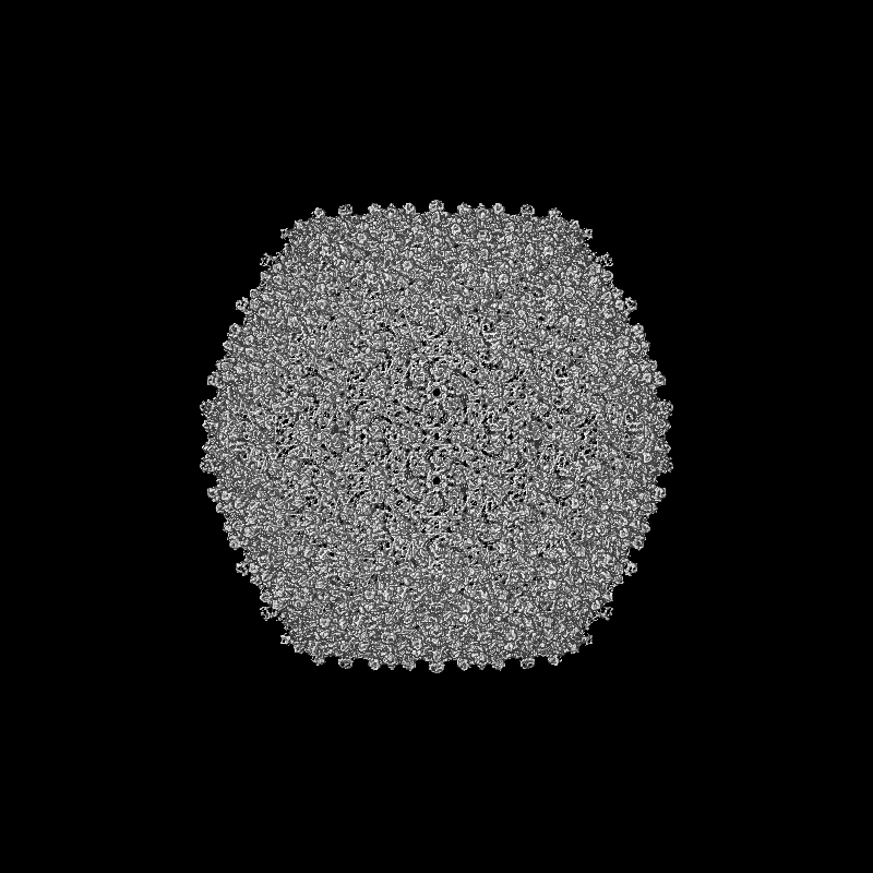
| File | Download / File: emd_46626.map.gz / Format: CCP4 / Size: 1.9 GB / Type: IMAGE STORED AS FLOATING POINT NUMBER (4 BYTES) | ||||||||||||||||||||||||||||||||||||
|---|---|---|---|---|---|---|---|---|---|---|---|---|---|---|---|---|---|---|---|---|---|---|---|---|---|---|---|---|---|---|---|---|---|---|---|---|---|
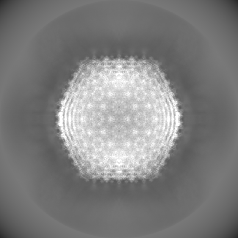
| Projections & slices | Image control
Images are generated by Spider. | ||||||||||||||||||||||||||||||||||||
| Voxel size | X=Y=Z: 1.668 Å | ||||||||||||||||||||||||||||||||||||
| Density |
| ||||||||||||||||||||||||||||||||||||
| Symmetry | Space group: 1 | ||||||||||||||||||||||||||||||||||||
| Details | EMDB XML:
|
 Z (Sec.)
 Y (Row.)
 X (Col.)


-Supplemental data
-Mask #1
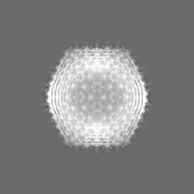
| File | emd_46626_msk_1.map | ||||||||||||
|---|---|---|---|---|---|---|---|---|---|---|---|---|---|
| Projections & Slices |
| ||||||||||||
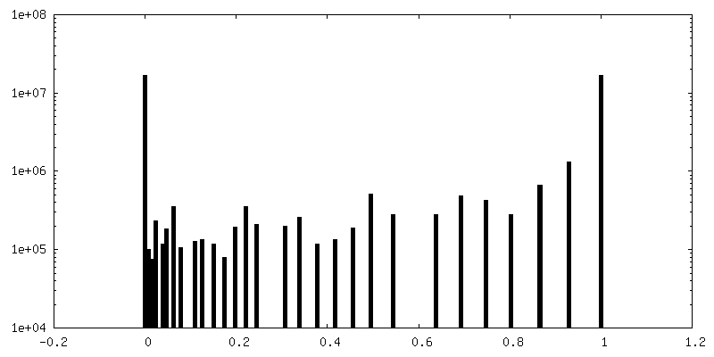
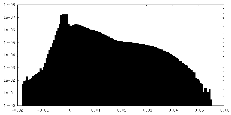
| Density Histograms |


-Half map: #2
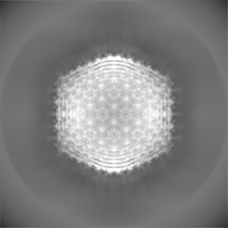
| File | emd_46626_half_map_1.map | ||||||||||||
|---|---|---|---|---|---|---|---|---|---|---|---|---|---|
| Projections & Slices |
| ||||||||||||
| Density Histograms |


-Half map: #1
| File | emd_46626_half_map_2.map | ||||||||||||
|---|---|---|---|---|---|---|---|---|---|---|---|---|---|
| Projections & Slices |
| ||||||||||||
| Density Histograms |


- Sample components
-Entire : Shigella phage B2
| Entire | Name: Shigella phage B2 (virus) |
|---|---|
| Components |
|
-Supramolecule #1: Shigella phage B2
| Supramolecule | Name: Shigella phage B2 / type: virus / ID: 1 / Parent: 0 / Macromolecule list: all / NCBI-ID: 2968270 / Sci species name: Shigella phage B2 / Virus type: VIRION / Virus isolate: OTHER / Virus enveloped: No / Virus empty: No |
|---|---|
| Host (natural) | Organism:  Shigella flexneri 2a str. 2457T (bacteria) Shigella flexneri 2a str. 2457T (bacteria) |
-Macromolecule #1: B2 Capsid
| Macromolecule | Name: B2 Capsid / type: protein_or_peptide / ID: 1 / Number of copies: 9 / Enantiomer: LEVO |
|---|---|
| Source (natural) | Organism: Shigella phage B2 (virus) |
| Molecular weight | Theoretical: 42.530359 KDa |
| Recombinant expression | Organism: Shigella flexneri 2a str. 2457T (bacteria) |
| Sequence | String: MKYNAPNTTP SSIGPQIRLD YYYKKALVDA AKEMYFGQLA EVTNMPKNMG KQIKLYHYVP LLDDRNVNDQ GIDAAGATIA NGNLYGSSK DIGTIPSKLP ALTENGGRVN RVGFTRIQLI GSIKKFGFFY EWTQEAMDFD TDEELDSHLI QEAVKGANEI T EDQLQIDL ...String: MKYNAPNTTP SSIGPQIRLD YYYKKALVDA AKEMYFGQLA EVTNMPKNMG KQIKLYHYVP LLDDRNVNDQ GIDAAGATIA NGNLYGSSK DIGTIPSKLP ALTENGGRVN RVGFTRIQLI GSIKKFGFFY EWTQEAMDFD TDEELDSHLI QEAVKGANEI T EDQLQIDL LNGAGVVRYP GAATSNADMT GEGTATVVTY EGLVKMGITL NDNLCPMQTK LIAGSLMTDT RTIQGARALY IG SELELQL RKMKDPFDNA AFIPVQQYAD AGNLLKGEIG SIASFRVIVV PKMLKWAGAG ATVTTNPGYY ATSGKYDVFP MLC VGSGSF TTIGFQTDGK TVKFTTYTKK PGIETVSYAD PYGEMGLTSI KWYYGSLILR PEWIALFKTV AAM |
-Macromolecule #2: B2 Dec Gp45
| Macromolecule | Name: B2 Dec Gp45 / type: protein_or_peptide / ID: 2 / Number of copies: 9 / Enantiomer: LEVO |
|---|---|
| Source (natural) | Organism: Shigella phage B2 (virus) |
| Molecular weight | Theoretical: 9.495771 KDa |
| Recombinant expression | Organism: Shigella flexneri 2a str. 2457T (bacteria) |
| Sequence | String: MAYSDVDAIL ADGKQAVAVK HGGGLVVVGE LGAQVLAAKD VSELPDGVGG TAPGAATTTT AGVVKQSTTQ AASVATDVAG VVTDLNALI TKLKAAGIMA |
-Experimental details
-Structure determination
| Method | cryo EM |
|---|---|
 Processing Processing | single particle reconstruction |
| Aggregation state | particle |
-Sample preparation
| Buffer | pH: 7.5 Component:
| |||||||||
|---|---|---|---|---|---|---|---|---|---|---|
| Vitrification | Cryogen name: ETHANE / Chamber humidity: 100 % / Chamber temperature: 277.15 K / Instrument: FEI VITROBOT MARK IV |
- Electron microscopy
| Microscope | FEI TITAN KRIOS |
|---|---|
| Image recording | Film or detector model: GATAN K3 BIOQUANTUM (6k x 4k) / Average electron dose: 40.0 e/Å2 |
| Electron beam | Acceleration voltage: 300 kV / Electron source:  FIELD EMISSION GUN FIELD EMISSION GUN |
| Electron optics | Illumination mode: FLOOD BEAM / Imaging mode: BRIGHT FIELD / Nominal defocus max: 3.5 µm / Nominal defocus min: 0.5 µm |
| Experimental equipment |  Model: Titan Krios / Image courtesy: FEI Company |

