 Movie
Movie Controller


+ Open data

- Basic information
| Entry |  | |||||||||||||||
|---|---|---|---|---|---|---|---|---|---|---|---|---|---|---|---|---|
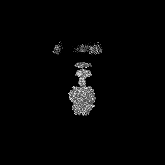
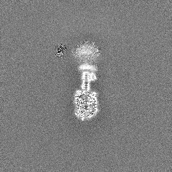
| Title | Shigella flexneri bacteriophage Moo19 Gp82 | |||||||||||||||
 Map data Map data | ||||||||||||||||
 Sample Sample |
| |||||||||||||||
 Keywords Keywords | Moo19 / Gp82 / VIRAL PROTEIN | |||||||||||||||
| Function / homology | Tail spike TSP1/Gp66, N-terminal domain / Tail spike TSP1/Gp66 receptor binding N-terminal domain / SGNH hydrolase-type esterase domain / GDSL-like Lipase/Acylhydrolase family / SGNH hydrolase superfamily / Tail fiber protein Function and homology information Function and homology information | |||||||||||||||
| Biological species |  Shigella virus Moo19 Shigella virus Moo19 | |||||||||||||||
| Method | single particle reconstruction / cryo EM / Resolution: 2.4 Å | |||||||||||||||
 Authors Authors | Subramanian S / Bergland Drarvik SM / Parent KN | |||||||||||||||
| Funding support |  United States, 4 items United States, 4 items
| |||||||||||||||
 Citation Citation | Journal: Sci Adv / Year: 2024 Title: Moo19 and B2: Structures of podophages with = 9 geometry and tailspikes with esterase activity. Authors: Sundharraman Subramanian / Silje M Bergland Drarvik / Kendal R Tinney / Sarah M Doore / Kristin N Parent / Abstract: Podophages are, by far, the least well studied of all the bacteriophages. Despite being classified together due to their short, noncontractile tails, there is a huge amount of diversity among members ...Podophages are, by far, the least well studied of all the bacteriophages. Despite being classified together due to their short, noncontractile tails, there is a huge amount of diversity among members of this group. Of the podophages, the N4-like family is the least well studied structurally and is quite divergent from well-characterized podophages such as T7 and P22. In this work, we isolate and fully characterize two members of the family by cryo-electron microscopy, genetics, and biochemistry. We describe the capsid features of Moo19 and B2, including a decoration protein. In addition, we have fully modeled the tail machinery for both phages and identify proteins with esterase activity. Genetic knockouts of the host reveal factors specific for host attachment including key modifications to the O-antigen on the lipopolysaccharide. Moo19 and B2 are both members, yet some distinct differences in the genome and structure place them into distinct clades. | |||||||||||||||
| History |
|
- Structure visualization
| Supplemental images |
|---|

- Downloads & links
-EMDB archive
| Map data | emd_46624.map.gz | 345.7 MB | EMDB map data format | |
|---|---|---|---|---|
| Header (meta data) | emd-46624-v30.xmlemd-46624.xml | 16.7 KB 16.7 KB | Display Display | EMDB header |
| FSC (resolution estimation) | emd_46624_fsc.xml | 18.4 KB | Display | FSC data file |
| Images |  emd_46624.png emd_46624.png | 122.9 KB | ||
| Masks | emd_46624_msk_1.map | 669.9 MB | Mask map | |
| Filedesc metadata | emd-46624.cif.gz | 6.1 KB | ||
| Others | emd_46624_half_map_1.map.gzemd_46624_half_map_2.map.gz | 622.2 MB 622.2 MB | ||
| Archive directory |  http://ftp.pdbj.org/pub/emdb/structures/EMD-46624ftp://ftp.pdbj.org/pub/emdb/structures/EMD-46624 http://ftp.pdbj.org/pub/emdb/structures/EMD-46624ftp://ftp.pdbj.org/pub/emdb/structures/EMD-46624 | HTTPS FTP |
-Related structure data
| Related structure data |  9d81MC  9d7zC  9d80C  9d82C  9d83C  9d84C M: atomic model generated by this map C: citing same article ( |
|---|---|
| Similar structure data |


-Links
| EMDB pages | EMDB (EBI/PDBe) / EMDataResource |
|---|
-Map
| File | Download / File: emd_46624.map.gz / Format: CCP4 / Size: 669.9 MB / Type: IMAGE STORED AS FLOATING POINT NUMBER (4 BYTES) | ||||||||||||||||||||||||||||||||||||
|---|---|---|---|---|---|---|---|---|---|---|---|---|---|---|---|---|---|---|---|---|---|---|---|---|---|---|---|---|---|---|---|---|---|---|---|---|---|
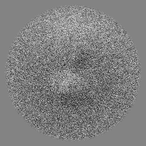
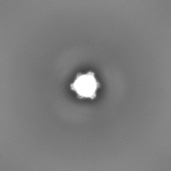
| Projections & slices | Image control
Images are generated by Spider. | ||||||||||||||||||||||||||||||||||||
| Voxel size | X=Y=Z: 0.886 Å | ||||||||||||||||||||||||||||||||||||
| Density |
| ||||||||||||||||||||||||||||||||||||
| Symmetry | Space group: 1 | ||||||||||||||||||||||||||||||||||||
| Details | EMDB XML:
|
 Z (Sec.)
 Y (Row.)
 X (Col.)


-Supplemental data
-Mask #1
| File | emd_46624_msk_1.map | ||||||||||||
|---|---|---|---|---|---|---|---|---|---|---|---|---|---|
| Projections & Slices |
| ||||||||||||
| Density Histograms |


-Half map: #1
| File | emd_46624_half_map_1.map | ||||||||||||
|---|---|---|---|---|---|---|---|---|---|---|---|---|---|
| Projections & Slices |
| ||||||||||||
| Density Histograms |


-Half map: #2
| File | emd_46624_half_map_2.map | ||||||||||||
|---|---|---|---|---|---|---|---|---|---|---|---|---|---|
| Projections & Slices |
| ||||||||||||
| Density Histograms |


- Sample components
-Entire : Shigella virus Moo19
| Entire | Name: Shigella virus Moo19 |
|---|---|
| Components |
|
-Supramolecule #1: Shigella virus Moo19
| Supramolecule | Name: Shigella virus Moo19 / type: virus / ID: 1 / Parent: 0 / Macromolecule list: all / NCBI-ID: 2886042 / Sci species name: Shigella virus Moo19 / Virus type: VIRION / Virus isolate: OTHER / Virus enveloped: No / Virus empty: No |
|---|---|
| Host (natural) | Organism:  Shigella flexneri 2a str. 2457T (bacteria) Shigella flexneri 2a str. 2457T (bacteria) |
-Macromolecule #1: Tail fiber protein
| Macromolecule | Name: Tail fiber protein / type: protein_or_peptide / ID: 1 / Number of copies: 1 / Enantiomer: LEVO |
|---|---|
| Source (natural) | Organism: Shigella virus Moo19 |
| Molecular weight | Theoretical: 118.004133 KDa |
| Recombinant expression | Organism: |
| Sequence | String: MGMNSHIPFD ADNDWTLDPY HCNRSNDPLV DKVIGNAYAV VRAVYCNLGN LKLLYDFLNT YGMVLGVKSE AELKKLNKLA KYARVYGFA DTGDRQVTDY LYVPDDTSGI RPDDQTATGS WIKVSTSGSG SGGTGGGSGS YIPYVYANGS ALGGETSFKV P AEALGVPF ...String: MGMNSHIPFD ADNDWTLDPY HCNRSNDPLV DKVIGNAYAV VRAVYCNLGN LKLLYDFLNT YGMVLGVKSE AELKKLNKLA KYARVYGFA DTGDRQVTDY LYVPDDTSGI RPDDQTATGS WIKVSTSGSG SGGTGGGSGS YIPYVYANGS ALGGETSFKV P AEALGVPF IIINGSVQYI GYGFSFNPAN STVTLSNPLV QGDEVIALTS AAPASPDNPN VSNWVQVNWL YNNGAAVGGE QV ITVPYNF KDVPAVYKNG ERYYKNLQTK SYVYDPSTRT VTMTELLAQG DRVIITLGGE SASLEITDRT TQEVARANNV KDT DVVLSS STNVVITDKK VLYDVNAQKY WDLPNLPPNV YIVKVEGNKL IYNPGAVVID LLEPANPLVI VEPVLSRLGA ETGN PMAGT FEKGATVDSA AKSVGSTMEG KLYRWEGALP KTVRAGDTPS SSGGIGSGKW VEITNATLRS QLASTGGAAM VKASD GRTV EQWLVQSDSA SFRAKNMAKL AWCDYQVHNR GSLKCCFLGD SMTAGFDRTS SDTIPAQDGD WATRASMNYP YRFASY LPE QSGCSVYITM RAISGYTAKQ AYEEALWQSN PNCDIVFIMY AINDSGGVAG ATLDLYMEYM EKLIRRYIDW GCAVVVQ RP SGGGQGAGNP AWLHWAKRMQ MVARVYGCPV FDAHEVMLNR HYAAVQSDGT HYNSMGYAIH GEKLASMLMA GGLLDTYK P VVNETTVWTG MMSDHIGWCD ARGNIGTGRS DGAYTRDKVT GVLQAGKATI CTFSFYLDAE AAHIYGKLDG LINTIYTNG YWWNNGNKPY YQYAVDIDNS FGASLQRVNK SANNYEGMPG SRKFVGRLIG RGWHTITLFT NLQGEALKDA FVNSITVQPI PIGLSTEQM WGQDEERRYR VVHTRRMPSP SGQGGTLPVA VALTGFQMRA PQSFLGTGPG TNAVPAPYFY NTVPGKLKVY N EKGDYIEW LVYKDGSSGL KWKGKVLTHS FADVASVPTL TAYMGTAKQN VIVAAGSSGA NQPLENIYDY NAGLQEQTGN PS TDLSWKG GIYLVFTLAW PSTAPTGYWT IELEGSDWFG NSESAVGCF UniProtKB: Tail fiber protein |
-Experimental details
-Structure determination
| Method | cryo EM |
|---|---|
 Processing Processing | single particle reconstruction |
| Aggregation state | particle |
-Sample preparation
| Buffer | pH: 7.5 Component:
| |||||||||
|---|---|---|---|---|---|---|---|---|---|---|
| Vitrification | Cryogen name: ETHANE / Chamber humidity: 100 % / Chamber temperature: 277.15 K / Instrument: FEI VITROBOT MARK IV |
- Electron microscopy
| Microscope | FEI TALOS ARCTICA |
|---|---|
| Image recording | Film or detector model: FEI FALCON IV (4k x 4k) / Average electron dose: 43.0 e/Å2 |
| Electron beam | Acceleration voltage: 200 kV / Electron source:  FIELD EMISSION GUN FIELD EMISSION GUN |
| Electron optics | Illumination mode: FLOOD BEAM / Imaging mode: BRIGHT FIELD / Nominal defocus max: 2.1 µm / Nominal defocus min: 0.7000000000000001 µm |
| Experimental equipment |  Model: Talos Arctica / Image courtesy: FEI Company |

