 Movie
Movie Controller

[English] 日本語
 Yorodumi
- EMDB-46622: Shigella flexneri bacteriophage Moo19 Icosahedral Reconstruction -
+ Open data

- Basic information
| Entry |  | |||||||||||||||
|---|---|---|---|---|---|---|---|---|---|---|---|---|---|---|---|---|
| Title | Shigella flexneri bacteriophage Moo19 Icosahedral Reconstruction | |||||||||||||||
 Map data Map data | ||||||||||||||||
 Sample Sample |
| |||||||||||||||
 Keywords Keywords | Moo19 / VIRUS | |||||||||||||||
| Function / homology | Phage capsid protein / Immunoglobulin-like domain superfamily / Immunoglobulin-like fold / Ig-like domain-containing protein / Major capsid protein Function and homology information Function and homology information | |||||||||||||||
| Biological species |  Shigella virus Moo19 Shigella virus Moo19 | |||||||||||||||
| Method | single particle reconstruction / cryo EM / Resolution: 3.6 Å | |||||||||||||||
 Authors Authors | Subramanian S / Bergland Drarvik SM / Parent KN | |||||||||||||||
| Funding support |  United States, 4 items United States, 4 items
| |||||||||||||||
 Citation Citation | Journal: Sci Adv / Year: 2024 Title: Moo19 and B2: Structures of podophages with = 9 geometry and tailspikes with esterase activity. Authors: Sundharraman Subramanian / Silje M Bergland Drarvik / Kendal R Tinney / Sarah M Doore / Kristin N Parent / Abstract: Podophages are, by far, the least well studied of all the bacteriophages. Despite being classified together due to their short, noncontractile tails, there is a huge amount of diversity among members ...Podophages are, by far, the least well studied of all the bacteriophages. Despite being classified together due to their short, noncontractile tails, there is a huge amount of diversity among members of this group. Of the podophages, the N4-like family is the least well studied structurally and is quite divergent from well-characterized podophages such as T7 and P22. In this work, we isolate and fully characterize two members of the family by cryo-electron microscopy, genetics, and biochemistry. We describe the capsid features of Moo19 and B2, including a decoration protein. In addition, we have fully modeled the tail machinery for both phages and identify proteins with esterase activity. Genetic knockouts of the host reveal factors specific for host attachment including key modifications to the O-antigen on the lipopolysaccharide. Moo19 and B2 are both members, yet some distinct differences in the genome and structure place them into distinct clades. | |||||||||||||||
| History |
|
- Structure visualization
| Supplemental images |
|---|

- Downloads & links
-EMDB archive
| Map data | emd_46622.map.gz | 265.9 MB | EMDB map data format | |
|---|---|---|---|---|
| Header (meta data) | emd-46622-v30.xmlemd-46622.xml | 17.4 KB 17.4 KB | Display Display | EMDB header |
| FSC (resolution estimation) | emd_46622_fsc.xml | 28 KB | Display | FSC data file |
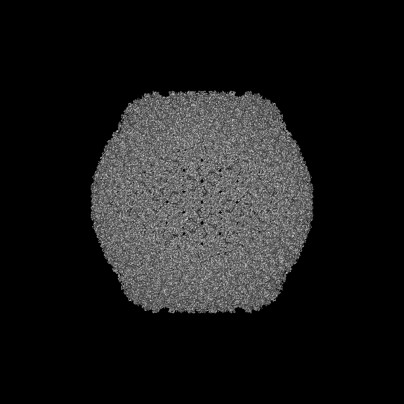
| Images |  emd_46622.png emd_46622.png | 304.1 KB | ||
| Masks | emd_46622_msk_1.map | 1.9 GB | Mask map | |
| Filedesc metadata | emd-46622.cif.gz | 5.8 KB | ||
| Others | emd_46622_half_map_1.map.gzemd_46622_half_map_2.map.gz | 1.5 GB 1.5 GB | ||
| Archive directory |  http://ftp.pdbj.org/pub/emdb/structures/EMD-46622ftp://ftp.pdbj.org/pub/emdb/structures/EMD-46622 http://ftp.pdbj.org/pub/emdb/structures/EMD-46622ftp://ftp.pdbj.org/pub/emdb/structures/EMD-46622 | HTTPS FTP |
-Related structure data
| Related structure data |  9d7zMC  9d80C  9d81C  9d82C  9d83C  9d84C M: atomic model generated by this map C: citing same article ( |
|---|---|
| Similar structure data |


-Links
| EMDB pages | EMDB (EBI/PDBe) / EMDataResource |
|---|
-Map
| File | Download / File: emd_46622.map.gz / Format: CCP4 / Size: 1.9 GB / Type: IMAGE STORED AS FLOATING POINT NUMBER (4 BYTES) | ||||||||||||||||||||||||||||||||||||
|---|---|---|---|---|---|---|---|---|---|---|---|---|---|---|---|---|---|---|---|---|---|---|---|---|---|---|---|---|---|---|---|---|---|---|---|---|---|
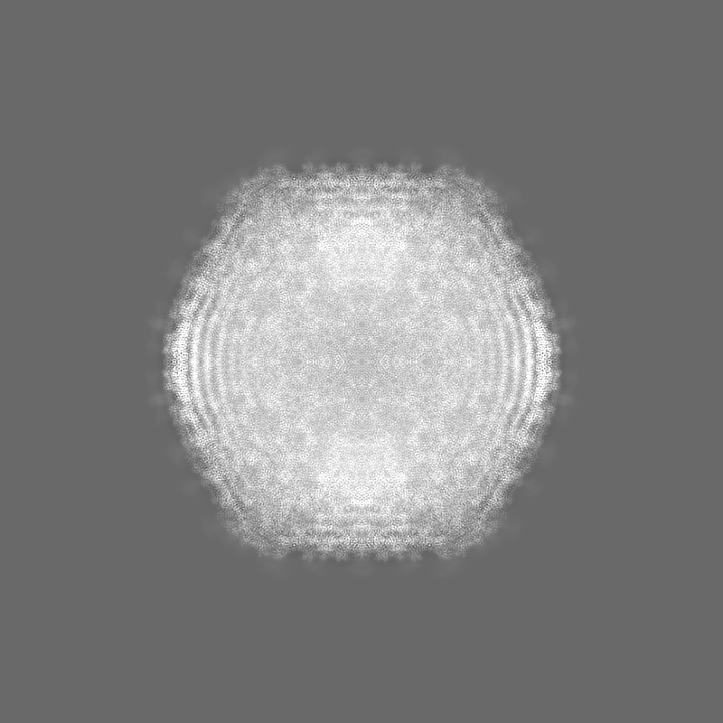
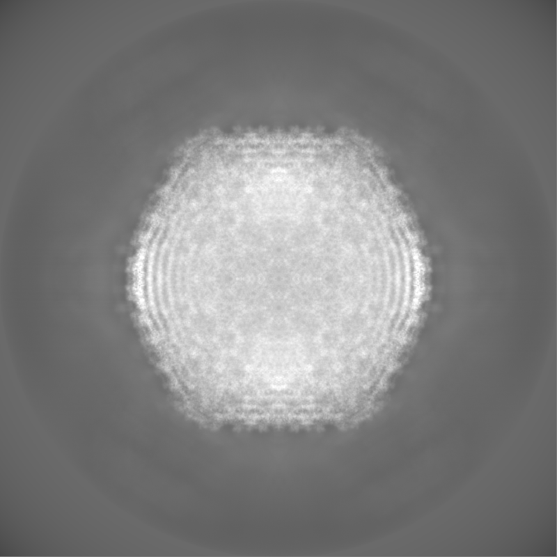
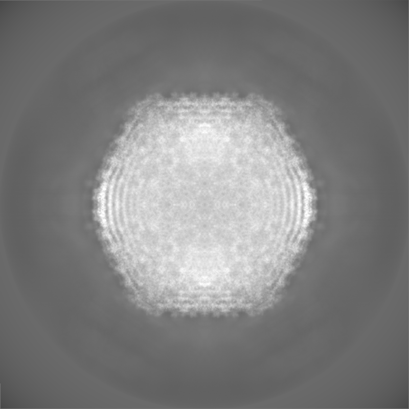
| Projections & slices | Image control
Images are generated by Spider. | ||||||||||||||||||||||||||||||||||||
| Voxel size | X=Y=Z: 1.632 Å | ||||||||||||||||||||||||||||||||||||
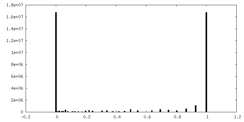
| Density |
| ||||||||||||||||||||||||||||||||||||
| Symmetry | Space group: 1 | ||||||||||||||||||||||||||||||||||||
| Details | EMDB XML:
|
 Z (Sec.)
 Y (Row.)
 X (Col.)


-Supplemental data
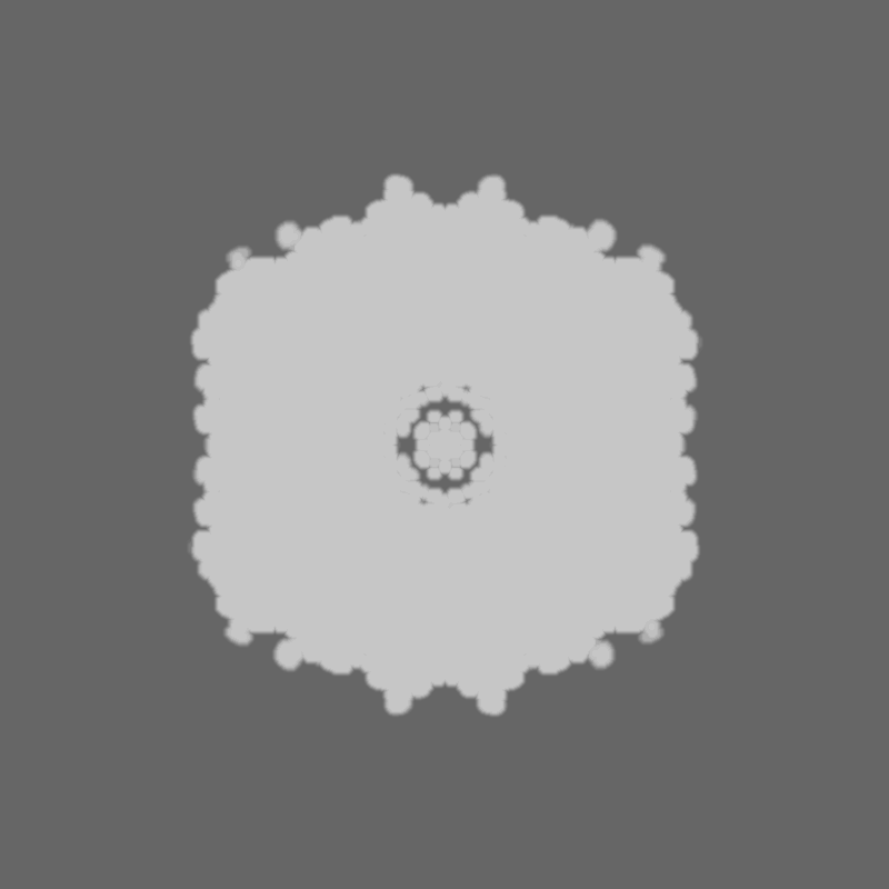
-Mask #1
| File | emd_46622_msk_1.map | ||||||||||||
|---|---|---|---|---|---|---|---|---|---|---|---|---|---|
| Projections & Slices |
| ||||||||||||
| Density Histograms |


-Half map: #1
| File | emd_46622_half_map_1.map | ||||||||||||
|---|---|---|---|---|---|---|---|---|---|---|---|---|---|
| Projections & Slices |
| ||||||||||||
| Density Histograms |


-Half map: #2
| File | emd_46622_half_map_2.map | ||||||||||||
|---|---|---|---|---|---|---|---|---|---|---|---|---|---|
| Projections & Slices |
| ||||||||||||
| Density Histograms |


- Sample components
-Entire : Shigella virus Moo19
| Entire | Name: Shigella virus Moo19 |
|---|---|
| Components |
|
-Supramolecule #1: Shigella virus Moo19
| Supramolecule | Name: Shigella virus Moo19 / type: virus / ID: 1 / Parent: 0 / Macromolecule list: all / NCBI-ID: 2886042 / Sci species name: Shigella virus Moo19 / Virus type: VIRION / Virus isolate: OTHER / Virus enveloped: No / Virus empty: No |
|---|---|
| Host (natural) | Organism:  Shigella flexneri 2a str. 2457T (bacteria) Shigella flexneri 2a str. 2457T (bacteria) |
-Macromolecule #1: Major capsid protein
| Macromolecule | Name: Major capsid protein / type: protein_or_peptide / ID: 1 / Number of copies: 9 / Enantiomer: LEVO |
|---|---|
| Source (natural) | Organism: Shigella virus Moo19 |
| Molecular weight | Theoretical: 43.971672 KDa |
| Recombinant expression | Organism: Shigella flexneri 2a str. 2457T (bacteria) |
| Sequence | String: MLNYNAPIDG QKSSIDGAGS DQMNTFYWLK KAIIQARKDQ YFMPLASVTN MPKNMGKTIK VYEYVPLLDD RNINDQGIDA NGAHIVNGN LYGSSKDIGT ITSKLPLLTE NGGRVNRVGF TRLSREGSIH KFGFFYEFTQ ESLDFDSDDQ LKEHLSRELM N GAVQITEA ...String: MLNYNAPIDG QKSSIDGAGS DQMNTFYWLK KAIIQARKDQ YFMPLASVTN MPKNMGKTIK VYEYVPLLDD RNINDQGIDA NGAHIVNGN LYGSSKDIGT ITSKLPLLTE NGGRVNRVGF TRLSREGSIH KFGFFYEFTQ ESLDFDSDDQ LKEHLSRELM N GAVQITEA VLQKDLLAAA GTVLYAGAAT SDATITGEGS TPSVITYKNL MRLDAILTDN RTPTQTTIIT GSRLVDTKVI GG TRVMYVG SELVPDLKAM KDLFGNKAFI EIQHYGDAGT LMNGEIGTID KFRIIQVPEM LHWAGAGAAA TDANPGYRTS TVN GTEHYD VYPVLVVGDD SFTTIGFQTD GKSVKFNVMT KMPGKETADR NDPYGETGFS SIKWYYGILV KRPERIAVMK AVAP L UniProtKB: Major capsid protein |
-Macromolecule #2: Ig-like domain-containing protein
| Macromolecule | Name: Ig-like domain-containing protein / type: protein_or_peptide / ID: 2 / Number of copies: 3 / Enantiomer: LEVO |
|---|---|
| Source (natural) | Organism: Shigella virus Moo19 |
| Molecular weight | Theoretical: 28.133709 KDa |
| Recombinant expression | Organism: Shigella flexneri 2a str. 2457T (bacteria) |
| Sequence | String: MPELKVAFNK DTYVATVLDA SGSVPSGSVN VGTFFHPDET YPDSYVIYHG VRELLYKRSE VDPAQPGFWP ENITNMQAVT IDNKATARL VLNTSLPRVV STIEGGKVTL SVVALGGKAP LKYKWEFRAP NASTWTAVSG QTTANLVLDN IDADKAGEYK V TVTDAAGT ...String: MPELKVAFNK DTYVATVLDA SGSVPSGSVN VGTFFHPDET YPDSYVIYHG VRELLYKRSE VDPAQPGFWP ENITNMQAVT IDNKATARL VLNTSLPRVV STIEGGKVTL SVVALGGKAP LKYKWEFRAP NASTWTAVSG QTTANLVLDN IDADKAGEYK V TVTDAAGT SVDSTALVAV GAYPPPALTG IKATPTSLSL SVATDAAGKT VALSAIPTDA ELGTLSIKTA PDSARATATI SG STLTVKP VAAGAATSVV VTNGKVDVTI TINVAA UniProtKB: Ig-like domain-containing protein |
-Experimental details
-Structure determination
| Method | cryo EM |
|---|---|
 Processing Processing | single particle reconstruction |
| Aggregation state | particle |
-Sample preparation
| Buffer | pH: 7.5 Component:
| |||||||||
|---|---|---|---|---|---|---|---|---|---|---|
| Vitrification | Cryogen name: ETHANE / Chamber humidity: 100 % / Chamber temperature: 277.15 K / Instrument: FEI VITROBOT MARK IV |
- Electron microscopy
| Microscope | FEI TITAN KRIOS |
|---|---|
| Image recording | Film or detector model: GATAN K3 BIOQUANTUM (6k x 4k) / Average electron dose: 33.0 e/Å2 |
| Electron beam | Acceleration voltage: 300 kV / Electron source:  FIELD EMISSION GUN FIELD EMISSION GUN |
| Electron optics | Illumination mode: FLOOD BEAM / Imaging mode: BRIGHT FIELD / Nominal defocus max: 3.5 µm / Nominal defocus min: 0.5 µm |
| Experimental equipment |  Model: Titan Krios / Image courtesy: FEI Company |

