 Movie
Movie Controller

[English] 日本語
 Yorodumi
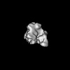
- EMDB-46606: Cryo-EM structure of BG505 DS-SOSIP.664 with 2 CH103 KN Fabs bound -
+ Open data

- Basic information
| Entry |  | ||||||||||||
|---|---|---|---|---|---|---|---|---|---|---|---|---|---|
| Title | Cryo-EM structure of BG505 DS-SOSIP.664 with 2 CH103 KN Fabs bound | ||||||||||||
 Map data Map data | |||||||||||||
 Sample Sample |
| ||||||||||||
 Keywords Keywords | Trimer / Env / BG505 / CH103 / VIRAL PROTEIN / VIRAL PROTEIN-IMMUNE SYSTEM complex | ||||||||||||
| Biological species |   Human immunodeficiency virus 1 / Human immunodeficiency virus 1 /  Homo sapiens (human) Homo sapiens (human) | ||||||||||||
| Method | single particle reconstruction / cryo EM / Resolution: 3.68 Å | ||||||||||||
 Authors Authors | Parsons RJ / Acharya P | ||||||||||||
| Funding support |  United States, 3 items United States, 3 items
| ||||||||||||
 Citation Citation | Journal: Acta Crystallogr D Struct Biol / Year: 2019 Title: Macromolecular structure determination using X-rays, neutrons and electrons: recent developments in Phenix. Authors: Dorothee Liebschner / Pavel V Afonine / Matthew L Baker / Gábor Bunkóczi / Vincent B Chen / Tristan I Croll / Bradley Hintze / Li Wei Hung / Swati Jain / Airlie J McCoy / Nigel W Moriarty ...Authors: Dorothee Liebschner / Pavel V Afonine / Matthew L Baker / Gábor Bunkóczi / Vincent B Chen / Tristan I Croll / Bradley Hintze / Li Wei Hung / Swati Jain / Airlie J McCoy / Nigel W Moriarty / Robert D Oeffner / Billy K Poon / Michael G Prisant / Randy J Read / Jane S Richardson / David C Richardson / Massimo D Sammito / Oleg V Sobolev / Duncan H Stockwell / Thomas C Terwilliger / Alexandre G Urzhumtsev / Lizbeth L Videau / Christopher J Williams / Paul D Adams /   Abstract: Diffraction (X-ray, neutron and electron) and electron cryo-microscopy are powerful methods to determine three-dimensional macromolecular structures, which are required to understand biological ...Diffraction (X-ray, neutron and electron) and electron cryo-microscopy are powerful methods to determine three-dimensional macromolecular structures, which are required to understand biological processes and to develop new therapeutics against diseases. The overall structure-solution workflow is similar for these techniques, but nuances exist because the properties of the reduced experimental data are different. Software tools for structure determination should therefore be tailored for each method. Phenix is a comprehensive software package for macromolecular structure determination that handles data from any of these techniques. Tasks performed with Phenix include data-quality assessment, map improvement, model building, the validation/rebuilding/refinement cycle and deposition. Each tool caters to the type of experimental data. The design of Phenix emphasizes the automation of procedures, where possible, to minimize repetitive and time-consuming manual tasks, while default parameters are chosen to encourage best practice. A graphical user interface provides access to many command-line features of Phenix and streamlines the transition between programs, project tracking and re-running of previous tasks. | ||||||||||||
| History |
|
- Structure visualization
| Supplemental images |
|---|

- Downloads & links
-EMDB archive
| Map data | emd_46606.map.gz | 118.1 MB |  EMDB map data format EMDB map data format | |
|---|---|---|---|---|
| Header (meta data) | emd-46606-v30.xmlemd-46606.xml | 24.5 KB 24.5 KB | Display Display | EMDB header |
| FSC (resolution estimation) | emd_46606_fsc.xml | 14.7 KB | Display | FSC data file |
| Images |  emd_46606.png emd_46606.png | 59.1 KB | ||
| Filedesc metadata | emd-46606.cif.gz | 7.9 KB | ||
| Others | emd_46606_half_map_1.map.gzemd_46606_half_map_2.map.gz | 115.8 MB 115.8 MB | ||
| Archive directory |  http://ftp.pdbj.org/pub/emdb/structures/EMD-46606ftp://ftp.pdbj.org/pub/emdb/structures/EMD-46606 http://ftp.pdbj.org/pub/emdb/structures/EMD-46606ftp://ftp.pdbj.org/pub/emdb/structures/EMD-46606 | HTTPS FTP |
-Related structure data
| Related structure data |  9d7iMC  9d7gC  9d7hC  9d7oC  9d7pC M: atomic model generated by this map C: citing same article ( |
|---|


-Links
| EMDB pages | EMDB (EBI/PDBe) / EMDataResource |
|---|
-Map
| File | Download / File: emd_46606.map.gz / Format: CCP4 / Size: 125 MB / Type: IMAGE STORED AS FLOATING POINT NUMBER (4 BYTES) | ||||||||||||||||||||||||||||||||||||
|---|---|---|---|---|---|---|---|---|---|---|---|---|---|---|---|---|---|---|---|---|---|---|---|---|---|---|---|---|---|---|---|---|---|---|---|---|---|
| Projections & slices | Image control
Images are generated by Spider. | ||||||||||||||||||||||||||||||||||||
| Voxel size | X=Y=Z: 1.08 Å | ||||||||||||||||||||||||||||||||||||
| Density |
| ||||||||||||||||||||||||||||||||||||
| Symmetry | Space group: 1 | ||||||||||||||||||||||||||||||||||||
| Details | EMDB XML:
|
 Z (Sec.)
 Y (Row.)
 X (Col.)


-Supplemental data
-Half map: #2
| File | emd_46606_half_map_1.map | ||||||||||||
|---|---|---|---|---|---|---|---|---|---|---|---|---|---|
| Projections & Slices |
| ||||||||||||
| Density Histograms |


-Half map: #1
| File | emd_46606_half_map_2.map | ||||||||||||
|---|---|---|---|---|---|---|---|---|---|---|---|---|---|
| Projections & Slices |
| ||||||||||||
| Density Histograms |


- Sample components
-Entire : BG505 HIV-1 Env with 2 CH103 K75 N76 Fabs bound
| Entire | Name: BG505 HIV-1 Env with 2 CH103 K75 N76 Fabs bound |
|---|---|
| Components |
|
-Supramolecule #1: BG505 HIV-1 Env with 2 CH103 K75 N76 Fabs bound
| Supramolecule | Name: BG505 HIV-1 Env with 2 CH103 K75 N76 Fabs bound / type: complex / ID: 1 / Parent: 0 / Macromolecule list: #1-#2, #4, #3 |
|---|---|
| Molecular weight | Theoretical: 250 KDa |
-Macromolecule #1: Surface protein gp120
| Macromolecule | Name: Surface protein gp120 / type: protein_or_peptide / ID: 1 / Number of copies: 3 / Enantiomer: LEVO |
|---|---|
| Source (natural) | Organism: Human immunodeficiency virus 1 |
| Molecular weight | Theoretical: 55.359906 KDa |
| Recombinant expression | Organism: Homo sapiens (human) |
| Sequence | String: MPMGSLQPLA TLYLLGMLVA SVLAAENLWV TVYYGVPVWK DAETTLFCAS DAKAYETEKH NVWATHACVP TDPNPQEIHL ENVTEEFNM WKNNMVEQMH TDIISLWDQS LKPCVKLTPL CVTLQCTNVT NNITDDMRGE LKNCSFNMTT ELRDKKQKVY S LFYRLDVV ...String: MPMGSLQPLA TLYLLGMLVA SVLAAENLWV TVYYGVPVWK DAETTLFCAS DAKAYETEKH NVWATHACVP TDPNPQEIHL ENVTEEFNM WKNNMVEQMH TDIISLWDQS LKPCVKLTPL CVTLQCTNVT NNITDDMRGE LKNCSFNMTT ELRDKKQKVY S LFYRLDVV QINENQGNRS NNSNKEYRLI NCNTSACTQA CPKVSFEPIP IHYCAPAGFA ILKCKDKKFN GTGPCPSVST VQ CTHGIKP VVSTQLLLNG SLAEEEVMIR SENITNNAKN ILVQFNTPVQ INCTRPNNNT RKSIRIGPGQ AFYATGDIIG DIR QAHCNV SKATWNETLG KVVKQLRKHF GNNTIIRFAN SSGGDLEVTT HSFNCGGEFF YCNTSGLFNS TWISNTSVQG SNST GSNDS ITLPCRIKQI INMWQRIGQC MYAPPIQGVI RCVSNITGLI LTRDGGSTNS TTETFRPGGG DMRDNWRSEL YKYKV VKIE PLGVAPTRCK RR |
-Macromolecule #2: Transmembrane protein gp41
| Macromolecule | Name: Transmembrane protein gp41 / type: protein_or_peptide / ID: 2 / Number of copies: 3 / Enantiomer: LEVO |
|---|---|
| Source (natural) | Organism: Human immunodeficiency virus 1 |
| Molecular weight | Theoretical: 18.344963 KDa |
| Recombinant expression | Organism: Homo sapiens (human) |
| Sequence | String: VVGRRRRRRA VGIGAVFLGF LGAAGSTMGA ASMTLTVQAR NLLSGIVQQQ SNLLRAPEAQ QHLLKLTVWG IKQLQARVLA VERYLRDQQ LLGIWGCSGK LICCTNVPWN SSWSNRNLSE IWDNMTWLQW DKEISNYTQI IYGLLEESQN QQEKNEQDLL A LD |
-Macromolecule #3: CH103 Fab light chain
| Macromolecule | Name: CH103 Fab light chain / type: protein_or_peptide / ID: 3 / Number of copies: 2 / Enantiomer: LEVO |
|---|---|
| Source (natural) | Organism: Homo sapiens (human) |
| Molecular weight | Theoretical: 24.530268 KDa |
| Recombinant expression | Organism: Homo sapiens (human) |
| Sequence | String: MGWSCIILFL VATATGSWAS YELTQPPSVS VSPGQTATIT CSGASTNVCW YQVKPGQSPE VVIFENYKRP SGIPDRFSGS KSGSTATLT IRGTQAIDEA DYYCQVWDSF STFVFGSGTQ VTVLGQPKAN PTVTLFPPSS EELQANKATL VCLISDFYPG A VTVAWKAD ...String: MGWSCIILFL VATATGSWAS YELTQPPSVS VSPGQTATIT CSGASTNVCW YQVKPGQSPE VVIFENYKRP SGIPDRFSGS KSGSTATLT IRGTQAIDEA DYYCQVWDSF STFVFGSGTQ VTVLGQPKAN PTVTLFPPSS EELQANKATL VCLISDFYPG A VTVAWKAD SSPVKAGVET TTPSKQSNNK YAASSYLSLT PEQWKSHRSY SCQVTHEGST VEKTVAPTEC S |
-Macromolecule #4: CH103 K75 N76 Fab heavy chain
| Macromolecule | Name: CH103 K75 N76 Fab heavy chain / type: protein_or_peptide / ID: 4 / Number of copies: 2 / Enantiomer: LEVO |
|---|---|
| Source (natural) | Organism: Homo sapiens (human) |
| Molecular weight | Theoretical: 25.885307 KDa |
| Recombinant expression | Organism: Homo sapiens (human) |
| Sequence | String: MGWSCIILFL VATATGVHSQ VQLQESGPGV VKSSETLSLT CTVSGGSMGG TYWSWLRLSP GKGLEWIGYI FHTGETNYSP SLKGRVSIS VDTSKNQFSL RLRSVTAADT AVYFCASLPR GQLVNAYFRN WGRGSLVSVT AASTKGPSVF PLAPSSKSTS G GTAALGCL ...String: MGWSCIILFL VATATGVHSQ VQLQESGPGV VKSSETLSLT CTVSGGSMGG TYWSWLRLSP GKGLEWIGYI FHTGETNYSP SLKGRVSIS VDTSKNQFSL RLRSVTAADT AVYFCASLPR GQLVNAYFRN WGRGSLVSVT AASTKGPSVF PLAPSSKSTS G GTAALGCL VKDYFPEPVT VSWNSGALTS GVHTFPAVLQ SSGLYSLSSV VTVPSSSLGT QTYICNVNHK PSNTKVDKKV EP KSCDK |
-Macromolecule #7: 2-acetamido-2-deoxy-beta-D-glucopyranose
| Macromolecule | Name: 2-acetamido-2-deoxy-beta-D-glucopyranose / type: ligand / ID: 7 / Number of copies: 30 / Formula: NAG |
|---|---|
| Molecular weight | Theoretical: 221.208 Da |
| Chemical component information |  ChemComp-NAG: |
-Experimental details
-Structure determination
| Method | cryo EM |
|---|---|
 Processing Processing | single particle reconstruction |
| Aggregation state | particle |
-Sample preparation
| Buffer | pH: 8 |
|---|---|
| Grid | Model: Quantifoil R1.2/1.3 / Material: COPPER / Mesh: 300 / Support film - Material: CARBON / Support film - topology: HOLEY / Pretreatment - Type: GLOW DISCHARGE / Pretreatment - Time: 15 sec. |
| Vitrification | Cryogen name: ETHANE / Chamber humidity: 95 % / Chamber temperature: 295.15 K / Instrument: LEICA EM GP / Details: Leica EM GP2. |
- Electron microscopy
| Microscope | TFS KRIOS |
|---|---|
| Specialist optics | Energy filter - Slit width: 20 eV |
| Image recording | Film or detector model: GATAN K3 BIOQUANTUM (6k x 4k) / Average electron dose: 59.1 e/Å2 |
| Electron beam | Acceleration voltage: 300 kV / Electron source:  FIELD EMISSION GUN FIELD EMISSION GUN |
| Electron optics | Illumination mode: FLOOD BEAM / Imaging mode: BRIGHT FIELD / Nominal defocus max: 1.8 µm / Nominal defocus min: 1.4000000000000001 µm |
| Experimental equipment |  Model: Titan Krios / Image courtesy: FEI Company |

