 Movie
Movie Controller

[English] 日本語
 Yorodumi
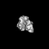
- PDB-9d7h: Cryo-EM structure of BG505 DS-SOSIP.664 with 1 CH103 KN Fab bound -
+ Open data

- Basic information
| Entry | Database: PDB / ID: 9d7h | ||||||||||||
|---|---|---|---|---|---|---|---|---|---|---|---|---|---|
| Title | Cryo-EM structure of BG505 DS-SOSIP.664 with 1 CH103 KN Fab bound | ||||||||||||
 Components Components |
| ||||||||||||
 Keywords Keywords | VIRAL PROTEIN/IMMUNE SYSTEM / Trimer / Env / BG505 / CH103 / VIRAL PROTEIN / VIRAL PROTEIN-IMMUNE SYSTEM complex | ||||||||||||
| Biological species |   Human immunodeficiency virus 1 Human immunodeficiency virus 1 Homo sapiens (human) Homo sapiens (human) | ||||||||||||
| Method | ELECTRON MICROSCOPY / single particle reconstruction / cryo EM / Resolution: 3.59 Å | ||||||||||||
 Authors Authors | Parsons, R.J. / Acharya, P. | ||||||||||||
| Funding support |  United States, 3items United States, 3items
| ||||||||||||
 Citation Citation | Journal: Structure / Year: 2025 Title: Acquisition of quaternary trimer interaction as a key step in the lineage maturation of a broad and potent HIV-1 neutralizing antibody. Authors: Qingbo Liu / Ruth J Parsons / Kevin Wiehe / Robert J Edwards / Kevin O Saunders / Peng Zhang / Huiyi Miao / Kedamawit Tilahun / Julia Jones / Yue Chen / Bhavna Hora / Wilton B Williams / ...Authors: Qingbo Liu / Ruth J Parsons / Kevin Wiehe / Robert J Edwards / Kevin O Saunders / Peng Zhang / Huiyi Miao / Kedamawit Tilahun / Julia Jones / Yue Chen / Bhavna Hora / Wilton B Williams / David Easterhoff / Xiao Huang / Katarzyna Janowska / Katayoun Mansouri / Barton F Haynes / Priyamvada Acharya / Paolo Lusso / Abstract: Although most broadly neutralizing antibodies (bNAbs) specific for the CD4-binding site (CD4-BS) of HIV-1 interact with a single gp120 protomer, a few mimic the quaternary binding mode of CD4, making ...Although most broadly neutralizing antibodies (bNAbs) specific for the CD4-binding site (CD4-BS) of HIV-1 interact with a single gp120 protomer, a few mimic the quaternary binding mode of CD4, making contact with a second protomer through elongated heavy chain framework 3 (FRH3) or complementarity-determining region 1 (CDRH1) loops. Here, we show that a CDRH3-dominated anti-CD4-BS bNAb, CH103, establishes quaternary interaction despite regular-length FRH3 and CDRH1. This quaternary interaction is critical for neutralization and is primarily mediated by two FRH3 acidic residues that were sequentially acquired and subjected to strong positive selection during CH103 maturation. Cryoelectron microscopy (cryo-EM) structures confirmed the role of the two FRH3 acidic residues in mediating quaternary contact and demonstrated that CH103 reaches the adjacent gp120 protomer by virtue of its unique angle of approach. Thus, the acquisition of quaternary interaction may constitute a key step in the lineage maturation of a broad and potent HIV-1 neutralizing antibody. #1: Journal: Acta Crystallogr D Struct Biol / Year: 2019 Title: Macromolecular structure determination using X-rays, neutrons and electrons: recent developments in Phenix. Authors: Dorothee Liebschner / Pavel V Afonine / Matthew L Baker / Gábor Bunkóczi / Vincent B Chen / Tristan I Croll / Bradley Hintze / Li Wei Hung / Swati Jain / Airlie J McCoy / Nigel W Moriarty ...Authors: Dorothee Liebschner / Pavel V Afonine / Matthew L Baker / Gábor Bunkóczi / Vincent B Chen / Tristan I Croll / Bradley Hintze / Li Wei Hung / Swati Jain / Airlie J McCoy / Nigel W Moriarty / Robert D Oeffner / Billy K Poon / Michael G Prisant / Randy J Read / Jane S Richardson / David C Richardson / Massimo D Sammito / Oleg V Sobolev / Duncan H Stockwell / Thomas C Terwilliger / Alexandre G Urzhumtsev / Lizbeth L Videau / Christopher J Williams / Paul D Adams /   Abstract: Diffraction (X-ray, neutron and electron) and electron cryo-microscopy are powerful methods to determine three-dimensional macromolecular structures, which are required to understand biological ...Diffraction (X-ray, neutron and electron) and electron cryo-microscopy are powerful methods to determine three-dimensional macromolecular structures, which are required to understand biological processes and to develop new therapeutics against diseases. The overall structure-solution workflow is similar for these techniques, but nuances exist because the properties of the reduced experimental data are different. Software tools for structure determination should therefore be tailored for each method. Phenix is a comprehensive software package for macromolecular structure determination that handles data from any of these techniques. Tasks performed with Phenix include data-quality assessment, map improvement, model building, the validation/rebuilding/refinement cycle and deposition. Each tool caters to the type of experimental data. The design of Phenix emphasizes the automation of procedures, where possible, to minimize repetitive and time-consuming manual tasks, while default parameters are chosen to encourage best practice. A graphical user interface provides access to many command-line features of Phenix and streamlines the transition between programs, project tracking and re-running of previous tasks. | ||||||||||||
| History |
|
- Structure visualization
| Structure viewer | Molecule:  MolmilJmol/JSmol MolmilJmol/JSmol |
|---|
- Downloads & links
-Download
| PDBx/mmCIF format | 9d7h.cif.gz | 386 KB | Display | PDBx/mmCIF format |
|---|---|---|---|---|
| PDB format | pdb9d7h.ent.gz | Display | PDB format | |
| PDBx/mmJSON format | 9d7h.json.gz | Tree view | PDBx/mmJSON format | |
| Others |  Other downloads Other downloads |
-Validation report
| Arichive directory | https://data.pdbj.org/pub/pdb/validation_reports/d7/9d7hftp://data.pdbj.org/pub/pdb/validation_reports/d7/9d7h | HTTPS FTP |
|---|
-Related structure data
| Related structure data |  46605MC  9d7gC  9d7iC  9d7oC  9d7pC M: map data used to model this data C: citing same article ( |
|---|


-Links
 PDBj
PDBj

- Assembly
| Deposited unit | 
|
|---|---|
| 1 |
|
-Components
-Protein , 2 types, 6 molecules ACEBDF
| #1: Protein | Mass: 55359.906 Da / Num. of mol.: 3 Source method: isolated from a genetically manipulated source Source: (gene. exp.) Human immunodeficiency virus 1 / Variant: BG505 / Production host: Homo sapiens (human)#2: Protein | Mass: 18344.963 Da / Num. of mol.: 3 Source method: isolated from a genetically manipulated source Source: (gene. exp.) Human immunodeficiency virus 1 / Variant: BG505 / Production host: Homo sapiens (human) |
|---|
-Antibody , 2 types, 2 molecules GH
| #3: Antibody | Mass: 24530.268 Da / Num. of mol.: 1 Source method: isolated from a genetically manipulated source Source: (gene. exp.) Homo sapiens (human) / Production host: Homo sapiens (human) |
|---|---|
| #4: Antibody | Mass: 25885.307 Da / Num. of mol.: 1 Source method: isolated from a genetically manipulated source Source: (gene. exp.) Homo sapiens (human) / Production host: Homo sapiens (human) |
-Sugars , 3 types, 53 molecules 
| #5: Polysaccharide | 2-acetamido-2-deoxy-beta-D-glucopyranose-(1-4)-2-acetamido-2-deoxy-beta-D-glucopyranose Source method: isolated from a genetically manipulated source #6: Polysaccharide | beta-D-mannopyranose-(1-4)-2-acetamido-2-deoxy-beta-D-glucopyranose-(1-4)-2-acetamido-2-deoxy-beta- ...beta-D-mannopyranose-(1-4)-2-acetamido-2-deoxy-beta-D-glucopyranose-(1-4)-2-acetamido-2-deoxy-beta-D-glucopyranose Source method: isolated from a genetically manipulated source #7: Sugar | ChemComp-NAG /  Type: D-saccharide, beta linking / Mass: 221.208 Da / Num. of mol.: 29 Type: D-saccharide, beta linking / Mass: 221.208 Da / Num. of mol.: 29Source method: isolated from a genetically manipulated source Formula: C8H15NO6 |
|---|
-Details
| Has ligand of interest | N |
|---|---|
| Has protein modification | Y |
-Experimental details
-Experiment
| Experiment | Method: ELECTRON MICROSCOPY |
|---|---|
| EM experiment | Aggregation state: PARTICLE / 3D reconstruction method: single particle reconstruction |
- Sample preparation
| Component | Name: BG505 HIV-1 Env with 1 CH103 K75 N76 Fab bound / Type: COMPLEX / Entity ID: #3, #1-#2, #4 / Source: MULTIPLE SOURCES |
|---|---|
| Molecular weight | Value: 0.25 MDa / Experimental value: NO |
| Buffer solution | pH: 8 |
| Specimen | Embedding applied: NO / Shadowing applied: NO / Staining applied: NO / Vitrification applied: YES |
| Specimen support | Grid material: COPPER / Grid mesh size: 300 divisions/in. / Grid type: Quantifoil R1.2/1.3 |
| Vitrification | Instrument: LEICA EM GP / Cryogen name: ETHANE / Humidity: 95 % / Chamber temperature: 295.15 K / Details: Leica EM GP2 |
- Electron microscopy imaging
| Experimental equipment |  Model: Titan Krios / Image courtesy: FEI Company |
|---|---|
| Microscopy | Model: TFS KRIOS |
| Electron gun | Electron source:  FIELD EMISSION GUN / Accelerating voltage: 300 kV / Illumination mode: FLOOD BEAM FIELD EMISSION GUN / Accelerating voltage: 300 kV / Illumination mode: FLOOD BEAM |
| Electron lens | Mode: BRIGHT FIELD / Nominal defocus max: 1800 nm / Nominal defocus min: 1400 nm |
| Image recording | Electron dose: 59.8 e/Å2 / Film or detector model: GATAN K3 BIOQUANTUM (6k x 4k) |
| EM imaging optics | Energyfilter slit width: 20 eV |
- Processing
| EM software | Name: PHENIX / Version: 1.21.1_5286 / Category: model refinement |
|---|---|
| Image processing | Details: The data were processed in cryoSPARCv4.01. Movies were aligned using Patch Motion Correction and the non-dose weighted aligned micrographs were used for the CTF correction with PatchCTF Estimation. |
| CTF correction | Details: CryoSPARCv4.01 Patch motion correction and CTF estimation jobs Type: PHASE FLIPPING AND AMPLITUDE CORRECTION |
| Particle selection | Num. of particles selected: 997941 |
| 3D reconstruction | Resolution: 3.59 Å / Resolution method: FSC 0.5 CUT-OFF / Num. of particles: 128185 / Symmetry type: POINT |
