[English] 日本語
 Yorodumi
- EMDB-44738: HIV Envelope BG505 SOSIP.664 in complex with one Fab of CH103 E75... -
+ Open data
- Basic information
| Entry |  | |||||||||
|---|---|---|---|---|---|---|---|---|---|---|
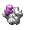
| Title | HIV Envelope BG505 SOSIP.664 in complex with one Fab of CH103 E75K/D76N mutant | |||||||||
 Map data Map data | HIV Envelope BG505 SOSIP.664 in complex with one Fab of CH103 E75K/D76N mutant | |||||||||
 Sample Sample |
| |||||||||
 Keywords Keywords | HIV Envelope Antibody Complex / VIRAL PROTEIN | |||||||||
| Biological species |  Human immunodeficiency virus 2 / Human immunodeficiency virus 2 /  Homo sapiens (human) Homo sapiens (human) | |||||||||
| Method | single particle reconstruction / negative staining / Resolution: 10.0 Å | |||||||||
 Authors Authors | Edwards RJ / Mansouri K | |||||||||
| Funding support |  United States, 1 items United States, 1 items
| |||||||||
 Citation Citation |  Journal: Structure / Year: 2025 Journal: Structure / Year: 2025Title: Acquisition of quaternary trimer interaction as a key step in the lineage maturation of a broad and potent HIV-1 neutralizing antibody. Authors: Qingbo Liu / Ruth J Parsons / Kevin Wiehe / Robert J Edwards / Kevin O Saunders / Peng Zhang / Huiyi Miao / Kedamawit Tilahun / Julia Jones / Yue Chen / Bhavna Hora / Wilton B Williams / ...Authors: Qingbo Liu / Ruth J Parsons / Kevin Wiehe / Robert J Edwards / Kevin O Saunders / Peng Zhang / Huiyi Miao / Kedamawit Tilahun / Julia Jones / Yue Chen / Bhavna Hora / Wilton B Williams / David Easterhoff / Xiao Huang / Katarzyna Janowska / Katayoun Mansouri / Barton F Haynes / Priyamvada Acharya / Paolo Lusso /  Abstract: Although most broadly neutralizing antibodies (bNAbs) specific for the CD4-binding site (CD4-BS) of HIV-1 interact with a single gp120 protomer, a few mimic the quaternary binding mode of CD4, making ...Although most broadly neutralizing antibodies (bNAbs) specific for the CD4-binding site (CD4-BS) of HIV-1 interact with a single gp120 protomer, a few mimic the quaternary binding mode of CD4, making contact with a second protomer through elongated heavy chain framework 3 (FRH3) or complementarity-determining region 1 (CDRH1) loops. Here, we show that a CDRH3-dominated anti-CD4-BS bNAb, CH103, establishes quaternary interaction despite regular-length FRH3 and CDRH1. This quaternary interaction is critical for neutralization and is primarily mediated by two FRH3 acidic residues that were sequentially acquired and subjected to strong positive selection during CH103 maturation. Cryoelectron microscopy (cryo-EM) structures confirmed the role of the two FRH3 acidic residues in mediating quaternary contact and demonstrated that CH103 reaches the adjacent gp120 protomer by virtue of its unique angle of approach. Thus, the acquisition of quaternary interaction may constitute a key step in the lineage maturation of a broad and potent HIV-1 neutralizing antibody. | |||||||||
| History |
|
- Structure visualization
| Supplemental images |
|---|
- Downloads & links
-EMDB archive
| Map data |  emd_44738.map.gz emd_44738.map.gz | 14.1 MB |  EMDB map data format EMDB map data format | |
|---|---|---|---|---|
| Header (meta data) |  emd-44738-v30.xml emd-44738-v30.xml emd-44738.xml emd-44738.xml | 19.2 KB 19.2 KB | Display Display |  EMDB header EMDB header |
| FSC (resolution estimation) |  emd_44738_fsc.xml emd_44738_fsc.xml | 5.8 KB | Display |  FSC data file FSC data file |
| Images |  emd_44738.png emd_44738.png | 51.9 KB | ||
| Filedesc metadata |  emd-44738.cif.gz emd-44738.cif.gz | 5 KB | ||
| Others |  emd_44738_half_map_1.map.gz emd_44738_half_map_1.map.gz emd_44738_half_map_2.map.gz emd_44738_half_map_2.map.gz | 11.8 MB 11.8 MB | ||
| Archive directory |  http://ftp.pdbj.org/pub/emdb/structures/EMD-44738 http://ftp.pdbj.org/pub/emdb/structures/EMD-44738 ftp://ftp.pdbj.org/pub/emdb/structures/EMD-44738 ftp://ftp.pdbj.org/pub/emdb/structures/EMD-44738 | HTTPS FTP |
-Related structure data
- Links
Links
| EMDB pages |  EMDB (EBI/PDBe) / EMDB (EBI/PDBe) /  EMDataResource EMDataResource |
|---|
- Map
Map
| File |  Download / File: emd_44738.map.gz / Format: CCP4 / Size: 15.6 MB / Type: IMAGE STORED AS FLOATING POINT NUMBER (4 BYTES) Download / File: emd_44738.map.gz / Format: CCP4 / Size: 15.6 MB / Type: IMAGE STORED AS FLOATING POINT NUMBER (4 BYTES) | ||||||||||||||||||||||||||||||||||||
|---|---|---|---|---|---|---|---|---|---|---|---|---|---|---|---|---|---|---|---|---|---|---|---|---|---|---|---|---|---|---|---|---|---|---|---|---|---|
| Annotation | HIV Envelope BG505 SOSIP.664 in complex with one Fab of CH103 E75K/D76N mutant | ||||||||||||||||||||||||||||||||||||
| Projections & slices | Image control
Images are generated by Spider. | ||||||||||||||||||||||||||||||||||||
| Voxel size | X=Y=Z: 2.16 Å | ||||||||||||||||||||||||||||||||||||
| Density |
| ||||||||||||||||||||||||||||||||||||
| Symmetry | Space group: 1 | ||||||||||||||||||||||||||||||||||||
| Details | EMDB XML:
|
-Supplemental data
-Half map: BG505-CH103 KN mutant half map
| File | emd_44738_half_map_1.map | ||||||||||||
|---|---|---|---|---|---|---|---|---|---|---|---|---|---|
| Annotation | BG505-CH103 KN mutant half map | ||||||||||||
| Projections & Slices |
| ||||||||||||
| Density Histograms |
-Half map: BG505-CH103 KN mutant half map
| File | emd_44738_half_map_2.map | ||||||||||||
|---|---|---|---|---|---|---|---|---|---|---|---|---|---|
| Annotation | BG505-CH103 KN mutant half map | ||||||||||||
| Projections & Slices |
| ||||||||||||
| Density Histograms |
- Sample components
-Entire : Complex of BG505 SOSIP.664 with two CH103 wildtype Fabs
| Entire | Name: Complex of BG505 SOSIP.664 with two CH103 wildtype Fabs |
|---|---|
| Components |
|
-Supramolecule #1: Complex of BG505 SOSIP.664 with two CH103 wildtype Fabs
| Supramolecule | Name: Complex of BG505 SOSIP.664 with two CH103 wildtype Fabs type: complex / ID: 1 / Parent: 0 |
|---|---|
| Molecular weight | Theoretical: 46 KDa |
-Supramolecule #2: BG505 SOSIP Trimer
| Supramolecule | Name: BG505 SOSIP Trimer / type: complex / ID: 2 / Parent: 1 |
|---|---|
| Source (natural) | Organism:  Human immunodeficiency virus 2 / Strain: BG505 Human immunodeficiency virus 2 / Strain: BG505 |
-Supramolecule #3: CH103 E75K/D76N mutant Fab
| Supramolecule | Name: CH103 E75K/D76N mutant Fab / type: complex / ID: 3 / Parent: 1 |
|---|---|
| Source (natural) | Organism:  Homo sapiens (human) / Strain: BG505 Homo sapiens (human) / Strain: BG505 |
-Experimental details
-Structure determination
| Method | negative staining |
|---|---|
 Processing Processing | single particle reconstruction |
| Aggregation state | particle |
- Sample preparation
| Concentration | 0.3 mg/mL | ||||||||||||
|---|---|---|---|---|---|---|---|---|---|---|---|---|---|
| Buffer | pH: 7.4 Component:
| ||||||||||||
| Staining | Type: NEGATIVE / Material: uranyl formate Details: Quenched sample was applied to a glow-discharged carbon-coated 300 mesh EM grid for 10 seconds, blotted and stained with 2 g/dL uranyl formate for 1 minute, blotted and air dried. | ||||||||||||
| Grid | Model: Homemade / Material: COPPER / Mesh: 300 / Support film - Material: CARBON / Support film - topology: CONTINUOUS / Support film - Film thickness: 4 / Pretreatment - Type: GLOW DISCHARGE / Pretreatment - Time: 20 sec. | ||||||||||||
| Details | Complex formed by incubating 20 micrograms trimer with 36 micrograms Fab overnight at 4 degrees C, followed by SEC purification over a Superose 6 Increase 10 300 column. Purified complex was concentrated to 1-2 mg/ml with a 100 kDA spin concentrator, then diluted with buffer containing 8 mM glutaraldehyde and incubated for 5 minutes. Excess glutaraldehyde was quenched by adding 1 M Tris buffer to a final Tris concentration of 80 mM. |
- Electron microscopy
| Microscope | FEI/PHILIPS EM420 |
|---|---|
| Image recording | Film or detector model: OTHER / Digitization - Dimensions - Width: 8736 pixel / Digitization - Dimensions - Height: 8740 pixel / Number grids imaged: 1 / Number real images: 130 / Average exposure time: 0.25 sec. / Average electron dose: 32.0 e/Å2 |
| Electron beam | Acceleration voltage: 120 kV / Electron source: LAB6 |
| Electron optics | Illumination mode: FLOOD BEAM / Imaging mode: BRIGHT FIELD / Cs: 2.0 mm / Nominal defocus max: 2.0 µm / Nominal defocus min: 0.2 µm / Nominal magnification: 49000 |
| Sample stage | Specimen holder model: SIDE ENTRY, EUCENTRIC |
+ Image processing
-Atomic model buiding 1
| Refinement | Protocol: RIGID BODY FIT |
|---|
 Movie
Movie Controller


 Z (Sec.)
 Y (Row.)
 X (Col.)


