 Movie
Movie Controller


+ Open data

- Basic information
| Entry | 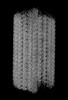 | |||||||||||||||||||||
|---|---|---|---|---|---|---|---|---|---|---|---|---|---|---|---|---|---|---|---|---|---|---|
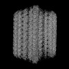
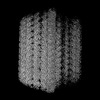
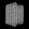
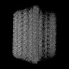
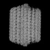
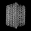
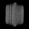
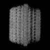
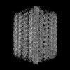
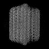
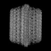
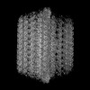
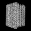
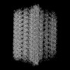
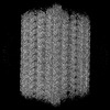
| Title | 48-nm doublet microtubule from Trichomonas vaginalis strain G3 | |||||||||||||||||||||
 Map data Map data | ||||||||||||||||||||||
 Sample Sample |
| |||||||||||||||||||||
 Keywords Keywords | doublet microtubule / tubulin / flagella / STRUCTURAL PROTEIN | |||||||||||||||||||||
| Function / homology |  Function and homology information Function and homology informationcilium-dependent cell motility / regulation of cilium beat frequency involved in ciliary motility / axonemal microtubule / nucleoside-diphosphate kinase / UTP biosynthetic process / CTP biosynthetic process / motile cilium / nucleoside diphosphate kinase activity / positive regulation of cell motility / GTP biosynthetic process ...cilium-dependent cell motility / regulation of cilium beat frequency involved in ciliary motility / axonemal microtubule / nucleoside-diphosphate kinase / UTP biosynthetic process / CTP biosynthetic process / motile cilium / nucleoside diphosphate kinase activity / positive regulation of cell motility / GTP biosynthetic process / mitotic cytokinesis / cilium assembly / axoneme / alpha-tubulin binding / Hsp70 protein binding / mitotic spindle organization / meiotic cell cycle / Hsp90 protein binding / structural constituent of cytoskeleton / microtubule cytoskeleton organization / mitotic spindle / mitotic cell cycle / cytoskeleton / microtubule / calmodulin binding / cilium / ciliary basal body / hydrolase activity / GTPase activity / GTP binding / ATP binding / metal ion binding / nucleus / cytoplasm Similarity search - Function | |||||||||||||||||||||
| Biological species |  Trichomonas vaginalis G3 (eukaryote) Trichomonas vaginalis G3 (eukaryote) | |||||||||||||||||||||
| Method | single particle reconstruction / cryo EM / Resolution: 4.2 Å | |||||||||||||||||||||
 Authors Authors | Stevens A / Zhou HZ / Kashyap S / Crofut EJ | |||||||||||||||||||||
| Funding support |  United States, 6 items United States, 6 items
| |||||||||||||||||||||
 Citation Citation | Journal: Nat Commun / Year: 2025 Title: Structures of Native Doublet Microtubules from Trichomonas vaginalis Reveal Parasite-Specific Proteins. Authors: Alexander Stevens / Saarang Kashyap / Ethan H Crofut / Shuqi E Wang / Katherine A Muratore / Patricia J Johnson / Z Hong Zhou / Abstract: Doublet microtubules (DMTs) are flagellar components required for the protist Trichomonas vaginalis (Tv) to swim through the human genitourinary tract to cause trichomoniasis, the most common non- ...Doublet microtubules (DMTs) are flagellar components required for the protist Trichomonas vaginalis (Tv) to swim through the human genitourinary tract to cause trichomoniasis, the most common non-viral sexually transmitted disease. Lack of structures of Tv's DMT (Tv-DMT) has prevented structure-guided drug design to manage Tv infection. Here, we determine the 16 nm, 32 nm, 48 nm and 96 nm-repeat structures of native Tv-DMT at resolution ranging from 3.4 to 4.4 Å by cryogenic electron microscopy (cryoEM) and built an atomic model for the entire Tv-DMT. These structures show that Tv-DMT is composed of 30 different proteins, including the α- and β-tubulin, 19 microtubule inner proteins (MIPs) and 9 microtubule outer proteins. While the A-tubule of Tv-DMT is simplistic compared to DMTs of other organisms, the B-tubule of Tv-DMT features parasite-specific proteins, such as TvFAP40 and TvFAP35. Notably, TvFAP40 and TvFAP35 form filaments near the inner and outer junctions, respectively, and interface with stabilizing MIPs. This atomic model of the Tv-DMT highlights diversity of eukaryotic motility machineries and provides a structural framework to inform rational design of therapeutics against trichomoniasis. | |||||||||||||||||||||
| History |
|
- Structure visualization
| Supplemental images |
|---|

- Downloads & links
-EMDB archive
| Map data | emd_46580.map.gz | 1.3 GB | EMDB map data format | |
|---|---|---|---|---|
| Header (meta data) | emd-46580-v30.xmlemd-46580.xml | 46.8 KB 46.8 KB | Display Display | EMDB header |
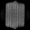
| Images |  emd_46580.png emd_46580.png | 147.9 KB | ||
| Filedesc metadata | emd-46580.cif.gz | 12.8 KB | ||
| Archive directory |  http://ftp.pdbj.org/pub/emdb/structures/EMD-46580ftp://ftp.pdbj.org/pub/emdb/structures/EMD-46580 http://ftp.pdbj.org/pub/emdb/structures/EMD-46580ftp://ftp.pdbj.org/pub/emdb/structures/EMD-46580 | HTTPS FTP |
-Related structure data
| Related structure data | 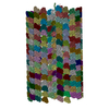 9d5nMC C: citing same article ( M: atomic model generated by this map |
|---|---|
| Similar structure data |


-Links
| EMDB pages | EMDB (EBI/PDBe) / EMDataResource |
|---|---|
| Related items in Molecule of the Month |


-Map
| File | Download / File: emd_46580.map.gz / Format: CCP4 / Size: 1.5 GB / Type: IMAGE STORED AS FLOATING POINT NUMBER (4 BYTES) | ||||||||||||||||||||||||||||||||||||
|---|---|---|---|---|---|---|---|---|---|---|---|---|---|---|---|---|---|---|---|---|---|---|---|---|---|---|---|---|---|---|---|---|---|---|---|---|---|
| Projections & slices | Image control
Images are generated by Spider. generated in cubic-lattice coordinate | ||||||||||||||||||||||||||||||||||||
| Voxel size | X=Y=Z: 1.1 Å | ||||||||||||||||||||||||||||||||||||
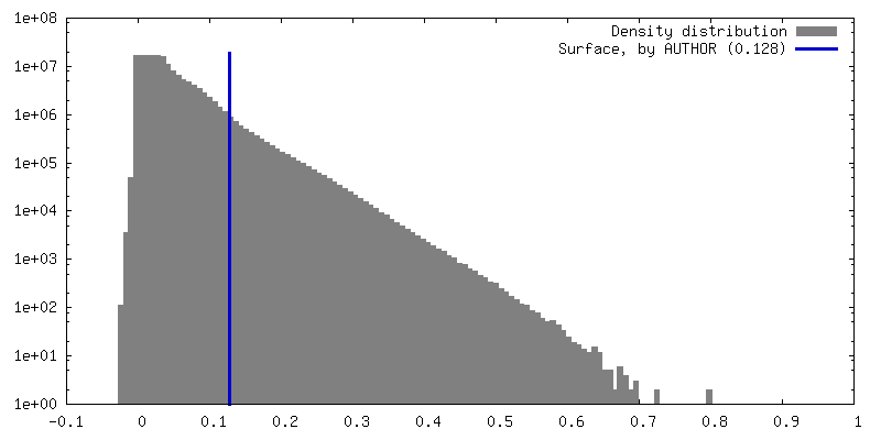
| Density |
| ||||||||||||||||||||||||||||||||||||
| Symmetry | Space group: 1 | ||||||||||||||||||||||||||||||||||||
| Details | EMDB XML:
|
 Z (Sec.)
 Y (Row.)
 X (Col.)


-Supplemental data
- Sample components
+Entire : doublet microtubule with microtubule inner proteins
+Supramolecule #1: doublet microtubule with microtubule inner proteins
+Macromolecule #1: Tubulin beta chain
+Macromolecule #2: FAP21
+Macromolecule #3: Tubulin alpha chain
+Macromolecule #4: IQ calmodulin-binding motif family protein
+Macromolecule #5: Flagellar protofilament ribbon protein, putative
+Macromolecule #6: TvFAP40
+Macromolecule #7: Parkin co-regulated protein
+Macromolecule #8: Cilia- and flagella-associated protein 45
+Macromolecule #9: Cilia- and flagella-associated protein 20
+Macromolecule #10: Cilia- and flagella-associated protein 53
+Macromolecule #11: Trichohyalin-plectin-homology domain-containing protein
+Macromolecule #12: Parkin co-regulated protein
+Macromolecule #13: Enkurin
+Macromolecule #14: FAP77
+Macromolecule #15: EF hand family protein
+Macromolecule #16: Cilia- and flagella-associated protein 52
+Macromolecule #17: Nucleoside diphosphate kinase
+Macromolecule #18: FAP161
+Macromolecule #19: RIIa domain-containing protein
+Macromolecule #20: FAP12
+Macromolecule #21: Meiosis-specific nuclear structural protein 1
+Macromolecule #22: MGC84469 protein, putative
+Macromolecule #23: GUANOSINE-5'-DIPHOSPHATE

+Macromolecule #24: GUANOSINE-5'-TRIPHOSPHATE

+Macromolecule #25: MAGNESIUM ION
-Experimental details
-Structure determination
| Method | cryo EM |
|---|---|
 Processing Processing | single particle reconstruction |
| Aggregation state | particle |
-Sample preparation
| Buffer | pH: 7.4 Component:
Details: 150 mM NaCl, 50 mM 325 Tris, 2 mM MgCl2, 1 mM DTT, 5 mM ATP 2 x complete protease inhibitor, pH 7.4 | |||||||||||||||
|---|---|---|---|---|---|---|---|---|---|---|---|---|---|---|---|---|
| Grid | Model: Quantifoil R2/1 / Material: COPPER / Mesh: 200 / Support film - Material: CARBON / Support film - topology: HOLEY / Pretreatment - Type: GLOW DISCHARGE / Pretreatment - Time: 30 sec. | |||||||||||||||
| Vitrification | Cryogen name: ETHANE-PROPANE / Chamber humidity: 100 % / Chamber temperature: 281 K / Instrument: FEI VITROBOT MARK IV |
- Electron microscopy
| Microscope | FEI TITAN KRIOS |
|---|---|
| Image recording | Film or detector model: GATAN K3 (6k x 4k) / Average electron dose: 45.0 e/Å2 |
| Electron beam | Acceleration voltage: 300 kV / Electron source:  FIELD EMISSION GUN FIELD EMISSION GUN |
| Electron optics | Calibrated defocus max: 2.0 µm / Calibrated defocus min: 1.2 µm / Illumination mode: FLOOD BEAM / Imaging mode: BRIGHT FIELD / Cs: 2.7 mm / Nominal defocus max: 2.0 µm / Nominal defocus min: 1.2 µm / Nominal magnification: 81000 |
| Sample stage | Specimen holder model: FEI TITAN KRIOS AUTOGRID HOLDER / Cooling holder cryogen: NITROGEN |
| Experimental equipment |  Model: Titan Krios / Image courtesy: FEI Company |
+Image processing
-Atomic model buiding 1
| Initial model | Chain - Source name: AlphaFold / Chain - Initial model type: in silico model |
|---|---|
| Refinement | Space: REAL / Protocol: FLEXIBLE FIT |
| Output model | PDB-9d5n: |
