Movie
Movie Controller


+ Open data

- Basic information
| Entry | Database: EMDB / ID: EMD-4615 | |||||||||
|---|---|---|---|---|---|---|---|---|---|---|
| Title | Dps-DNA crystal structure determined in vitro | |||||||||
 Map data Map data | ||||||||||
 Sample Sample |
| |||||||||
| Biological species |  | |||||||||
| Method | subtomogram averaging / cryo EM / Resolution: 13.5 Å | |||||||||
 Authors Authors | Chesnokov YM / Kamyshinsky RA / Orekhov AS / Vasiliev AL | |||||||||
| Funding support |  Russian Federation, 1 items Russian Federation, 1 items
| |||||||||
 Citation Citation | Journal: FEBS Lett / Year: 2019 Title: Protective Dps-DNA co-crystallization in stressed cells: an in vitro structural study by small-angle X-ray scattering and cryo-electron tomography. Authors: Liubov A Dadinova / Yurii M Chesnokov / Roman A Kamyshinsky / Ivan A Orlov / Maxim V Petoukhov / Andrey A Mozhaev / Ekaterina Yu Soshinskaya / Vassili N Lazarev / Valentin A Manuvera / Anton ...Authors: Liubov A Dadinova / Yurii M Chesnokov / Roman A Kamyshinsky / Ivan A Orlov / Maxim V Petoukhov / Andrey A Mozhaev / Ekaterina Yu Soshinskaya / Vassili N Lazarev / Valentin A Manuvera / Anton S Orekhov / Alexander L Vasiliev / Eleonora V Shtykova /  Abstract: Under severe or prolonged stress, bacteria produce a nonspecific DNA-binding protein (Dps), which effectively protects DNA against damaging agents both in vitro and in vivo by forming intracellular ...Under severe or prolonged stress, bacteria produce a nonspecific DNA-binding protein (Dps), which effectively protects DNA against damaging agents both in vitro and in vivo by forming intracellular biocrystals. The phenomenon of protective crystallization of DNA in living cells has been intensively investigated during the last two decades; however, the results of studies are somewhat contradictory, and up to now, there has been no direct determination of a Dps-DNA crystal structure. Here, we report the in vitro analysis of the vital process of Dps-DNA co-crystallization using two complementary structural methods: synchrotron small-angle X-ray scattering in solution and cryo-electron tomography. Importantly, for the first time, the DNA in the co-crystals was visualized, and the lattice parameters of the crystalline Dps-DNA complex were determined. | |||||||||
| History |
|
- Structure visualization
| Movie |
 Movie viewer Movie viewer |
|---|---|
| Structure viewer | EM map: SurfViewMolmilJmol/JSmol |
| Supplemental images |

- Downloads & links
-EMDB archive
| Map data | emd_4615.map.gz | 1.5 MB | EMDB map data format | |
|---|---|---|---|---|
| Header (meta data) | emd-4615-v30.xmlemd-4615.xml | 17 KB 17 KB | Display Display | EMDB header |
| FSC (resolution estimation) | emd_4615_fsc.xml | 3 KB | Display | FSC data file |
| Images | 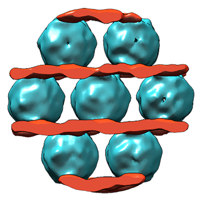 emd_4615.png emd_4615.png | 202.4 KB | ||
| Masks | emd_4615_msk_1.map | 2 MB | Mask map | |
| Others | emd_4615_half_map_1.map.gzemd_4615_half_map_2.map.gz | 1.4 MB 1.4 MB | ||
| Archive directory |  http://ftp.pdbj.org/pub/emdb/structures/EMD-4615ftp://ftp.pdbj.org/pub/emdb/structures/EMD-4615 http://ftp.pdbj.org/pub/emdb/structures/EMD-4615ftp://ftp.pdbj.org/pub/emdb/structures/EMD-4615 | HTTPS FTP |
-Validation report
| Summary document | emd_4615_validation.pdf.gz | 418.4 KB | Display | EMDB validaton report |
|---|---|---|---|---|
| Full document | emd_4615_full_validation.pdf.gz | 417.5 KB | Display | |
| Data in XML | emd_4615_validation.xml.gz | 8.4 KB | Display | |
| Arichive directory | https://ftp.pdbj.org/pub/emdb/validation_reports/EMD-4615ftp://ftp.pdbj.org/pub/emdb/validation_reports/EMD-4615 | HTTPS FTP |
-Related structure data
| Similar structure data |
|---|


-Links
| EMDB pages | EMDB (EBI/PDBe) / EMDataResource |
|---|
-Map
| File | Download / File: emd_4615.map.gz / Format: CCP4 / Size: 2 MB / Type: IMAGE STORED AS FLOATING POINT NUMBER (4 BYTES) | ||||||||||||||||||||||||||||||||||||||||||||||||||||||||||||||||||||
|---|---|---|---|---|---|---|---|---|---|---|---|---|---|---|---|---|---|---|---|---|---|---|---|---|---|---|---|---|---|---|---|---|---|---|---|---|---|---|---|---|---|---|---|---|---|---|---|---|---|---|---|---|---|---|---|---|---|---|---|---|---|---|---|---|---|---|---|---|---|
| Projections & slices | Image control
Images are generated by Spider. | ||||||||||||||||||||||||||||||||||||||||||||||||||||||||||||||||||||
| Voxel size | X=Y=Z: 3.7 Å | ||||||||||||||||||||||||||||||||||||||||||||||||||||||||||||||||||||
| Density |
| ||||||||||||||||||||||||||||||||||||||||||||||||||||||||||||||||||||
| Symmetry | Space group: 1 | ||||||||||||||||||||||||||||||||||||||||||||||||||||||||||||||||||||
| Details | EMDB XML:
CCP4 map header:
| ||||||||||||||||||||||||||||||||||||||||||||||||||||||||||||||||||||
 Z (Sec.)
 Y (Row.)
 X (Col.)


-Supplemental data
-Mask #1
| File | emd_4615_msk_1.map | ||||||||||||
|---|---|---|---|---|---|---|---|---|---|---|---|---|---|
| Projections & Slices |
| ||||||||||||
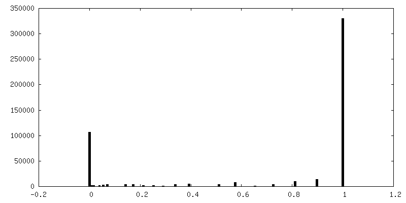
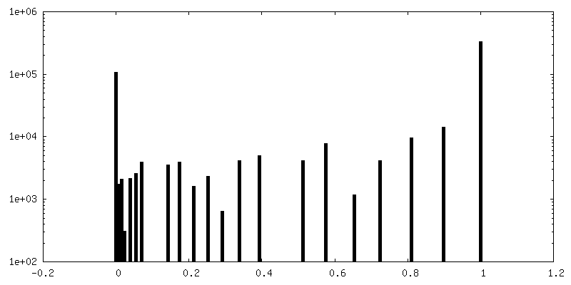
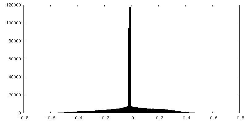
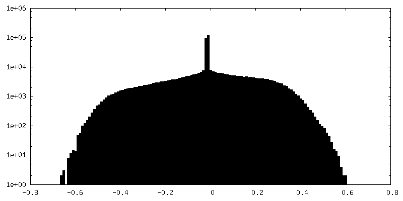
| Density Histograms |


-Half map: #1
| File | emd_4615_half_map_1.map | ||||||||||||
|---|---|---|---|---|---|---|---|---|---|---|---|---|---|
| Projections & Slices |
| ||||||||||||
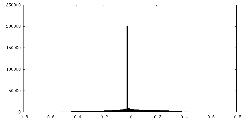
| Density Histograms |


-Half map: #2
| File | emd_4615_half_map_2.map | ||||||||||||
|---|---|---|---|---|---|---|---|---|---|---|---|---|---|
| Projections & Slices |
| ||||||||||||
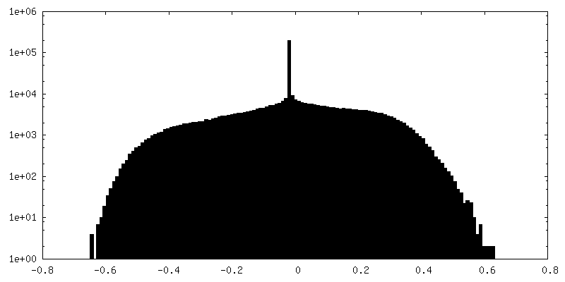
| Density Histograms |


- Sample components
-Entire : Dps-DNA co-crystal
| Entire | Name: Dps-DNA co-crystal |
|---|---|
| Components |
|
-Supramolecule #1: Dps-DNA co-crystal
| Supramolecule | Name: Dps-DNA co-crystal / type: complex / ID: 1 / Parent: 0 Details: Dps protein and DNA were mixed in the concentrations corresponding to the Dps/DNA ratio 1Dps dodecamer/60 bp of DNA. A ring vector pcDNA-hIRR-GFP 9900 bp was used as DNA sample. |
|---|---|
| Source (natural) | Organism: |
| Recombinant expression | Organism: |
| Molecular weight | Theoretical: 224 KDa |
-Experimental details
-Structure determination
| Method | cryo EM |
|---|---|
 Processing Processing | subtomogram averaging |
| Aggregation state | 3D array |
-Sample preparation
| Concentration | 3.1 mg/mL | ||||||||
|---|---|---|---|---|---|---|---|---|---|
| Buffer | pH: 8 Component:
| ||||||||
| Grid | Material: COPPER / Mesh: 200 / Support film - Material: CARBON / Support film - topology: LACEY / Pretreatment - Type: GLOW DISCHARGE / Pretreatment - Atmosphere: AIR / Pretreatment - Pressure: 0.026 kPa / Details: Pelco EasiGlow ( 25 mA) | ||||||||
| Vitrification | Cryogen name: ETHANE / Chamber humidity: 95 % / Chamber temperature: 283 K / Instrument: FEI VITROBOT MARK IV / Details: blotting for 1.5 seconds. |
- Electron microscopy
| Microscope | FEI TITAN KRIOS |
|---|---|
| Specialist optics | Spherical aberration corrector: Cs image corrector (CEOS, Germany) |
| Image recording | Film or detector model: FEI FALCON II (4k x 4k) / Detector mode: INTEGRATING / Digitization - Dimensions - Width: 4096 pixel / Digitization - Dimensions - Height: 4096 pixel / Digitization - Sampling interval: 14.0 µm / Number grids imaged: 1 / Average exposure time: 1.0 sec. / Average electron dose: 1.02 e/Å2 |
| Electron beam | Acceleration voltage: 300 kV / Electron source:  FIELD EMISSION GUN FIELD EMISSION GUN |
| Electron optics | C2 aperture diameter: 100.0 µm / Calibrated magnification: 37837 / Illumination mode: FLOOD BEAM / Imaging mode: BRIGHT FIELD / Cs: 0.01 mm / Nominal defocus max: 4.5 µm / Nominal defocus min: 3.0 µm / Nominal magnification: 18000 |
| Sample stage | Specimen holder model: FEI TITAN KRIOS AUTOGRID HOLDER / Cooling holder cryogen: NITROGEN |
| Experimental equipment |  Model: Titan Krios / Image courtesy: FEI Company |
+Image processing

-Atomic model buiding 1
| Initial model | PDB ID: |
|---|---|
| Refinement | Space: REAL / Protocol: RIGID BODY FIT / Target criteria: Cross-correlation coefficient |