National Institutes of Health/National Institute of Neurological Disorders and Stroke (NIH/NINDS)
P01NS092525
United States
National Institutes of Health/National Institute of General Medical Sciences (NIH/NIGMS)
5P41GM103832
United States
National Institutes of Health/National Institute on Aging (NIH/NIA)
PO1AG054407
United States
National Institutes of Health/National Institute of Neurological Disorders and Stroke (NIH/NINDS)
F32NS086253
United States
National Institutes of Health/National Institute of General Medical Sciences (NIH/NIGMS)
S10OD021600
United States
Citation
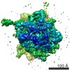
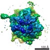
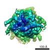
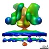
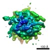
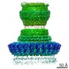
Journal: Structure / Year: 2020 Title: Multi-scale 3D Cryo-Correlative Microscopy for Vitrified Cells. Authors: Gong-Her Wu / Patrick G Mitchell / Jesus G Galaz-Montoya / Corey W Hecksel / Emily M Sontag / Vimal Gangadharan / Jeffrey Marshman / David Mankus / Margaret E Bisher / Abigail K R Lytton- ...Authors: Gong-Her Wu / Patrick G Mitchell / Jesus G Galaz-Montoya / Corey W Hecksel / Emily M Sontag / Vimal Gangadharan / Jeffrey Marshman / David Mankus / Margaret E Bisher / Abigail K R Lytton-Jean / Judith Frydman / Kirk Czymmek / Wah Chiu / Abstract: Three-dimensional (3D) visualization of vitrified cells can uncover structures of subcellular complexes without chemical fixation or staining. Here, we present a pipeline integrating three imaging ...Three-dimensional (3D) visualization of vitrified cells can uncover structures of subcellular complexes without chemical fixation or staining. Here, we present a pipeline integrating three imaging modalities to visualize the same specimen at cryogenic temperature at different scales: cryo-fluorescence confocal microscopy, volume cryo-focused ion beam scanning electron microscopy, and transmission cryo-electron tomography. Our proof-of-concept benchmark revealed the 3D distribution of organelles and subcellular structures in whole heat-shocked yeast cells, including the ultrastructure of protein inclusions that recruit fluorescently-labeled chaperone Hsp104. Since our workflow efficiently integrates imaging at three different scales and can be applied to other types of cells, it could be used for large-scale phenotypic studies of frozen-hydrated specimens in a variety of healthy and diseased conditions with and without treatments.
History
Deposition
May 10, 2020
-
Header (metadata) release
Sep 2, 2020
-
Map release
Sep 2, 2020
-
Update
Sep 2, 2020
-
Current status
Sep 2, 2020
Processing site: RCSB / Status: Released
-
Structure visualization
Movie
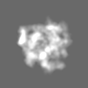
Surface view with section colored by density value
In the structure databanks used in Yorodumi, some data are registered as the other names, "COVID-19 virus" and "2019-nCoV". Here are the details of the virus and the list of structure data.
Jan 31, 2019. EMDB accession codes are about to change! (news from PDBe EMDB page)
EMDB accession codes are about to change! (news from PDBe EMDB page)
The allocation of 4 digits for EMDB accession codes will soon come to an end. Whilst these codes will remain in use, new EMDB accession codes will include an additional digit and will expand incrementally as the available range of codes is exhausted. The current 4-digit format prefixed with “EMD-” (i.e. EMD-XXXX) will advance to a 5-digit format (i.e. EMD-XXXXX), and so on. It is currently estimated that the 4-digit codes will be depleted around Spring 2019, at which point the 5-digit format will come into force.
The EM Navigator/Yorodumi systems omit the EMD- prefix.
Related info.:Q: What is EMD? / ID/Accession-code notation in Yorodumi/EM Navigator
Yorodumi is a browser for structure data from EMDB, PDB, SASBDB, etc.
This page is also the successor to EM Navigator detail page, and also detail information page/front-end page for Omokage search.
The word "yorodu" (or yorozu) is an old Japanese word meaning "ten thousand". "mi" (miru) is to see.
Related info.:EMDB / PDB / SASBDB / Comparison of 3 databanks / Yorodumi Search / Aug 31, 2016. New EM Navigator & Yorodumi / Yorodumi Papers / Jmol/JSmol / Function and homology information / Changes in new EM Navigator and Yorodumi
 Movie
Movie Controller

 Yorodumi
 Open data
 Basic information
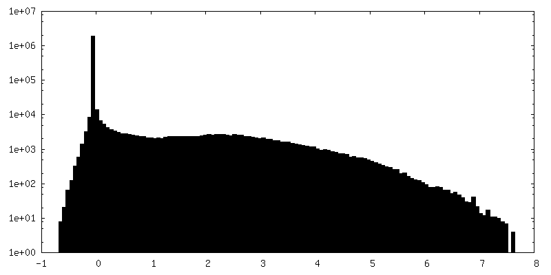
 Map data
 Sample
 Authors
 United States, 5 items
  Citation
 Structure visualization
 Movie viewer
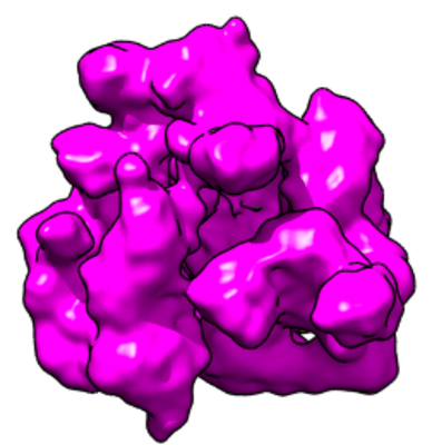
 Downloads & links
 emd_21952.png
 http://ftp.pdbj.org/pub/emdb/structures/EMD-21952

 Z (Sec.)
 Y (Row.)
 X (Col.)


 Sample components
 Processing
 Electron microscopy
 FIELD EMISSION GUN
