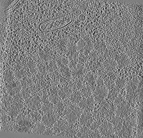
- EMDB-21953: Downsampled and filtered tomogram of a region from a cryoFIB-SEM-... -
+
Open data
ID or keywords:
Loading...
-
Basic information
Entry
Database: EMDB / ID: EMD-21953
Title
Downsampled and filtered tomogram of a region from a cryoFIB-SEM-generated lamellae of yeast cells under heat shock stress showing large protein aggregates
Map data
Binned-by-8 and filtered tomogram of a region from a cryoFIB-SEM-generated lamellae of yeast cells under heat shock stress showing large protein aggregates.
Sample
Cell: Downsamples and filtered tomogram of a region from a cryoFIB-SEM-generated lamellae of yeast cells under heat shock stress showing large protein aggregates
National Institutes of Health/National Institute of Neurological Disorders and Stroke (NIH/NINDS)
P01NS092525
United States
National Institutes of Health/National Institute of General Medical Sciences (NIH/NIGMS)
5P41GM103832
United States
National Institutes of Health/National Institute on Aging (NIH/NIA)
PO1AG054407
United States
National Institutes of Health/National Institute of Neurological Disorders and Stroke (NIH/NINDS)
F32NS086253
United States
National Institutes of Health/National Institute of General Medical Sciences (NIH/NIGMS)
S10OD021600
United States
Department of Energy (DOE, United States)
BERFWP 100463
United States
Citation
Journal: Structure / Year: 2020 Title: Multi-scale 3D Cryo-Correlative Microscopy for Vitrified Cells. Authors: Gong-Her Wu / Patrick G Mitchell / Jesus G Galaz-Montoya / Corey W Hecksel / Emily M Sontag / Vimal Gangadharan / Jeffrey Marshman / David Mankus / Margaret E Bisher / Abigail K R Lytton- ...Authors: Gong-Her Wu / Patrick G Mitchell / Jesus G Galaz-Montoya / Corey W Hecksel / Emily M Sontag / Vimal Gangadharan / Jeffrey Marshman / David Mankus / Margaret E Bisher / Abigail K R Lytton-Jean / Judith Frydman / Kirk Czymmek / Wah Chiu / Abstract: Three-dimensional (3D) visualization of vitrified cells can uncover structures of subcellular complexes without chemical fixation or staining. Here, we present a pipeline integrating three imaging ...Three-dimensional (3D) visualization of vitrified cells can uncover structures of subcellular complexes without chemical fixation or staining. Here, we present a pipeline integrating three imaging modalities to visualize the same specimen at cryogenic temperature at different scales: cryo-fluorescence confocal microscopy, volume cryo-focused ion beam scanning electron microscopy, and transmission cryo-electron tomography. Our proof-of-concept benchmark revealed the 3D distribution of organelles and subcellular structures in whole heat-shocked yeast cells, including the ultrastructure of protein inclusions that recruit fluorescently-labeled chaperone Hsp104. Since our workflow efficiently integrates imaging at three different scales and can be applied to other types of cells, it could be used for large-scale phenotypic studies of frozen-hydrated specimens in a variety of healthy and diseased conditions with and without treatments.
Download / File: emd_21953.map.gz / Format: CCP4 / Size: 174.3 MB / Type: IMAGE STORED AS FLOATING POINT NUMBER (4 BYTES)
Annotation
Binned-by-8 and filtered tomogram of a region from a cryoFIB-SEM-generated lamellae of yeast cells under heat shock stress showing large protein aggregates.
A: 13251.054 Å / B: 12808.43 Å / C: 5698.783 Å α=β=γ: 90.0 °
CCP4 map header:
mode
Image stored as Reals
Å/pix. X/Y/Z
27.663995824635
27.663995680346
27.663995145631
M x/y/z
479
463
206
origin x/y/z
0.000
0.000
0.000
length x/y/z
13251.054
12808.430
5698.783
α/β/γ
90.000
90.000
90.000
start NX/NY/NZ
79
74
0
NX/NY/NZ
93
103
213
MAP C/R/S
1
2
3
start NC/NR/NS
0
0
-103
NC/NR/NS
479
463
206
D min/max/mean
-3.000
3.000
-0.000
-
Supplemental data
-
Sample components
-
Entire : Downsamples and filtered tomogram of a region from a cryoFIB-SEM-...
Entire
Name: Downsamples and filtered tomogram of a region from a cryoFIB-SEM-generated lamellae of yeast cells under heat shock stress showing large protein aggregates
Components
Cell: Downsamples and filtered tomogram of a region from a cryoFIB-SEM-generated lamellae of yeast cells under heat shock stress showing large protein aggregates
-
Supramolecule #1: Downsamples and filtered tomogram of a region from a cryoFIB-SEM-...
Supramolecule
Name: Downsamples and filtered tomogram of a region from a cryoFIB-SEM-generated lamellae of yeast cells under heat shock stress showing large protein aggregates type: cell / ID: 1 / Parent: 0
Focused ion beam - Instrument: OTHER / Focused ion beam - Ion: OTHER / Focused ion beam - Voltage: 30 kV / Focused ion beam - Current: 20 nA / Focused ion beam - Duration: 60 sec. / Focused ion beam - Temperature: 100 K / Focused ion beam - Initial thickness: 1000 nm / Focused ion beam - Final thickness: 520 nm Focused ion beam - Details: The value given for _emd_sectioning_focused_ion_beam.instrument is Zeiss Crossbeam 540 FIB-SEM. This is not in a list of allowed values set(['DB235', 'OTHER']) so OTHER is ...Focused ion beam - Details: The value given for _emd_sectioning_focused_ion_beam.instrument is Zeiss Crossbeam 540 FIB-SEM. This is not in a list of allowed values set(['DB235', 'OTHER']) so OTHER is written into the XML file.
-
Electron microscopy
Microscope
TFS KRIOS
Image recording
Film or detector model: GATAN K2 SUMMIT (4k x 4k) / Detector mode: COUNTING / Average electron dose: 0.8264 e/Å2
Electron beam
Acceleration voltage: 300 kV / Electron source: FIELD EMISSION GUN
Electron optics
Illumination mode: FLOOD BEAM / Imaging mode: BRIGHT FIELD
Experimental equipment
Model: Titan Krios / Image courtesy: FEI Company
-
Image processing
Final reconstruction
Algorithm: SIMULTANEOUS ITERATIVE (SIRT) / Software - Name: IMOD Details: The SIRT, downsampled, and filtered tomogram was computed for visualization only. The related EMDB entry showcasing a ribosome subtomogram average used similar tomograms but reconstructed ...Details: The SIRT, downsampled, and filtered tomogram was computed for visualization only. The related EMDB entry showcasing a ribosome subtomogram average used similar tomograms but reconstructed with weighted-back-projection, at full-size, and unfiltered. Number images used: 121
CTF correction
Software - Name: IMOD / Details: 3D CTF correction
+
About Yorodumi
-
News
-
Feb 9, 2022. New format data for meta-information of EMDB entries
New format data for meta-information of EMDB entries
Version 3 of the EMDB header file is now the official format.
The previous official version 1.9 will be removed from the archive.
In the structure databanks used in Yorodumi, some data are registered as the other names, "COVID-19 virus" and "2019-nCoV". Here are the details of the virus and the list of structure data.
Jan 31, 2019. EMDB accession codes are about to change! (news from PDBe EMDB page)
EMDB accession codes are about to change! (news from PDBe EMDB page)
The allocation of 4 digits for EMDB accession codes will soon come to an end. Whilst these codes will remain in use, new EMDB accession codes will include an additional digit and will expand incrementally as the available range of codes is exhausted. The current 4-digit format prefixed with “EMD-” (i.e. EMD-XXXX) will advance to a 5-digit format (i.e. EMD-XXXXX), and so on. It is currently estimated that the 4-digit codes will be depleted around Spring 2019, at which point the 5-digit format will come into force.
The EM Navigator/Yorodumi systems omit the EMD- prefix.
Related info.:Q: What is EMD? / ID/Accession-code notation in Yorodumi/EM Navigator
Yorodumi is a browser for structure data from EMDB, PDB, SASBDB, etc.
This page is also the successor to EM Navigator detail page, and also detail information page/front-end page for Omokage search.
The word "yorodu" (or yorozu) is an old Japanese word meaning "ten thousand". "mi" (miru) is to see.
Related info.:EMDB / PDB / SASBDB / Comparison of 3 databanks / Yorodumi Search / Aug 31, 2016. New EM Navigator & Yorodumi / Yorodumi Papers / Jmol/JSmol / Function and homology information / Changes in new EM Navigator and Yorodumi
 Movie
Movie Controller

 Yorodumi
 Open data
 Basic information
 Map data
 Sample
 Authors
 United States, 6 items
  Citation
 Structure visualization
 Movie viewer
 Downloads & links
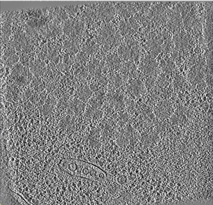 emd_21953.png
 http://ftp.pdbj.org/pub/emdb/structures/EMD-21953
 Z (Sec.)
 Y (Row.)
 X (Col.)


 Sample components
 Processing
 Electron microscopy
 FIELD EMISSION GUN
