Movie
Movie Controller

[English] 日本語
 Yorodumi
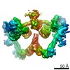
- EMDB-45850: Focused map of XRCC4/Ligase IV on the active side of ligation com... -
+ Open data

- Basic information
| Entry |  | |||||||||
|---|---|---|---|---|---|---|---|---|---|---|
| Title | Focused map of XRCC4/Ligase IV on the active side of ligation complex in the NHEJ pathway | |||||||||
 Map data Map data | Focused map of active XRCC4/Ligase IV complex in the ligation complex I | |||||||||
 Sample Sample |
| |||||||||
 Keywords Keywords | NHEJ / ligation / XLF / PAXX / DNA repair / Ligase IV / DNA BINDING PROTEIN | |||||||||
| Biological species |  Homo sapiens (human) Homo sapiens (human) | |||||||||
| Method | single particle reconstruction / cryo EM / Resolution: 3.9 Å | |||||||||
 Authors Authors | Li J / Liu L / Gellert M / Yang W | |||||||||
| Funding support |  United States, 1 items United States, 1 items
| |||||||||
 Citation Citation | Journal: To Be Published Title: The ligation complex in the NHEJ pathway Authors: Li J / Liu L / Gellert M / Yang W | |||||||||
| History |
|
- Structure visualization
| Supplemental images |
|---|

- Downloads & links
-EMDB archive
| Map data | emd_45850.map.gz | 252 MB |  EMDB map data format EMDB map data format | |
|---|---|---|---|---|
| Header (meta data) | emd-45850-v30.xmlemd-45850.xml | 16.6 KB 16.6 KB | Display Display | EMDB header |
| FSC (resolution estimation) | emd_45850_fsc.xml | 18.2 KB | Display | FSC data file |
| Images |  emd_45850.png emd_45850.png | 81.2 KB | ||
| Masks | emd_45850_msk_1.map | 512 MB | Mask map | |
| Filedesc metadata | emd-45850.cif.gz | 5.6 KB | ||
| Others | emd_45850_half_map_1.map.gzemd_45850_half_map_2.map.gz | 474.8 MB 474.8 MB | ||
| Archive directory |  http://ftp.pdbj.org/pub/emdb/structures/EMD-45850ftp://ftp.pdbj.org/pub/emdb/structures/EMD-45850 http://ftp.pdbj.org/pub/emdb/structures/EMD-45850ftp://ftp.pdbj.org/pub/emdb/structures/EMD-45850 | HTTPS FTP |
-Validation report
| Summary document | emd_45850_validation.pdf.gz | 967.8 KB | Display | EMDB validaton report |
|---|---|---|---|---|
| Full document | emd_45850_full_validation.pdf.gz | 967.3 KB | Display | |
| Data in XML | emd_45850_validation.xml.gz | 26 KB | Display | |
| Data in CIF | emd_45850_validation.cif.gz | 34.6 KB | Display | |
| Arichive directory | https://ftp.pdbj.org/pub/emdb/validation_reports/EMD-45850ftp://ftp.pdbj.org/pub/emdb/validation_reports/EMD-45850 | HTTPS FTP |
-Related structure data
| Related structure data | C: citing same article ( |
|---|


-Links
| EMDB pages | EMDB (EBI/PDBe) / EMDataResource |
|---|
-Map
| File | Download / File: emd_45850.map.gz / Format: CCP4 / Size: 512 MB / Type: IMAGE STORED AS FLOATING POINT NUMBER (4 BYTES) | ||||||||||||||||||||||||||||||||||||
|---|---|---|---|---|---|---|---|---|---|---|---|---|---|---|---|---|---|---|---|---|---|---|---|---|---|---|---|---|---|---|---|---|---|---|---|---|---|
| Annotation | Focused map of active XRCC4/Ligase IV complex in the ligation complex I | ||||||||||||||||||||||||||||||||||||
| Projections & slices | Image control
Images are generated by Spider. | ||||||||||||||||||||||||||||||||||||
| Voxel size | X=Y=Z: 0.833 Å | ||||||||||||||||||||||||||||||||||||
| Density |
| ||||||||||||||||||||||||||||||||||||
| Symmetry | Space group: 1 | ||||||||||||||||||||||||||||||||||||
| Details | EMDB XML:
|
 Z (Sec.)
 Y (Row.)
 X (Col.)


-Supplemental data
-Mask #1
| File | emd_45850_msk_1.map | ||||||||||||
|---|---|---|---|---|---|---|---|---|---|---|---|---|---|
| Projections & Slices |
| ||||||||||||
| Density Histograms |


-Half map: half 2 map
| File | emd_45850_half_map_1.map | ||||||||||||
|---|---|---|---|---|---|---|---|---|---|---|---|---|---|
| Annotation | half 2 map | ||||||||||||
| Projections & Slices |
| ||||||||||||
| Density Histograms |


-Half map: half 1 map
| File | emd_45850_half_map_2.map | ||||||||||||
|---|---|---|---|---|---|---|---|---|---|---|---|---|---|
| Annotation | half 1 map | ||||||||||||
| Projections & Slices |
| ||||||||||||
| Density Histograms |


- Sample components
-Entire : Ligation complex in the NHEJ pathway
| Entire | Name: Ligation complex in the NHEJ pathway |
|---|---|
| Components |
|
-Supramolecule #1: Ligation complex in the NHEJ pathway
| Supramolecule | Name: Ligation complex in the NHEJ pathway / type: complex / ID: 1 / Parent: 0 / Macromolecule list: all |
|---|---|
| Source (natural) | Organism: Homo sapiens (human) |
| Molecular weight | Theoretical: 854 KDa |
-Macromolecule #1: human XRCC4
| Macromolecule | Name: human XRCC4 / type: protein_or_peptide / ID: 1 / Enantiomer: LEVO |
|---|---|
| Source (natural) | Organism: Homo sapiens (human) |
| Recombinant expression | Organism: Homo sapiens (human) |
| Sequence | String: MERKISRIHL VSEPSITHFL QVSWEKTLES GFVITLTDGH SAWTGTVSES EISQEADDMA MEKGKYVGE LRKALLSGAG PADVYTFNFS KESCYFFFEK NLKDVSFRLG SFNLEKVENP A EVIRELIC YCLDTIAENQ AKNEHLQKEN ERLLRDWNDV QGRFEKCVSA ...String: MERKISRIHL VSEPSITHFL QVSWEKTLES GFVITLTDGH SAWTGTVSES EISQEADDMA MEKGKYVGE LRKALLSGAG PADVYTFNFS KESCYFFFEK NLKDVSFRLG SFNLEKVENP A EVIRELIC YCLDTIAENQ AKNEHLQKEN ERLLRDWNDV QGRFEKCVSA KEALETDLYK RF ILVLNEK KTKIRSLHNK LLNAAQEREK DIKQEGETAI CSEMTADRDP VYDESTDEES ENQ TDLSGL ASAAVSKDDS IISSLDVTDI APSRKRRQRM QRNLGTEPKM APQENQLQEK ENSR PDSSL PETSKKEHIS AENMSLETLR NSSPEDLFDE I |
-Macromolecule #2: human Ligase IV
| Macromolecule | Name: human Ligase IV / type: protein_or_peptide / ID: 2 / Enantiomer: LEVO |
|---|---|
| Source (natural) | Organism: Homo sapiens (human) |
| Recombinant expression | Organism: Homo sapiens (human) |
| Sequence | String: GPVMAASQTS QTVASHVPFA DLCSTLERIQ KSKGRAEKIR HFREFLDSWR KFHDALHKNH KDV TDSFYP AMRLILPQLE RERMAYGIKE TMLAKLYIEL LNLPRDGKDA LKLLNYRTPT GTHGDAGDFA MIAYFVLKPR CLQKGSLTIQ QVNDLLDSIA SNNSAKRKDL ...String: GPVMAASQTS QTVASHVPFA DLCSTLERIQ KSKGRAEKIR HFREFLDSWR KFHDALHKNH KDV TDSFYP AMRLILPQLE RERMAYGIKE TMLAKLYIEL LNLPRDGKDA LKLLNYRTPT GTHGDAGDFA MIAYFVLKPR CLQKGSLTIQ QVNDLLDSIA SNNSAKRKDL IKKSLLQLIT QSSALEQKWL IRMIIKDLKL GVSQQTIFSV FHNDAAELHN VTTDLEKVCR QLHDPSVGLS DISI TLFSA FKPMLAAIAD IEHIEKDMKH QSFYIETKLD GERMQMHKDG DVYKYFSRNG YNYTD QFGA SPTEGSLTPF IHNAFKADIQ ICILDGEMMA YNPNTQTFMQ KGTKFDIKRM VEDSDL QTC YCVFDVLMVN NKKLGHETLR KRYEILSSIF TPIPGRIEIV QKTQAHTKNE VIDALNE AI DKREEGIMVK QPLSIYKPDK RGEGWLKIKP EYVSGLMDEL DILIVGGYWG KGSRGGMM S HFLCAVAEKP PPGEKPSVFH TLSRVGSGCT MKELYDLGLK LAKYWKPFHR KAPPSSILC GTEKPEVYIE PCNSVIVQIK AAEIVPSDMY KTGCTLRFPR IEKIRDDKEW HECMTLDDLE QLRGKASGK LASKHLYIGG DDEPQEKKRK AAPKMKKVIG IIEHLKAPNL TNVNKISNIF E DVEFCVMS GTDSQPKPDL ENRIAEFGGY IVQNPGPDTY CVIAGSENIR VKNIILSNKH DV VKPAWLL ECFKTKSFVP WQPRFMIHMC PSTKEHFARE YDCYGDSYFI DTDLNQLKEV FSG IKNSNE QTPEEMASLI ADLEYRYSWD CSPLSMFRRH TVYLDSYAVI NDLSTKNEGT RLAI KALEL RFHGAKVVSC LAEGVSHVII GEDHSRVADF KAFRRTFKRK FKILKESWVT DSIDK CELQ EENQYLI |
-Experimental details
-Structure determination
| Method | cryo EM |
|---|---|
 Processing Processing | single particle reconstruction |
| Aggregation state | particle |
-Sample preparation
| Concentration | 0.35 mg/mL |
|---|---|
| Buffer | pH: 7.9 |
| Vitrification | Cryogen name: ETHANE / Chamber humidity: 100 % / Chamber temperature: 277 K |
- Electron microscopy
| Microscope | FEI TITAN KRIOS |
|---|---|
| Image recording | Film or detector model: GATAN K3 BIOQUANTUM (6k x 4k) / Average electron dose: 42.2 e/Å2 |
| Electron beam | Acceleration voltage: 300 kV / Electron source:  FIELD EMISSION GUN FIELD EMISSION GUN |
| Electron optics | Illumination mode: FLOOD BEAM / Imaging mode: BRIGHT FIELD / Nominal defocus max: 1.5 µm / Nominal defocus min: 0.5 µm |
| Experimental equipment |  Model: Titan Krios / Image courtesy: FEI Company |