 Movie
Movie Controller


+ Open data

- Basic information
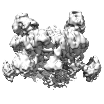
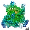
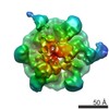
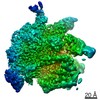
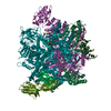
| Entry | Database: EMDB / ID: EMD-4554 | |||||||||
|---|---|---|---|---|---|---|---|---|---|---|
| Title | Truncated human R2TP complex, structure 1 | |||||||||
 Map data Map data | ||||||||||
 Sample Sample |
| |||||||||
| Biological species |  Homo sapiens (human) Homo sapiens (human) | |||||||||
| Method | single particle reconstruction / cryo EM / Resolution: 3.97 Å | |||||||||
 Authors Authors | Munoz-Hernandez H / Rodriguez CF / Llorca O | |||||||||
 Citation Citation | Journal: Sci Adv / Year: 2019 Title: Structural mechanism for regulation of the AAA-ATPases RUVBL1-RUVBL2 in the R2TP co-chaperone revealed by cryo-EM. Authors: Hugo Muñoz-Hernández / Mohinder Pal / Carlos F Rodríguez / Rafael Fernandez-Leiro / Chrisostomos Prodromou / Laurence H Pearl / Oscar Llorca /   Abstract: The human R2TP complex (RUVBL1-RUVBL2-RPAP3-PIH1D1) is an HSP90 co-chaperone required for the maturation of several essential multiprotein complexes, including RNA polymerase II, small nucleolar ...The human R2TP complex (RUVBL1-RUVBL2-RPAP3-PIH1D1) is an HSP90 co-chaperone required for the maturation of several essential multiprotein complexes, including RNA polymerase II, small nucleolar ribonucleoproteins, and PIKK complexes such as mTORC1 and ATR-ATRIP. RUVBL1-RUVBL2 AAA-ATPases are also primary components of other essential complexes such as INO80 and Tip60 remodelers. Despite recent efforts, the molecular mechanisms regulating RUVBL1-RUVBL2 in these complexes remain elusive. Here, we report cryo-EM structures of R2TP and show how access to the nucleotide-binding site of RUVBL2 is coupled to binding of the client recruitment component of R2TP (PIH1D1) to its DII domain. This interaction induces conformational rearrangements that lead to the destabilization of an N-terminal segment of RUVBL2 that acts as a gatekeeper to nucleotide exchange. This mechanism couples protein-induced motions of the DII domains with accessibility of the nucleotide-binding site in RUVBL1-RUVBL2, and it is likely a general mechanism shared with other RUVBL1-RUVBL2-containing complexes. | |||||||||
| History |
|
- Structure visualization
| Movie |
 Movie viewer Movie viewer |
|---|---|
| Structure viewer | EM map: SurfViewMolmilJmol/JSmol |
| Supplemental images |
- Downloads & links
-EMDB archive
| Map data | emd_4554.map.gz | 5.9 MB | EMDB map data format | |
|---|---|---|---|---|
| Header (meta data) | emd-4554-v30.xmlemd-4554.xml | 13.6 KB 13.6 KB | Display Display | EMDB header |
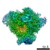
| Images |  emd_4554.png emd_4554.png | 64.3 KB | ||
| Archive directory |  http://ftp.pdbj.org/pub/emdb/structures/EMD-4554ftp://ftp.pdbj.org/pub/emdb/structures/EMD-4554 http://ftp.pdbj.org/pub/emdb/structures/EMD-4554ftp://ftp.pdbj.org/pub/emdb/structures/EMD-4554 | HTTPS FTP |
-Related structure data
| Related structure data |  4552C  4553C  4555C  4556C  4557C  6qi8C  6qi9C C: citing same article ( |
|---|---|
| Similar structure data |
-Links
| EMDB pages | EMDB (EBI/PDBe) / EMDataResource |
|---|
-Map
| File | Download / File: emd_4554.map.gz / Format: CCP4 / Size: 67 MB / Type: IMAGE STORED AS FLOATING POINT NUMBER (4 BYTES) | ||||||||||||||||||||||||||||||||||||||||||||||||||||||||||||||||||||
|---|---|---|---|---|---|---|---|---|---|---|---|---|---|---|---|---|---|---|---|---|---|---|---|---|---|---|---|---|---|---|---|---|---|---|---|---|---|---|---|---|---|---|---|---|---|---|---|---|---|---|---|---|---|---|---|---|---|---|---|---|---|---|---|---|---|---|---|---|---|
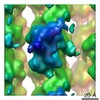
| Projections & slices | Image control
Images are generated by Spider. | ||||||||||||||||||||||||||||||||||||||||||||||||||||||||||||||||||||
| Voxel size | X=Y=Z: 1.07 Å | ||||||||||||||||||||||||||||||||||||||||||||||||||||||||||||||||||||
| Density |
| ||||||||||||||||||||||||||||||||||||||||||||||||||||||||||||||||||||
| Symmetry | Space group: 1 | ||||||||||||||||||||||||||||||||||||||||||||||||||||||||||||||||||||
| Details | EMDB XML:
CCP4 map header:
| ||||||||||||||||||||||||||||||||||||||||||||||||||||||||||||||||||||
 Z (Sec.)
 Y (Row.)
 X (Col.)


-Supplemental data
- Sample components
-Entire : Truncated human R2TP complex, structure 1
| Entire | Name: Truncated human R2TP complex, structure 1 |
|---|---|
| Components |
|
-Supramolecule #1: Truncated human R2TP complex, structure 1
| Supramolecule | Name: Truncated human R2TP complex, structure 1 / type: complex / ID: 1 / Parent: 0 / Macromolecule list: all |
|---|---|
| Source (natural) | Organism: Homo sapiens (human) |
| Recombinant expression | Organism:  |
-Macromolecule #1: RuvB-like 1
| Macromolecule | Name: RuvB-like 1 / type: protein_or_peptide / ID: 1 / Enantiomer: LEVO |
|---|---|
| Source (natural) | Organism: Homo sapiens (human) |
| Recombinant expression | Organism: |
| Sequence | String: MKIEEVKSTT KTQRIASHSH VKGLGLDESG LAKQAASGLV GQENAREACG VIVELIKSKK MAGRAVLLA GPPGTGKTAL ALAIAQELGS KVPFCPMVGS EVYSTEIKKT EVLMENFRRA I GLRIKETK EVYEGEVTEL TPCETENPMG GYGKTISHVI IGLKTAKGTK ...String: MKIEEVKSTT KTQRIASHSH VKGLGLDESG LAKQAASGLV GQENAREACG VIVELIKSKK MAGRAVLLA GPPGTGKTAL ALAIAQELGS KVPFCPMVGS EVYSTEIKKT EVLMENFRRA I GLRIKETK EVYEGEVTEL TPCETENPMG GYGKTISHVI IGLKTAKGTK QLKLDPSIFE SL QKERVEA GDVIYIEANS GAVKRQGRCD TYATEFDLEA EEYVPLPKGD VHKKKEIIQD VTL HDLDVA NARPQGGQDI LSMMGQLMKP KKTEITDKLR GEINKVVNKY IDQGIAELVP GVLF VDEVH MLDIECFTYL HRALESSIAP IVIFASNRGN CVIRGTEDIT SPHGIPLDLL DRVMI IRTM LYTPQEMKQI IKIRAQTEGI NISEEALNHL GEIGTKTTLR YSVQLLTPAN LLAKIN GKD SIEKEHVEEI SELFYDAKSS AKILADQQDK YMK |
-Macromolecule #2: RuvB-like 2
| Macromolecule | Name: RuvB-like 2 / type: protein_or_peptide / ID: 2 / Enantiomer: LEVO |
|---|---|
| Source (natural) | Organism: Homo sapiens (human) |
| Recombinant expression | Organism: |
| Sequence | String: MATVTATTKV PEIRDVTRIE RIGAHSHIRG LGLDDALEPR QASQGMVGQL AARRAAGVVL EMIREGKIA GRAVLIAGQP GTGKTAIAMG MAQALGPDTP FTAIAGSEIF SLEMSKTEAL T QAFRRSIG VRIKEETEII EGEVVEIQID RPATGTGSKV GKLTLKTTEM ...String: MATVTATTKV PEIRDVTRIE RIGAHSHIRG LGLDDALEPR QASQGMVGQL AARRAAGVVL EMIREGKIA GRAVLIAGQP GTGKTAIAMG MAQALGPDTP FTAIAGSEIF SLEMSKTEAL T QAFRRSIG VRIKEETEII EGEVVEIQID RPATGTGSKV GKLTLKTTEM ETIYDLGTKM IE SLTKDKV QAGDVITIDK ATGKISKLGR SFTRARDYDA MGSQTKFVQC PDGELQKRKE VVH TVSLHE IDVINSRTQG FLALFSGDTG EIKSEVREQI NAKVAEWREE GKAEIIPGVL FIDE VHMLD IESFSFLNRA LESDMAPVLI MATNRGITRI RGTSYQSPHG IPIDLLDRLL IVSTT PYSE KDTKQILRIR CEEEDVEMSE DAYTVLTRIG LETSLRYAIQ LITAASLVCR KRKGTE VQV DDIKRVYSLF LDESRSTQYM KEYQDAFLFN ELKGETMDTS |
-Macromolecule #3: PIH1 domain-containing protein 1
| Macromolecule | Name: PIH1 domain-containing protein 1 / type: protein_or_peptide / ID: 3 / Enantiomer: LEVO |
|---|---|
| Source (natural) | Organism: Homo sapiens (human) |
| Recombinant expression | Organism: |
| Sequence | String: MANPKLLGMG LSEAEAIGAD SARFEELLLQ ASKELQQAQT TRPESTQIQP QPGFCIKTNS SEGKVFINI CHSPSIPPPA DVTEEELLQM LEEDQAGFRI PMSLGEPHAE LDAKGQGCTA Y DVAVNSDF YRRMQNSDFL RELVITIARE GLEDKYNLQL NPEWRMMKNR ...String: MANPKLLGMG LSEAEAIGAD SARFEELLLQ ASKELQQAQT TRPESTQIQP QPGFCIKTNS SEGKVFINI CHSPSIPPPA DVTEEELLQM LEEDQAGFRI PMSLGEPHAE LDAKGQGCTA Y DVAVNSDF YRRMQNSDFL RELVITIARE GLEDKYNLQL NPEWRMMKNR PFMGSISQQN IR SEQRPRI QELGDLYTPA PGRAESGPEK PHLNLWLEAP DLLLAEVDLP KLDGALGLSL EIG ENRLVM GGPQQLYHLD AYIPLQINSH ESKAAFHRKR KQLMVAMPLL PVPS |
-Macromolecule #4: RNA polymerase II-associated protein 3
| Macromolecule | Name: RNA polymerase II-associated protein 3 / type: protein_or_peptide / ID: 4 / Enantiomer: LEVO |
|---|---|
| Source (natural) | Organism: Homo sapiens (human) |
| Recombinant expression | Organism: |
| Sequence | String: KGHWDDVFLD STQRQNVVKP IDNPPHPGST KPLKKVIIEE TGNLIQTIDV PDSTTAAAPE NNPINLANV IAATGTTSKK NSSQDDLFPT SDTPRAKVLK IEEVSDTSSL QPQASLKQDV C QSYSEKMP IEIEQKPAQF ATTVLPPIPA NSFQLESDFR QLKSSPDMLY ...String: KGHWDDVFLD STQRQNVVKP IDNPPHPGST KPLKKVIIEE TGNLIQTIDV PDSTTAAAPE NNPINLANV IAATGTTSKK NSSQDDLFPT SDTPRAKVLK IEEVSDTSSL QPQASLKQDV C QSYSEKMP IEIEQKPAQF ATTVLPPIPA NSFQLESDFR QLKSSPDMLY QYLKQIEPSL YP KLFQKNL DPDVFNQIVK ILHDFYIEKE KPLLIFEILQ RLSELKRFDM AVMFMSETEK KIA RALFNH IDKSGLKDSS VEELKKRYGG |
-Experimental details
-Structure determination
| Method | cryo EM |
|---|---|
 Processing Processing | single particle reconstruction |
| Aggregation state | particle |
-Sample preparation
| Buffer | pH: 7.8 |
|---|---|
| Grid | Model: Quantifoil R1.2/1.3 / Material: COPPER / Mesh: 300 |
| Vitrification | Cryogen name: ETHANE / Chamber humidity: 100 % / Chamber temperature: 277 K / Instrument: FEI VITROBOT MARK IV |
- Electron microscopy
| Microscope | FEI TITAN KRIOS |
|---|---|
| Image recording | Film or detector model: GATAN K2 SUMMIT (4k x 4k) / Detector mode: COUNTING / Average electron dose: 1.18 e/Å2 |
| Electron beam | Acceleration voltage: 300 kV / Electron source:  FIELD EMISSION GUN FIELD EMISSION GUN |
| Electron optics | Illumination mode: FLOOD BEAM / Imaging mode: BRIGHT FIELD |
| Experimental equipment |  Model: Titan Krios / Image courtesy: FEI Company |
+Image processing
-Atomic model buiding 1
| Initial model | PDB ID: |
|---|---|
| Refinement | Space: REAL / Protocol: RIGID BODY FIT |

